 Movie
Movie Controller

[English] 日本語
 Yorodumi
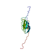
- PDB-5j7n: Crystal structure of a small heat-shock protein from Xylella fast... -
+ Open data

- Basic information
| Entry | Database: PDB / ID: 5j7n | ||||||
|---|---|---|---|---|---|---|---|
| Title | Crystal structure of a small heat-shock protein from Xylella fastidiosa reveals a distinct high order structure | ||||||
 Components Components | Low molecular weight heat shock protein | ||||||
 Keywords Keywords |  CHAPERONE / Small heat shock protein / Xylella fastidiosa / alpha-crystallin domain / citrus variegated chlorosis CHAPERONE / Small heat shock protein / Xylella fastidiosa / alpha-crystallin domain / citrus variegated chlorosis | ||||||
| Function / homology |  Function and homology information Function and homology information: / Immunoglobulin-like - #790 / Hsp20/alpha crystallin family / Small heat shock protein (sHSP) domain profile. / Alpha crystallin/Hsp20 domain / HSP20-like chaperone / Immunoglobulin-like / Sandwich / Mainly BetaSimilarity search - Domain/homology | ||||||
| Biological species |  Xylella fastidiosa (bacteria) Xylella fastidiosa (bacteria) | ||||||
| Method | X-RAY DIFFRACTION / SYNCHROTRON / MOLECULAR REPLACEMENT / Resolution: 2.9 Å | ||||||
 Authors Authors | Fonseca, E.M.B. / Scorsato, V. / dos Santos, C.A. / Tomazini Jr., A. / Aparicio, R. / Polikarpov, I. | ||||||
 Citation Citation | Journal: Acta Crystallogr F Struct Biol Commun / Year: 2017 Title: Crystal structure of a small heat-shock protein from Xylella fastidiosa reveals a distinct high-order structure. Authors: Fonseca, E.M. / Scorsato, V. / Dos Santos, M.L. / Junior, A.T. / Tada, S.F. / Dos Santos, C.A. / de Toledo, M.A. / de Souza, A.P. / Polikarpov, I. / Aparicio, R. | ||||||
| History |
|
- Structure visualization
| Structure viewer | Molecule: MolmilJmol/JSmol |
|---|
- Downloads & links
-Download
| PDBx/mmCIF format | 5j7n.cif.gz | 58 KB | Display | PDBx/mmCIF format |
|---|---|---|---|---|
| PDB format | pdb5j7n.ent.gz | 41.9 KB | Display | PDB format |
| PDBx/mmJSON format | 5j7n.json.gz | Tree view | PDBx/mmJSON format | |
| Others |  Other downloads Other downloads |
-Validation report
| Arichive directory | https://data.pdbj.org/pub/pdb/validation_reports/j7/5j7nftp://data.pdbj.org/pub/pdb/validation_reports/j7/5j7n | HTTPS FTP |
|---|
-Related structure data
| Related structure data |  3glaS S: Starting model for refinement |
|---|---|
| Similar structure data |


-Links
 PDBj
PDBj


- Assembly
| Deposited unit | 
| ||||||||
|---|---|---|---|---|---|---|---|---|---|
| 1 |
| ||||||||
| 2 | 
| ||||||||
| 3 | 
| ||||||||
| Unit cell |
|
-Components
| #1: Protein | Mass: 17880.926 Da / Num. of mol.: 1 Source method: isolated from a genetically manipulated source Source: (gene. exp.) Xylella fastidiosa (strain 9a5c) (bacteria)Strain: 9a5c / Gene: XF_2234 / Production host: Escherichia coli (E. coli) / References: UniProt: Q9PBB0 |
|---|
-Experimental details
-Experiment
| Experiment | Method: X-RAY DIFFRACTION / Number of used crystals: 1 |
|---|
- Sample preparation
| Crystal | Density Matthews: 2.41 Å3/Da / Density % sol: 48.93 % |
|---|---|
| Crystal grow | Temperature: 291 K / Method: vapor diffusion, hanging drop Details: 0.1 M HEPES sodium salt pH 7.5, 0.3 M sodium citrate, 20% 2-propanol, 15% ethylene glycol |
-Data collection
| Diffraction | Mean temperature: 100 K |
|---|---|
| Diffraction source | Source: SYNCHROTRON / Site: LNLS  / Beamline: W01B-MX2 / Wavelength: 1.4586 Å / Beamline: W01B-MX2 / Wavelength: 1.4586 Å |
| Detector | Type: MARMOSAIC 225 mm CCD / Detector: CCD / Date: Sep 7, 2009 |
| Radiation | Protocol: SINGLE WAVELENGTH / Monochromatic (M) / Laue (L): M / Scattering type: x-ray |
| Radiation wavelength | Wavelength: 1.4586 Å / Relative weight: 1 |
| Reflection | Resolution: 2.9→68.94 Å / Num. obs: 4089 / % possible obs: 98 % / Redundancy: 18.6 % / Rmerge(I) obs: 0.115 / Net I/σ(I): 17.3 |
| Reflection shell | Resolution: 2.9→3 Å / Redundancy: 19.3 % / Rmerge(I) obs: 0.673 / Mean I/σ(I) obs: 3.5 / % possible all: 97.5 |
- Processing
| Software |
| |||||||||||||||||||||||||||||||||||||||||||||||||||||||||||||||||||||||||||||||||||||||||||||||||||||||||||||||||||||||||||||
|---|---|---|---|---|---|---|---|---|---|---|---|---|---|---|---|---|---|---|---|---|---|---|---|---|---|---|---|---|---|---|---|---|---|---|---|---|---|---|---|---|---|---|---|---|---|---|---|---|---|---|---|---|---|---|---|---|---|---|---|---|---|---|---|---|---|---|---|---|---|---|---|---|---|---|---|---|---|---|---|---|---|---|---|---|---|---|---|---|---|---|---|---|---|---|---|---|---|---|---|---|---|---|---|---|---|---|---|---|---|---|---|---|---|---|---|---|---|---|---|---|---|---|---|---|---|---|
| Refinement | Method to determine structure: MOLECULAR REPLACEMENT Starting model: 3GLA Resolution: 2.9→40.452 Å / SU ML: 0 / Cross valid method: THROUGHOUT / σ(F): 1.39 / Phase error: 20.33 / Stereochemistry target values: ML
| |||||||||||||||||||||||||||||||||||||||||||||||||||||||||||||||||||||||||||||||||||||||||||||||||||||||||||||||||||||||||||||
| Solvent computation | Shrinkage radii: 0.9 Å / VDW probe radii: 1.11 Å / Solvent model: FLAT BULK SOLVENT MODEL | |||||||||||||||||||||||||||||||||||||||||||||||||||||||||||||||||||||||||||||||||||||||||||||||||||||||||||||||||||||||||||||
| Refinement step | Cycle: LAST / Resolution: 2.9→40.452 Å
| |||||||||||||||||||||||||||||||||||||||||||||||||||||||||||||||||||||||||||||||||||||||||||||||||||||||||||||||||||||||||||||
| Refine LS restraints |
| |||||||||||||||||||||||||||||||||||||||||||||||||||||||||||||||||||||||||||||||||||||||||||||||||||||||||||||||||||||||||||||
| Refinement TLS params. | Method: refined / Refine-ID: X-RAY DIFFRACTION
| |||||||||||||||||||||||||||||||||||||||||||||||||||||||||||||||||||||||||||||||||||||||||||||||||||||||||||||||||||||||||||||
| Refinement TLS group |
|
