 Movie
Movie Controller

[English] 日本語
 Yorodumi
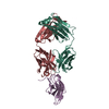
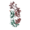
- PDB-5fcu: CRYSTAL STRUCTURE OF THE INNER DOMAIN OF CLADE A/E HIV-1 GP120 IN... -
+ Open data

- Basic information
| Entry | Database: PDB / ID: 5fcu | ||||||
|---|---|---|---|---|---|---|---|
| Title | CRYSTAL STRUCTURE OF THE INNER DOMAIN OF CLADE A/E HIV-1 GP120 IN COMPLEX WITH THE ADCC-POTENT RHESUS MACAQUE ANTIBODY JR4 | ||||||
 Components Components |
| ||||||
 Keywords Keywords |  VIRAL PROTEIN/IMMUNE SYSTEM / HIV-1 GP120 / VIRAL PROTEIN / VIRAL PROTEIN-IMMUNE SYSTEM COMPLEX VIRAL PROTEIN/IMMUNE SYSTEM / HIV-1 GP120 / VIRAL PROTEIN / VIRAL PROTEIN-IMMUNE SYSTEM COMPLEX | ||||||
| Function / homology | Gp120 core superfamily / Envelope glycoprotein GP120 / Human immunodeficiency virus 1, envelope glycoprotein Gp120 / viral envelope / Immunoglobulins / Immunoglobulin-like / Sandwich / Mainly Beta / clade A/E 93TH057 HIV-1 gp120 core Function and homology information Function and homology information | ||||||
| Biological species |  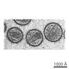 Human immunodeficiency virus 1 Human immunodeficiency virus 1 Macaca mulatta (Rhesus monkey) Macaca mulatta (Rhesus monkey) | ||||||
| Method | X-RAY DIFFRACTION / SYNCHROTRON / MOLECULAR REPLACEMENT / Resolution: 1.85 Å | ||||||
 Authors Authors | Gohain, N. / Tolbert, W.D. / Pazgier, M. | ||||||
| Funding support |  United States, 1items United States, 1items
| ||||||
 Citation Citation | Journal: Structure / Year: 2016 Title: Paring Down HIV Env: Design and Crystal Structure of a Stabilized Inner Domain of HIV-1 gp120 Displaying a Major ADCC Target of the A32 Region. Authors: Tolbert, W.D. / Gohain, N. / Veillette, M. / Chapleau, J.P. / Orlandi, C. / Visciano, M.L. / Ebadi, M. / DeVico, A.L. / Fouts, T.R. / Finzi, A. / Lewis, G.K. / Pazgier, M. | ||||||
| History |
|
- Structure visualization
| Structure viewer | Molecule: MolmilJmol/JSmol |
|---|
- Downloads & links
-Download
| PDBx/mmCIF format | 5fcu.cif.gz | 232 KB | Display | PDBx/mmCIF format |
|---|---|---|---|---|
| PDB format | pdb5fcu.ent.gz | 181.7 KB | Display | PDB format |
| PDBx/mmJSON format | 5fcu.json.gz | Tree view | PDBx/mmJSON format | |
| Others |  Other downloads Other downloads |
-Validation report
| Arichive directory | https://data.pdbj.org/pub/pdb/validation_reports/fc/5fcuftp://data.pdbj.org/pub/pdb/validation_reports/fc/5fcu | HTTPS FTP |
|---|
-Related structure data
| Related structure data |  4yblC  4yc2C  2rfeS S: Starting model for refinement C: citing same article ( |
|---|---|
| Similar structure data |


-Links
 PDBj
PDBj


- Assembly
| Deposited unit | 
| ||||||||
|---|---|---|---|---|---|---|---|---|---|
| 1 |
| ||||||||
| Unit cell |
| ||||||||
| Components on special symmetry positions |
|
-Components
-Antibody , 2 types, 2 molecules HL
| #2: Antibody | Mass: 24847.766 Da / Num. of mol.: 1 Source method: isolated from a genetically manipulated source Source: (gene. exp.) Macaca mulatta (Rhesus monkey) / Cell line (production host): HEK 293 / Production host:  HOMO SAPIENS (human) HOMO SAPIENS (human) |
|---|---|
| #3: Antibody | Mass: 22719.137 Da / Num. of mol.: 1 Source method: isolated from a genetically manipulated source Source: (gene. exp.) Macaca mulatta (Rhesus monkey) / Cell line (production host): HEK 293 / Production host: HOMO SAPIENS (human) |
-Protein / Sugars , 2 types, 2 molecules G
| #1: Protein | Mass: 18802.348 Da / Num. of mol.: 1 / Fragment: UNP residues 1-140 Source method: isolated from a genetically manipulated source Source: (gene. exp.) Human immunodeficiency virus 1 / Gene: HIV-1 Env / Cell line (production host): HEK 293 / Production host: HOMO SAPIENS (human) / References: UniProt: A0A0M3KKW9 |
|---|---|
| #4: Sugar | ChemComp-NAG / N-Acetylglucosamine Type: D-saccharide, beta linking / Mass: 221.208 Da / Num. of mol.: 1 Type: D-saccharide, beta linking / Mass: 221.208 Da / Num. of mol.: 1Source method: isolated from a genetically manipulated source Formula: C8H15NO6 |
-Non-polymers , 3 types, 382 molecules 


| #5: Chemical | Sulfate Mass: 96.063 Da / Num. of mol.: 3 / Source method: obtained synthetically / Formula: SO4 Mass: 96.063 Da / Num. of mol.: 3 / Source method: obtained synthetically / Formula: SO4#6: Chemical | Chloride Mass: 35.453 Da / Num. of mol.: 3 / Source method: obtained synthetically / Formula: Cl Mass: 35.453 Da / Num. of mol.: 3 / Source method: obtained synthetically / Formula: Cl#7: Water | ChemComp-HOH / | WaterMass: 18.015 Da / Num. of mol.: 376 / Source method: isolated from a natural source / Formula: H2O |
|---|
-Experimental details
-Experiment
| Experiment | Method: X-RAY DIFFRACTION / Number of used crystals: 1 |
|---|
- Sample preparation
| Crystal | Density Matthews: 2.4 Å3/Da / Density % sol: 48.75 % |
|---|---|
| Crystal grow | Temperature: 294 K / Method: vapor diffusion, hanging drop / pH: 7.5 Details: 20% PEG MONOMETHYL ETHER 5000, 0.2M AMMONIUM SULFATE, 0.1M TRIS-HCL PH 7.5, VAPOR DIFFUSION, HANGING DROP, TEMPERATURE 294K PH range: 7.5 |
-Data collection
| Diffraction | Mean temperature: 100 K |
|---|---|
| Diffraction source | Source: SYNCHROTRON / Site: SSRL / Beamline: BL12-2 / Wavelength: 0.9795 Å |
| Detector | Type: DECTRIS PILATUS 6M / Detector: PIXEL / Date: Jan 31, 2014 / Details: RH COATED FLAT MIRROR |
| Radiation | Monochromator: SI(111) / Protocol: SINGLE WAVELENGTH / Monochromatic (M) / Laue (L): M / Scattering type: x-ray |
| Radiation wavelength | Wavelength: 0.9795 Å / Relative weight: 1 |
| Reflection | Resolution: 1.85→50 Å / Num. obs: 51569 / % possible obs: 94.9 % / Observed criterion σ(I): 0 / Redundancy: 5.8 % / Rmerge(I) obs: 0.107 / Net I/σ(I): 15 |
| Reflection shell | Resolution: 1.85→1.88 Å / Redundancy: 5.7 % / Rmerge(I) obs: 1 / Mean I/σ(I) obs: 1.1 / % possible all: 91.4 |
- Processing
| Software |
| ||||||||||||||||||||||||||||||||||||||||||||||||||||||||||||||||||||||||||||||||||||||||||||||||||||||||||||||||||||||||||||||||||||||||||||||||||||||||||||||||||||||||||||||||||||||
|---|---|---|---|---|---|---|---|---|---|---|---|---|---|---|---|---|---|---|---|---|---|---|---|---|---|---|---|---|---|---|---|---|---|---|---|---|---|---|---|---|---|---|---|---|---|---|---|---|---|---|---|---|---|---|---|---|---|---|---|---|---|---|---|---|---|---|---|---|---|---|---|---|---|---|---|---|---|---|---|---|---|---|---|---|---|---|---|---|---|---|---|---|---|---|---|---|---|---|---|---|---|---|---|---|---|---|---|---|---|---|---|---|---|---|---|---|---|---|---|---|---|---|---|---|---|---|---|---|---|---|---|---|---|---|---|---|---|---|---|---|---|---|---|---|---|---|---|---|---|---|---|---|---|---|---|---|---|---|---|---|---|---|---|---|---|---|---|---|---|---|---|---|---|---|---|---|---|---|---|---|---|---|---|
| Refinement | Method to determine structure: MOLECULAR REPLACEMENT Starting model: 2RFE Resolution: 1.85→50 Å / Cor.coef. Fo:Fc: 0.962 / Cor.coef. Fo:Fc free: 0.946 / SU B: 7.555 / SU ML: 0.113 / Cross valid method: THROUGHOUT / σ(F): 0 / ESU R: 0.134 / ESU R Free: 0.129 / Stereochemistry target values: MAXIMUM LIKELIHOOD / Details: HYDROGENS HAVE BEEN ADDED IN THE RIDING POSITIONS
| ||||||||||||||||||||||||||||||||||||||||||||||||||||||||||||||||||||||||||||||||||||||||||||||||||||||||||||||||||||||||||||||||||||||||||||||||||||||||||||||||||||||||||||||||||||||
| Solvent computation | Ion probe radii: 0.8 Å / Shrinkage radii: 0.8 Å / VDW probe radii: 1.2 Å / Solvent model: MASK | ||||||||||||||||||||||||||||||||||||||||||||||||||||||||||||||||||||||||||||||||||||||||||||||||||||||||||||||||||||||||||||||||||||||||||||||||||||||||||||||||||||||||||||||||||||||
| Displacement parameters | Biso mean: 44.5 Å2
| ||||||||||||||||||||||||||||||||||||||||||||||||||||||||||||||||||||||||||||||||||||||||||||||||||||||||||||||||||||||||||||||||||||||||||||||||||||||||||||||||||||||||||||||||||||||
| Refinement step | Cycle: LAST / Resolution: 1.85→50 Å
| ||||||||||||||||||||||||||||||||||||||||||||||||||||||||||||||||||||||||||||||||||||||||||||||||||||||||||||||||||||||||||||||||||||||||||||||||||||||||||||||||||||||||||||||||||||||
| Refine LS restraints |
|