 Movie
Movie Controller

[English] 日本語
 Yorodumi
- PDB-3vhu: Mineralocorticoid receptor ligand-binding domain with spironolactone -
+ Open data

- Basic information
| Entry | Database: PDB / ID: 3vhu | ||||||
|---|---|---|---|---|---|---|---|
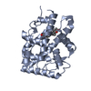
| Title | Mineralocorticoid receptor ligand-binding domain with spironolactone | ||||||
 Components Components | Mineralocorticoid receptor | ||||||
 Keywords Keywords | TRANSCRIPTION/INHIBITOR / NUCLEAR RECEPTOR / TRANSCRIPTION FACTOR / ACTIVATING MUTATION / HYPERTENSION / ANTAGONIST / SPIRONOLACTONE / TRANSCRIPTION-INHIBITOR complex | ||||||
| Function / homology |  Function and homology information Function and homology informationnuclear steroid receptor activity / estrogen response element binding / intracellular steroid hormone receptor signaling pathway / HSP90 chaperone cycle for steroid hormone receptors (SHR) in the presence of ligand / TBP-class protein binding / steroid binding / SUMOylation of intracellular receptors / Nuclear Receptor transcription pathway / positive regulation of non-canonical NF-kappaB signal transduction / nuclear receptor activity ...nuclear steroid receptor activity / estrogen response element binding / intracellular steroid hormone receptor signaling pathway / HSP90 chaperone cycle for steroid hormone receptors (SHR) in the presence of ligand / TBP-class protein binding / steroid binding / SUMOylation of intracellular receptors / Nuclear Receptor transcription pathway / positive regulation of non-canonical NF-kappaB signal transduction / nuclear receptor activity / sequence-specific double-stranded DNA binding / receptor complex / DNA-binding transcription factor activity, RNA polymerase II-specific / DNA-binding transcription factor activity / chromatin / endoplasmic reticulum membrane / regulation of transcription by RNA polymerase II / signal transduction / zinc ion binding / nucleoplasm / cytosolSimilarity search - Function | ||||||
| Biological species |  Homo sapiens (human) Homo sapiens (human) | ||||||
| Method | X-RAY DIFFRACTION / SYNCHROTRON / MOLECULAR REPLACEMENT / Resolution: 2.11 Å | ||||||
 Authors Authors | Sogabe, S. / Habuka, N. | ||||||
 Citation Citation | Journal: J.Med.Chem. / Year: 2011 Title: Identification of Benzoxazin-3-one Derivatives as Novel, Potent, and Selective Nonsteroidal Mineralocorticoid Receptor Antagonists Authors: Hasui, T. / Matsunaga, N. / Ora, T. / Ohyabu, N. / Nishigaki, N. / Imura, Y. / Igata, Y. / Matsui, H. / Motoyaji, T. / Tanaka, T. / Habuka, N. / Sogabe, S. / Ono, M. / Siedem, C.S. / Tang, T. ...Authors: Hasui, T. / Matsunaga, N. / Ora, T. / Ohyabu, N. / Nishigaki, N. / Imura, Y. / Igata, Y. / Matsui, H. / Motoyaji, T. / Tanaka, T. / Habuka, N. / Sogabe, S. / Ono, M. / Siedem, C.S. / Tang, T.P. / Gauthier, C. / De Meese, L.A. / Boyd, S.A. / Fukumoto, S. | ||||||
| History |
|
- Structure visualization
| Structure viewer | Molecule: MolmilJmol/JSmol |
|---|
- Downloads & links
-Download
| PDBx/mmCIF format | 3vhu.cif.gz | 121.4 KB | Display | PDBx/mmCIF format |
|---|---|---|---|---|
| PDB format | pdb3vhu.ent.gz | 93.5 KB | Display | PDB format |
| PDBx/mmJSON format | 3vhu.json.gz | Tree view | PDBx/mmJSON format | |
| Others |  Other downloads Other downloads |
-Validation report
| Arichive directory | https://data.pdbj.org/pub/pdb/validation_reports/vh/3vhuftp://data.pdbj.org/pub/pdb/validation_reports/vh/3vhu | HTTPS FTP |
|---|
-Related structure data
| Related structure data |  3vhvC  2ab2S S: Starting model for refinement C: citing same article ( |
|---|---|
| Similar structure data |


-Links
 PDBj
PDBj

- Assembly
| Deposited unit | 
| ||||||||
|---|---|---|---|---|---|---|---|---|---|
| 1 |
| ||||||||
| Unit cell |
|
-Components
| #1: Protein | / MR / Nuclear receptor subfamily 3 group C member 2 Mass: 34006.219 Da / Num. of mol.: 1 / Fragment: LIGAND-BINDING DOMAIN, UNP residues 712-984 / Mutation: C808S, S810L Source method: isolated from a genetically manipulated source Source: (gene. exp.) Homo sapiens (human) / Gene: NR3C2, MCR, MLR / Production host:  Escherichia coli (E. coli) / References: UniProt: P08235 Escherichia coli (E. coli) / References: UniProt: P08235 |
|---|---|
| #2: Chemical | ChemComp-SNL / Spironolactone  Mass: 416.573 Da / Num. of mol.: 1 / Source method: obtained synthetically / Formula: C24H32O4S / Comment: medication*YM Mass: 416.573 Da / Num. of mol.: 1 / Source method: obtained synthetically / Formula: C24H32O4S / Comment: medication*YM |
| #3: Water | ChemComp-HOH / Water Mass: 18.015 Da / Num. of mol.: 62 / Source method: isolated from a natural source / Formula: H2O Mass: 18.015 Da / Num. of mol.: 62 / Source method: isolated from a natural source / Formula: H2O |
-Experimental details
-Experiment
| Experiment | Method: X-RAY DIFFRACTION / Number of used crystals: 1 |
|---|
- Sample preparation
| Crystal | Density Matthews: 2.44 Å3/Da / Density % sol: 49.67 % |
|---|---|
| Crystal grow | Temperature: 293 K / Method: vapor diffusion, sitting drop / pH: 7 Details: 0.1M HEPES pH 7.0, 1.26M Lithium sulfate, 6% PEG MME 2000, VAPOR DIFFUSION, SITTING DROP, temperature 293K |
-Data collection
| Diffraction | Mean temperature: 100 K |
|---|---|
| Diffraction source | Source: SYNCHROTRON / Site: ALS  / Beamline: 5.0.3 / Wavelength: 1 Å / Beamline: 5.0.3 / Wavelength: 1 Å |
| Detector | Type: ADSC QUANTUM 210r / Detector: CCD / Date: Nov 19, 2005 |
| Radiation | Monochromator: Si(111) / Protocol: SINGLE WAVELENGTH / Monochromatic (M) / Laue (L): M / Scattering type: x-ray |
| Radiation wavelength | Wavelength: 1 Å / Relative weight: 1 |
| Reflection | Resolution: 2.11→50 Å / Num. obs: 19366 / % possible obs: 98.6 % / Observed criterion σ(I): 1 / Redundancy: 6 % / Rsym value: 0.059 / Net I/σ(I): 26.2 |
| Reflection shell | Resolution: 2.11→2.2 Å / Mean I/σ(I) obs: 2.4 / Rsym value: 0.515 / % possible all: 90.3 |
- Processing
| Software |
| |||||||||||||||||||||||||||||||||||||||||||||||||||||||||||||||||
|---|---|---|---|---|---|---|---|---|---|---|---|---|---|---|---|---|---|---|---|---|---|---|---|---|---|---|---|---|---|---|---|---|---|---|---|---|---|---|---|---|---|---|---|---|---|---|---|---|---|---|---|---|---|---|---|---|---|---|---|---|---|---|---|---|---|---|
| Refinement | Method to determine structure: MOLECULAR REPLACEMENT Starting model: PDB ENTRY 2AB2 Resolution: 2.11→40 Å / Cor.coef. Fo:Fc: 0.961 / Cor.coef. Fo:Fc free: 0.94 / SU B: 11.761 / SU ML: 0.136 / Cross valid method: THROUGHOUT / ESU R Free: 0.181 / Stereochemistry target values: MAXIMUM LIKELIHOOD / Details: HYDROGENS HAVE BEEN ADDED IN THE RIDING POSITIONS
| |||||||||||||||||||||||||||||||||||||||||||||||||||||||||||||||||
| Solvent computation | Ion probe radii: 0.8 Å / Shrinkage radii: 0.8 Å / VDW probe radii: 1.4 Å / Solvent model: MASK | |||||||||||||||||||||||||||||||||||||||||||||||||||||||||||||||||
| Displacement parameters | Biso mean: 52.646 Å2
| |||||||||||||||||||||||||||||||||||||||||||||||||||||||||||||||||
| Refinement step | Cycle: LAST / Resolution: 2.11→40 Å
| |||||||||||||||||||||||||||||||||||||||||||||||||||||||||||||||||
| Refine LS restraints |
| |||||||||||||||||||||||||||||||||||||||||||||||||||||||||||||||||
| LS refinement shell | Resolution: 2.109→2.164 Å / Total num. of bins used: 20
| |||||||||||||||||||||||||||||||||||||||||||||||||||||||||||||||||
| Refinement TLS params. | Method: refined / Origin x: 24.6682 Å / Origin y: 21.9858 Å / Origin z: 3.0734 Å
| |||||||||||||||||||||||||||||||||||||||||||||||||||||||||||||||||
| Refinement TLS group |
|
