 Movie
Movie Controller


+ Open data

- Basic information
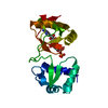
| Entry | Database: PDB / ID: 3nti | ||||||
|---|---|---|---|---|---|---|---|
| Title | Crystal structure of Tudor and Aubergine [R15(me2s)] complex | ||||||
 Components Components |
| ||||||
 Keywords Keywords |  TRANSCRIPTION / Tudor domain / OB-fold / germ cell formation TRANSCRIPTION / Tudor domain / OB-fold / germ cell formation | ||||||
| Function / homology |  Function and homology information Function and homology informationpositive regulation of oskar mRNA translation / pole cell development / maintenance of pole plasm mRNA location / positive regulation of post-transcriptional gene silencing by RNA / regulation of oskar mRNA translation / retrotransposon silencing by mRNA destabilization / P granule assembly / pole plasm protein localization / oocyte karyosome formation / methylation-dependent protein binding ...positive regulation of oskar mRNA translation / pole cell development / maintenance of pole plasm mRNA location / positive regulation of post-transcriptional gene silencing by RNA / regulation of oskar mRNA translation / retrotransposon silencing by mRNA destabilization / P granule assembly / pole plasm protein localization / oocyte karyosome formation / methylation-dependent protein binding / pole plasm / secondary piRNA processing / regulation of pole plasm oskar mRNA localization / P granule organization / piRNA binding / pole plasm assembly / segmentation / RNA endonuclease activity, producing 5'-phosphomonoesters / pole cell formation / dorsal appendage formation / piRNA processing / global gene silencing by mRNA cleavage / intracellular mRNA localization / regulatory ncRNA-mediated post-transcriptional gene silencing / retrotransposon silencing / oocyte maturation / regulatory ncRNA-mediated gene silencing / P granule / positive regulation of innate immune response / mitotic chromosome condensation / positive regulation of nuclear-transcribed mRNA poly(A) tail shortening / oogenesis / germ cell development / heterochromatin formation / RNA endonuclease activity / spermatogenesis / defense response to Gram-negative bacterium / mitochondrion / nucleus / cytosol / cytoplasmSimilarity search - Function | ||||||
| Biological species |  Drosophila melanogaster (fruit fly) Drosophila melanogaster (fruit fly) | ||||||
| Method | X-RAY DIFFRACTION / SYNCHROTRON / MOLECULAR REPLACEMENT / Resolution: 2.8 Å | ||||||
 Authors Authors | Liu, H.P. / Huang, Y. / Li, Z.Z. / Gong, W.M. / Xu, R.M. | ||||||
 Citation Citation | Journal: Genes Dev. / Year: 2010 Title: Structural basis for methylarginine-dependent recognition of Aubergine by Tudor Authors: Liu, H.P. / Wang, J.Y. / Huang, Y. / Li, Z.Z. / Gong, W.M. / Lehmann, R. / Xu, R.M. | ||||||
| History |
|
- Structure visualization
| Structure viewer | Molecule: MolmilJmol/JSmol |
|---|
- Downloads & links
-Download
| PDBx/mmCIF format | 3nti.cif.gz | 48 KB | Display | PDBx/mmCIF format |
|---|---|---|---|---|
| PDB format | pdb3nti.ent.gz | 32.6 KB | Display | PDB format |
| PDBx/mmJSON format | 3nti.json.gz | Tree view | PDBx/mmJSON format | |
| Others |  Other downloads Other downloads |
-Validation report
| Arichive directory | https://data.pdbj.org/pub/pdb/validation_reports/nt/3ntiftp://data.pdbj.org/pub/pdb/validation_reports/nt/3nti | HTTPS FTP |
|---|
-Related structure data
| Related structure data |  3nthC  3ntkSC C: citing same article ( S: Starting model for refinement |
|---|---|
| Similar structure data |


-Links
 PDBj
PDBj
- Assembly
| Deposited unit | 
| ||||||||
|---|---|---|---|---|---|---|---|---|---|
| 1 |
| ||||||||
| Unit cell |
|
-Components
| #1: Protein | Mother Mass: 19568.021 Da / Num. of mol.: 1 / Fragment: the last extended Tudor domain Source method: isolated from a genetically manipulated source Source: (gene. exp.) Drosophila melanogaster (fruit fly) / Gene: tud / Plasmid: pET-Smt3 / Production host:  Escherichia coli (E. coli) / Strain (production host): Rosetta / References: UniProt: P25823 Escherichia coli (E. coli) / Strain (production host): Rosetta / References: UniProt: P25823 |
|---|---|
| #2: Protein/peptide | / Aub[R15(me2s)] Mass: 1468.755 Da / Num. of mol.: 1 / Source method: obtained synthetically / Details: This sequence occurs naturally in drosophila. / Source: (synth.) Drosophila melanogaster (fruit fly) / References: UniProt: O76922 |
| #3: Water | ChemComp-HOH / Water Mass: 18.015 Da / Num. of mol.: 18 / Source method: isolated from a natural source / Formula: H2O Mass: 18.015 Da / Num. of mol.: 18 / Source method: isolated from a natural source / Formula: H2O |
-Experimental details
-Experiment
| Experiment | Method: X-RAY DIFFRACTION / Number of used crystals: 1 |
|---|
- Sample preparation
| Crystal | Density Matthews: 2.16 Å3/Da / Density % sol: 42.94 % |
|---|---|
| Crystal grow | Temperature: 289 K / Method: vapor diffusion, hanging drop / pH: 6 Details: 30% PEG8000, 0.2M sodium acetate trihytrate, 0.1M sodium cacodylate trihydrate, pH 6.0, VAPOR DIFFUSION, HANGING DROP, temperature 289K |
-Data collection
| Diffraction | Mean temperature: 100 K |
|---|---|
| Diffraction source | Source: SYNCHROTRON / Site: SSRF  / Beamline: BL17U / Wavelength: 0.9793 Å / Beamline: BL17U / Wavelength: 0.9793 Å |
| Detector | Type: MARMOSAIC 225 mm CCD / Detector: CCD / Date: Mar 11, 2010 |
| Radiation | Protocol: SINGLE WAVELENGTH / Monochromatic (M) / Laue (L): M / Scattering type: x-ray |
| Radiation wavelength | Wavelength: 0.9793 Å / Relative weight: 1 |
| Reflection | Resolution: 2.8→50 Å / Num. obs: 5026 / % possible obs: 99.9 % / Redundancy: 5.4 % / Rmerge(I) obs: 0.062 |
| Reflection shell | Resolution: 2.8→2.9 Å / Redundancy: 5.5 % / Rmerge(I) obs: 0.326 / Mean I/σ(I) obs: 4 / Num. unique all: 484 / % possible all: 100 |
- Processing
| Software |
| ||||||||||||||||||||||||||||||||||||||||||||||||||||||||||||||||||
|---|---|---|---|---|---|---|---|---|---|---|---|---|---|---|---|---|---|---|---|---|---|---|---|---|---|---|---|---|---|---|---|---|---|---|---|---|---|---|---|---|---|---|---|---|---|---|---|---|---|---|---|---|---|---|---|---|---|---|---|---|---|---|---|---|---|---|---|
| Refinement | Method to determine structure: MOLECULAR REPLACEMENT Starting model: 3NTK Resolution: 2.8→50 Å / Occupancy max: 1 / Occupancy min: 1 / FOM work R set: 0.8147 / σ(F): 0
| ||||||||||||||||||||||||||||||||||||||||||||||||||||||||||||||||||
| Solvent computation | Bsol: 48.3909 Å2 | ||||||||||||||||||||||||||||||||||||||||||||||||||||||||||||||||||
| Displacement parameters | Biso max: 100 Å2 / Biso mean: 69.2995 Å2 / Biso min: 38.22 Å2
| ||||||||||||||||||||||||||||||||||||||||||||||||||||||||||||||||||
| Refinement step | Cycle: LAST / Resolution: 2.8→50 Å
| ||||||||||||||||||||||||||||||||||||||||||||||||||||||||||||||||||
| Refine LS restraints |
| ||||||||||||||||||||||||||||||||||||||||||||||||||||||||||||||||||
| LS refinement shell | Refine-ID: X-RAY DIFFRACTION / Total num. of bins used: 10
| ||||||||||||||||||||||||||||||||||||||||||||||||||||||||||||||||||
| Xplor file |
|
