 Movie
Movie Controller


+ Open data

- Basic information
| Entry | Database: PDB / ID: 3lwk | ||||||
|---|---|---|---|---|---|---|---|
| Title | Crystal structure of human Beta-crystallin A4 (CRYBA4) | ||||||
 Components Components | Beta-crystallin A4 | ||||||
 Keywords Keywords |  STRUCTURAL PROTEIN / Beta-crystallin / Structural Genomics Consortium / SGC / Cataract / Disease mutation / Eye lens protein / Microphthalmia / Oxidation / Phosphoprotein STRUCTURAL PROTEIN / Beta-crystallin / Structural Genomics Consortium / SGC / Cataract / Disease mutation / Eye lens protein / Microphthalmia / Oxidation / Phosphoprotein | ||||||
| Function / homology |  Function and homology information Function and homology informationstructural constituent of eye lens / camera-type eye development / lens development in camera-type eye / visual perception / identical protein bindingSimilarity search - Function | ||||||
| Biological species |  Homo sapiens (human) Homo sapiens (human) | ||||||
| Method | X-RAY DIFFRACTION / SYNCHROTRON / MOLECULAR REPLACEMENT / Resolution: 1.7 Å | ||||||
 Authors Authors | Chaikuad, A. / Shafqat, N. / Krojer, T. / Yue, W.W. / Cocking, R. / Vollmar, M. / Muniz, J.R.C. / Pike, A.C.W. / Arrowsmith, C.H. / Weigelt, J. ...Chaikuad, A. / Shafqat, N. / Krojer, T. / Yue, W.W. / Cocking, R. / Vollmar, M. / Muniz, J.R.C. / Pike, A.C.W. / Arrowsmith, C.H. / Weigelt, J. / Edwards, A.M. / Bountra, C. / Oppermann, U. / Structural Genomics Consortium (SGC) | ||||||
 Citation Citation | Journal: To be Published Title: Crystal structure of human Beta-crystallin A4 (CRYBA4) Authors: Chaikuad, A. / Shafqat, N. / Krojer, T. / Yue, W.W. / Cocking, R. / Vollmar, M. / Muniz, J.R.C. / Pike, A.C.W. / von Delft, F. / Arrowsmith, C.H. / Weigelt, J. / Edwards, A.M. / Bountra, C. / Oppermann, U. | ||||||
| History |
|
- Structure visualization
| Structure viewer | Molecule: MolmilJmol/JSmol |
|---|
- Downloads & links
-Download
| PDBx/mmCIF format | 3lwk.cif.gz | 58.7 KB | Display | PDBx/mmCIF format |
|---|---|---|---|---|
| PDB format | pdb3lwk.ent.gz | 42.2 KB | Display | PDB format |
| PDBx/mmJSON format | 3lwk.json.gz | Tree view | PDBx/mmJSON format | |
| Others |  Other downloads Other downloads |
-Validation report
| Arichive directory | https://data.pdbj.org/pub/pdb/validation_reports/lw/3lwkftp://data.pdbj.org/pub/pdb/validation_reports/lw/3lwk | HTTPS FTP |
|---|
-Related structure data
| Related structure data | 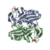 1okiS S: Starting model for refinement |
|---|---|
| Similar structure data |


-Links
 PDBj
PDBj
- Assembly
| Deposited unit | 
| ||||||||||||||||||||||||
|---|---|---|---|---|---|---|---|---|---|---|---|---|---|---|---|---|---|---|---|---|---|---|---|---|---|
| 1 | 
| ||||||||||||||||||||||||
| Unit cell |
| ||||||||||||||||||||||||
| Components on special symmetry positions |
| ||||||||||||||||||||||||
| Details | AUTHOR STATES THAT THE BIOLOGICAL ASSEMBLY IS UNKNOWN. |
-Components
| #1: Protein | Mass: 21812.057 Da / Num. of mol.: 1 / Fragment: residues 8-196 Source method: isolated from a genetically manipulated source Source: (gene. exp.) Homo sapiens (human) / Gene: CRYBA4 / Plasmid: pNIC28-Bsa4 / Production host:  Escherichia coli (E. coli) / Strain (production host): BL21(DE3)-R3-pRARE2 / References: UniProt: P53673 Escherichia coli (E. coli) / Strain (production host): BL21(DE3)-R3-pRARE2 / References: UniProt: P53673 | ||
|---|---|---|---|
| #2: Chemical | ChemComp-PO4 / Phosphate  Mass: 94.971 Da / Num. of mol.: 1 / Source method: obtained synthetically / Formula: PO4 Mass: 94.971 Da / Num. of mol.: 1 / Source method: obtained synthetically / Formula: PO4 | ||
| #3: Chemical | ChemComp-GOL / Glycerol  Mass: 92.094 Da / Num. of mol.: 5 / Source method: obtained synthetically / Formula: C3H8O3 Mass: 92.094 Da / Num. of mol.: 5 / Source method: obtained synthetically / Formula: C3H8O3#4: Water | ChemComp-HOH / | Water Mass: 18.015 Da / Num. of mol.: 190 / Source method: isolated from a natural source / Formula: H2O Mass: 18.015 Da / Num. of mol.: 190 / Source method: isolated from a natural source / Formula: H2O |
-Experimental details
-Experiment
| Experiment | Method: X-RAY DIFFRACTION / Number of used crystals: 1 |
|---|
- Sample preparation
| Crystal | Density Matthews: 2.32 Å3/Da / Density % sol: 47.04 % |
|---|---|
| Crystal grow | Temperature: 293.15 K / Method: vapor diffusion, sitting drop / pH: 9 Details: 1.5M Ammonium Phosphate, 5% Glycerol, 0.1M Tris, pH 9.0, VAPOR DIFFUSION, SITTING DROP, temperature 293.15K |
-Data collection
| Diffraction | Mean temperature: 100 K |
|---|---|
| Diffraction source | Source: SYNCHROTRON / Site: Diamond  / Beamline: I02 / Wavelength: 0.9796 Å / Beamline: I02 / Wavelength: 0.9796 Å |
| Detector | Type: ADSC QUANTUM 315 / Detector: CCD / Date: Sep 25, 2009 / Details: Kirkpatrick Baez bimorph mirror pair |
| Radiation | Monochromator: Si (111) double crystal monochromator / Protocol: SINGLE WAVELENGTH / Monochromatic (M) / Laue (L): M / Scattering type: x-ray |
| Radiation wavelength | Wavelength: 0.9796 Å / Relative weight: 1 |
| Reflection | Resolution: 1.7→67.03 Å / Num. all: 22916 / Num. obs: 22910 / % possible obs: 100 % / Observed criterion σ(F): 0 / Observed criterion σ(I): 0 / Redundancy: 7.8 % / Biso Wilson estimate: 17 Å2 / Rmerge(I) obs: 0.104 / Net I/σ(I): 13.6 |
| Reflection shell | Resolution: 1.7→1.79 Å / Redundancy: 8 % / Rmerge(I) obs: 0.72 / Mean I/σ(I) obs: 2.8 / Num. unique all: 3284 / % possible all: 100 |
- Processing
| Software |
| ||||||||||||||||||||||||||||||||||||||||||||||||||||||||||||||||||||||||||||||||||||||||||||||||||||
|---|---|---|---|---|---|---|---|---|---|---|---|---|---|---|---|---|---|---|---|---|---|---|---|---|---|---|---|---|---|---|---|---|---|---|---|---|---|---|---|---|---|---|---|---|---|---|---|---|---|---|---|---|---|---|---|---|---|---|---|---|---|---|---|---|---|---|---|---|---|---|---|---|---|---|---|---|---|---|---|---|---|---|---|---|---|---|---|---|---|---|---|---|---|---|---|---|---|---|---|---|---|
| Refinement | Method to determine structure: MOLECULAR REPLACEMENT Starting model: PDB ENTRY 1oki Resolution: 1.7→37.42 Å / Cor.coef. Fo:Fc: 0.96 / Cor.coef. Fo:Fc free: 0.948 / SU B: 3.585 / SU ML: 0.054 / TLS residual ADP flag: LIKELY RESIDUAL / Cross valid method: THROUGHOUT / σ(F): 0 / ESU R: 0.096 / ESU R Free: 0.093 / Stereochemistry target values: MAXIMUM LIKELIHOOD / Details: HYDROGENS HAVE BEEN ADDED IN THE RIDING POSITIONS
| ||||||||||||||||||||||||||||||||||||||||||||||||||||||||||||||||||||||||||||||||||||||||||||||||||||
| Solvent computation | Ion probe radii: 0.8 Å / Shrinkage radii: 0.8 Å / VDW probe radii: 1.4 Å / Solvent model: MASK | ||||||||||||||||||||||||||||||||||||||||||||||||||||||||||||||||||||||||||||||||||||||||||||||||||||
| Displacement parameters | Biso mean: 11.119 Å2
| ||||||||||||||||||||||||||||||||||||||||||||||||||||||||||||||||||||||||||||||||||||||||||||||||||||
| Refine analyze | Luzzati coordinate error obs: 0.187 Å | ||||||||||||||||||||||||||||||||||||||||||||||||||||||||||||||||||||||||||||||||||||||||||||||||||||
| Refinement step | Cycle: LAST / Resolution: 1.7→37.42 Å
| ||||||||||||||||||||||||||||||||||||||||||||||||||||||||||||||||||||||||||||||||||||||||||||||||||||
| Refine LS restraints |
| ||||||||||||||||||||||||||||||||||||||||||||||||||||||||||||||||||||||||||||||||||||||||||||||||||||
| LS refinement shell | Resolution: 1.7→1.744 Å / Total num. of bins used: 20
| ||||||||||||||||||||||||||||||||||||||||||||||||||||||||||||||||||||||||||||||||||||||||||||||||||||
| Refinement TLS params. | Method: refined / Refine-ID: X-RAY DIFFRACTION
| ||||||||||||||||||||||||||||||||||||||||||||||||||||||||||||||||||||||||||||||||||||||||||||||||||||
| Refinement TLS group |
|
