 Movie
Movie Controller


+ Open data

- Basic information
| Entry | Database: PDB / ID: 1oki | ||||||
|---|---|---|---|---|---|---|---|
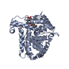
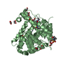
| Title | Crystal structure of truncated human beta-B1-crystallin | ||||||
 Components Components | BETA CRYSTALLIN B1 | ||||||
 Keywords Keywords |  EYE LENS PROTEIN / CRYSTALLIN / CATARACT EYE LENS PROTEIN / CRYSTALLIN / CATARACT | ||||||
| Function / homology |  Function and homology information Function and homology informationstructural constituent of eye lens / lens development in camera-type eye / visual perceptionSimilarity search - Function | ||||||
| Biological species |  HOMO SAPIENS (human) HOMO SAPIENS (human) | ||||||
| Method | X-RAY DIFFRACTION / SYNCHROTRON / MOLECULAR REPLACEMENT / Resolution: 1.4 Å | ||||||
 Authors Authors | van Montfort, R.L.M. / Bateman, O.A. / Lubsen, N.H. / Slingsby, C. | ||||||
 Citation Citation | Journal: Protein Sci. / Year: 2003 Title: Crystal Structure of Truncated Human Beta-B1-Crystallin Authors: Van Montfort, R.L.M. / Bateman, O.A. / Lubsen, N.H. / Slingsby, C. | ||||||
| History |
|
- Structure visualization
| Structure viewer | Molecule: MolmilJmol/JSmol |
|---|
- Downloads & links
-Download
| PDBx/mmCIF format | 1oki.cif.gz | 97.2 KB | Display | PDBx/mmCIF format |
|---|---|---|---|---|
| PDB format | pdb1oki.ent.gz | 73.9 KB | Display | PDB format |
| PDBx/mmJSON format | 1oki.json.gz | Tree view | PDBx/mmJSON format | |
| Others |  Other downloads Other downloads |
-Validation report
| Arichive directory | https://data.pdbj.org/pub/pdb/validation_reports/ok/1okiftp://data.pdbj.org/pub/pdb/validation_reports/ok/1oki | HTTPS FTP |
|---|
-Related structure data
| Related structure data |  2bb2S S: Starting model for refinement |
|---|---|
| Similar structure data |


-Links
 PDBj
PDBj
- Assembly
| Deposited unit | 
| ||||||||
|---|---|---|---|---|---|---|---|---|---|
| 1 |
| ||||||||
| Unit cell |
| ||||||||
| Noncrystallographic symmetry (NCS) | NCS oper: (Code: given Matrix: (-0.0001, -1, -0.0003), Vector : |
-Components
| #1: Protein | Mass: 24252.088 Da / Num. of mol.: 2 / Fragment: CRYSTALLIN DOMAINS, RESIDUES 42-251 Source method: isolated from a genetically manipulated source Source: (gene. exp.) HOMO SAPIENS (human) / Tissue: EYE LENSLens (anatomy) / Production host:  Escherichia coli BL21(DE3) (bacteria) / References: UniProt: P53674 Escherichia coli BL21(DE3) (bacteria) / References: UniProt: P53674#2: Water | ChemComp-HOH / | Water Mass: 18.015 Da / Num. of mol.: 423 / Source method: isolated from a natural source / Formula: H2O Mass: 18.015 Da / Num. of mol.: 423 / Source method: isolated from a natural source / Formula: H2OCompound details | CRYSTALLINS FORM THE DOMINANT STRUCTURAL COMPONENTS OF THE VERTEBRATE EYE LENS. THE PROTEIN EXISTS ...CRYSTALLIN | |
|---|
-Experimental details
-Experiment
| Experiment | Method: X-RAY DIFFRACTION / Number of used crystals: 1 |
|---|
- Sample preparation
| Crystal | Density Matthews: 1.8 Å3/Da / Density % sol: 31 % | ||||||||||||||||||||||||
|---|---|---|---|---|---|---|---|---|---|---|---|---|---|---|---|---|---|---|---|---|---|---|---|---|---|
| Crystal grow | pH: 8.5 Details: 24-30% PEG4000, 0.1M TRIS/HCL PH 7.0-8.5, 0.2M MGCL2 | ||||||||||||||||||||||||
| Crystal grow | *PLUS pH: 8 / Method: batch method / Details: Bateman, O.A., (2001) Exp. Eye Res., 73, 321. | ||||||||||||||||||||||||
| Components of the solutions | *PLUS
|
-Data collection
| Diffraction | Mean temperature: 100 K |
|---|---|
| Diffraction source | Source: SYNCHROTRON / Site: ESRF  / Beamline: ID14-3 / Wavelength: 0.931 / Beamline: ID14-3 / Wavelength: 0.931 |
| Detector | Type: MARRESEARCH / Detector: CCD |
| Radiation | Monochromator: SI(111) / Protocol: SINGLE WAVELENGTH / Monochromatic (M) / Laue (L): M / Scattering type: x-ray |
| Radiation wavelength | Wavelength: 0.931 Å / Relative weight: 1 |
| Reflection | Resolution: 1.4→35 Å / Num. obs: 60531 / % possible obs: 85.6 % / Redundancy: 2 % / Rmerge(I) obs: 0.043 / Net I/σ(I): 8.9 |
| Reflection | *PLUS Highest resolution: 1.4 Å / Num. measured all: 315295 / Rmerge(I) obs: 0.043 |
- Processing
| Software |
| ||||||||||||||||||||||||||||||||||||||||||||||||||||||||||||||||||||||||||||||||||||||||||||||||||||||||||||||||||||||||||||||||||||||||||||||||||||||||||||||||||||||||||||||||||||||
|---|---|---|---|---|---|---|---|---|---|---|---|---|---|---|---|---|---|---|---|---|---|---|---|---|---|---|---|---|---|---|---|---|---|---|---|---|---|---|---|---|---|---|---|---|---|---|---|---|---|---|---|---|---|---|---|---|---|---|---|---|---|---|---|---|---|---|---|---|---|---|---|---|---|---|---|---|---|---|---|---|---|---|---|---|---|---|---|---|---|---|---|---|---|---|---|---|---|---|---|---|---|---|---|---|---|---|---|---|---|---|---|---|---|---|---|---|---|---|---|---|---|---|---|---|---|---|---|---|---|---|---|---|---|---|---|---|---|---|---|---|---|---|---|---|---|---|---|---|---|---|---|---|---|---|---|---|---|---|---|---|---|---|---|---|---|---|---|---|---|---|---|---|---|---|---|---|---|---|---|---|---|---|---|
| Refinement | Method to determine structure: MOLECULAR REPLACEMENT Starting model: PDB ENTRY 2BB2 Resolution: 1.4→45.17 Å / Cor.coef. Fo:Fc: 0.973 / Cor.coef. Fo:Fc free: 0.967 / SU B: 1.26 / SU ML: 0.05 / Cross valid method: THROUGHOUT / ESU R: 0.074 / ESU R Free: 0.075 / Stereochemistry target values: MAXIMUM LIKELIHOOD Details: HYDROGENS HAVE BEEN ADDED IN THE RIDING POSITIONS. INITIAL DATASET PROCESSED IN P43212. TYR130 AT THE CRYSTALLOGRAPHIC TWOFOLD CLASHED WITH TYR130 OF A SYMMETRY RELATED MOLECULE. AS MOLECULE ...Details: HYDROGENS HAVE BEEN ADDED IN THE RIDING POSITIONS. INITIAL DATASET PROCESSED IN P43212. TYR130 AT THE CRYSTALLOGRAPHIC TWOFOLD CLASHED WITH TYR130 OF A SYMMETRY RELATED MOLECULE. AS MOLECULE IS A DIMER IN SOLULTION, LOWERED SPACE GROUP TO P43 WITH TWOFOLD NCS SYMMETRY. TYR130 POSSIBLY IN TWO CONFORMATIONS. PRIMARY CONFORMATION MODELLED.
| ||||||||||||||||||||||||||||||||||||||||||||||||||||||||||||||||||||||||||||||||||||||||||||||||||||||||||||||||||||||||||||||||||||||||||||||||||||||||||||||||||||||||||||||||||||||
| Solvent computation | Ion probe radii: 0.8 Å / Shrinkage radii: 0.8 Å / VDW probe radii: 1.4 Å / Solvent model: BABINET MODEL WITH MASK | ||||||||||||||||||||||||||||||||||||||||||||||||||||||||||||||||||||||||||||||||||||||||||||||||||||||||||||||||||||||||||||||||||||||||||||||||||||||||||||||||||||||||||||||||||||||
| Displacement parameters | Biso mean: 17.61 Å2
| ||||||||||||||||||||||||||||||||||||||||||||||||||||||||||||||||||||||||||||||||||||||||||||||||||||||||||||||||||||||||||||||||||||||||||||||||||||||||||||||||||||||||||||||||||||||
| Refinement step | Cycle: LAST / Resolution: 1.4→45.17 Å
| ||||||||||||||||||||||||||||||||||||||||||||||||||||||||||||||||||||||||||||||||||||||||||||||||||||||||||||||||||||||||||||||||||||||||||||||||||||||||||||||||||||||||||||||||||||||
| Refine LS restraints |
|