 Movie
Movie Controller

[English] 日本語
 Yorodumi
- PDB-3lk5: Crystal structure of putative Geranylgeranyl pyrophosphate syntha... -
+ Open data

- Basic information
| Entry | Database: PDB / ID: 3lk5 | ||||||
|---|---|---|---|---|---|---|---|
| Title | Crystal structure of putative Geranylgeranyl pyrophosphate synthase from Corynebacterium glutamicum | ||||||
 Components Components | Geranylgeranyl pyrophosphate synthase | ||||||
 Keywords Keywords |  TRANSFERASE / STRUCTURAL GENOMICS / PROTEIN STRUCTURE INITIATIVE / Geranylgeranyl pyrophosphate synthase / Isoprene biosynthesis / PSI-2 / New York SGX Research Center for Structural Genomics / NYSGXRC TRANSFERASE / STRUCTURAL GENOMICS / PROTEIN STRUCTURE INITIATIVE / Geranylgeranyl pyrophosphate synthase / Isoprene biosynthesis / PSI-2 / New York SGX Research Center for Structural Genomics / NYSGXRC | ||||||
| Function / homology |  Function and homology informationdimethylallyltranstransferase / dimethylallyltranstransferase activity / isoprenoid biosynthetic process Function and homology informationdimethylallyltranstransferase / dimethylallyltranstransferase activity / isoprenoid biosynthetic processSimilarity search - Function | ||||||
| Biological species |  Corynebacterium glutamicum (bacteria) Corynebacterium glutamicum (bacteria) | ||||||
| Method | X-RAY DIFFRACTION / SYNCHROTRON / SAD / Resolution: 1.9 Å | ||||||
 Authors Authors | Malashkevich, V.N. / Toro, R. / Patskovsky, Y. / Sauder, J.M. / Burley, S.K. / Almo, S.C. / New York SGX Research Center for Structural Genomics (NYSGXRC) | ||||||
 Citation Citation | Journal: To be Published Title: Crystal structure of putative Geranylgeranyl pyrophosphate synthase from Corynebacterium glutamicum Atcc 13032 Authors: Malashkevich, V.N. / Toro, R. / Patskovsky, Y. / Sauder, J.M. / Burley, S.K. / Almo, S.C. | ||||||
| History |
|
- Structure visualization
| Structure viewer | Molecule: MolmilJmol/JSmol |
|---|
- Downloads & links
-Download
| PDBx/mmCIF format | 3lk5.cif.gz | 78.5 KB | Display | PDBx/mmCIF format |
|---|---|---|---|---|
| PDB format | pdb3lk5.ent.gz | 61.6 KB | Display | PDB format |
| PDBx/mmJSON format | 3lk5.json.gz | Tree view | PDBx/mmJSON format | |
| Others |  Other downloads Other downloads |
-Validation report
| Arichive directory | https://data.pdbj.org/pub/pdb/validation_reports/lk/3lk5ftp://data.pdbj.org/pub/pdb/validation_reports/lk/3lk5 | HTTPS FTP |
|---|
-Related structure data
| Similar structure data | |
|---|---|
| Other databases |


-Links
 PDBj
PDBj
- Assembly
| Deposited unit | 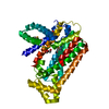
| ||||||||
|---|---|---|---|---|---|---|---|---|---|
| 1 | 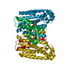
| ||||||||
| Unit cell |
|
-Components
| #1: Protein | Mass: 41478.656 Da / Num. of mol.: 1 Source method: isolated from a genetically manipulated source Source: (gene. exp.) Corynebacterium glutamicum (bacteria) / Strain: ATCC 13032 / Gene: Cgl2172 / Plasmid: BC-PSGX3(BC) / Production host: Escherichia coli (E. coli) / Strain (production host): BL21(DE3)CODON+RIL / References: UniProt: Q8NNM1, dimethylallyltranstransferase |
|---|---|
| #2: Chemical | ChemComp-GOL / Glycerol  Mass: 92.094 Da / Num. of mol.: 1 / Source method: obtained synthetically / Formula: C3H8O3 Mass: 92.094 Da / Num. of mol.: 1 / Source method: obtained synthetically / Formula: C3H8O3 |
| #3: Water | ChemComp-HOH / Water Mass: 18.015 Da / Num. of mol.: 194 / Source method: isolated from a natural source / Formula: H2O Mass: 18.015 Da / Num. of mol.: 194 / Source method: isolated from a natural source / Formula: H2O |
-Experimental details
-Experiment
| Experiment | Method: X-RAY DIFFRACTION / Number of used crystals: 1 |
|---|
- Sample preparation
| Crystal | Density Matthews: 2.85 Å3/Da / Density % sol: 56.85 % |
|---|---|
| Crystal grow | Temperature: 298 K / Method: vapor diffusion, sitting drop / pH: 7.5 Details: 30% PEG4000, 0.2M ammonium sulfate, pH 7.5, VAPOR DIFFUSION, SITTING DROP, temperature 298K |
-Data collection
| Diffraction | Mean temperature: 100 K |
|---|---|
| Diffraction source | Source: SYNCHROTRON / Site: NSLS  / Beamline: X29A / Wavelength: 0.9791 Å / Beamline: X29A / Wavelength: 0.9791 Å |
| Detector | Type: ADSC QUANTUM 315 / Detector: CCD / Date: Jan 23, 2010 |
| Radiation | Protocol: SINGLE WAVELENGTH / Scattering type: x-ray |
| Radiation wavelength | Wavelength: 0.9791 Å / Relative weight: 1 |
| Reflection | Resolution: 1.9→50 Å / Num. obs: 37221 / % possible obs: 99.7 % / Redundancy: 5.5 % / Rmerge(I) obs: 0.055 / Χ2: 0.983 / Net I/σ(I): 9.4 |
| Reflection shell | Resolution: 1.9→1.93 Å / Redundancy: 3.5 % / Rmerge(I) obs: 0.609 / Num. unique all: 4613 / Χ2: 0.81 / % possible all: 95.6 |
-Phasing
| Phasing | Method: SAD |
|---|
- Processing
| Software |
| |||||||||||||||||||||||||||||||||||||||||||||||||||||||||||||||||
|---|---|---|---|---|---|---|---|---|---|---|---|---|---|---|---|---|---|---|---|---|---|---|---|---|---|---|---|---|---|---|---|---|---|---|---|---|---|---|---|---|---|---|---|---|---|---|---|---|---|---|---|---|---|---|---|---|---|---|---|---|---|---|---|---|---|---|
| Refinement | Method to determine structure: SAD / Resolution: 1.9→19.7 Å / Cor.coef. Fo:Fc: 0.97 / Cor.coef. Fo:Fc free: 0.961 / Occupancy max: 1 / Occupancy min: 0.5 / SU B: 5.115 / SU ML: 0.069 / TLS residual ADP flag: LIKELY RESIDUAL / Cross valid method: THROUGHOUT / σ(F): 0 / ESU R: 0.109 / ESU R Free: 0.107 / Stereochemistry target values: MAXIMUM LIKELIHOOD Details: HYDROGENS HAVE BEEN ADDED IN THE RIDING POSITIONS, U VALUES RESIDUAL ONLY
| |||||||||||||||||||||||||||||||||||||||||||||||||||||||||||||||||
| Solvent computation | Ion probe radii: 0.8 Å / Shrinkage radii: 0.8 Å / VDW probe radii: 1.4 Å / Solvent model: BABINET MODEL WITH MASK | |||||||||||||||||||||||||||||||||||||||||||||||||||||||||||||||||
| Displacement parameters | Biso max: 89.93 Å2 / Biso mean: 34.455 Å2 / Biso min: 17.31 Å2
| |||||||||||||||||||||||||||||||||||||||||||||||||||||||||||||||||
| Refinement step | Cycle: LAST / Resolution: 1.9→19.7 Å
| |||||||||||||||||||||||||||||||||||||||||||||||||||||||||||||||||
| Refine LS restraints |
| |||||||||||||||||||||||||||||||||||||||||||||||||||||||||||||||||
| LS refinement shell | Resolution: 1.9→1.949 Å / Total num. of bins used: 20
| |||||||||||||||||||||||||||||||||||||||||||||||||||||||||||||||||
| Refinement TLS params. | Method: refined / Origin x: -1.4547 Å / Origin y: 94.4191 Å / Origin z: 1.0118 Å
| |||||||||||||||||||||||||||||||||||||||||||||||||||||||||||||||||
| Refinement TLS group |
|
