 Movie
Movie Controller

[English] 日本語
 Yorodumi
- PDB-2vw2: Crystal structure of the NanB sialidase from Streptococcus pneumoniae -
+ Open data

- Basic information
| Entry | Database: PDB / ID: 2vw2 | ||||||
|---|---|---|---|---|---|---|---|
| Title | Crystal structure of the NanB sialidase from Streptococcus pneumoniae | ||||||
 Components Components | SIALIDASE B | ||||||
 Keywords Keywords |  HYDROLASE / SIALIDASE / GLYCOSIDASE / DRUG DESIGN / NEURAMINIDASE / VIRULENCE FACTOR HYDROLASE / SIALIDASE / GLYCOSIDASE / DRUG DESIGN / NEURAMINIDASE / VIRULENCE FACTOR | ||||||
| Function / homology |  Function and homology information Function and homology informationexo-alpha-(2->3)-sialidase activity / exo-alpha-(2->6)-sialidase activity / exo-alpha-(2->8)-sialidase activity / exo-alpha-sialidase / carbohydrate metabolic processSimilarity search - Function | ||||||
| Biological species |   STREPTOCOCCUS PNEUMONIAE (bacteria) STREPTOCOCCUS PNEUMONIAE (bacteria) | ||||||
| Method | X-RAY DIFFRACTION / MOLECULAR REPLACEMENT / Resolution: 1.7 Å | ||||||
 Authors Authors | Xu, G. / Potter, J.A. / Russell, R.J.M. / Oggioni, M.R. / Andrew, P.W. / Taylor, G.L. | ||||||
 Citation Citation | Journal: J.Mol.Biol. / Year: 2008 Title: Crystal Structure of the Nanb Sialidase from Streptococcus Pneumoniae Authors: Xu, G. / Potter, J.A. / Russell, R.J.M. / Oggioni, M.R. / Andrew, P.W. / Taylor, G.L. | ||||||
| History |
| ||||||
| Remark 700 | SHEET THE SHEET STRUCTURE OF THIS MOLECULE IS BIFURCATED. IN ORDER TO REPRESENT THIS FEATURE IN ... SHEET THE SHEET STRUCTURE OF THIS MOLECULE IS BIFURCATED. IN ORDER TO REPRESENT THIS FEATURE IN THE SHEET RECORDS BELOW, TWO SHEETS ARE DEFINED. |
- Structure visualization
| Structure viewer | Molecule: MolmilJmol/JSmol |
|---|
- Downloads & links
-Download
| PDBx/mmCIF format | 2vw2.cif.gz | 161 KB | Display | PDBx/mmCIF format |
|---|---|---|---|---|
| PDB format | pdb2vw2.ent.gz | 124.1 KB | Display | PDB format |
| PDBx/mmJSON format | 2vw2.json.gz | Tree view | PDBx/mmJSON format | |
| Others |  Other downloads Other downloads |
-Validation report
| Arichive directory | https://data.pdbj.org/pub/pdb/validation_reports/vw/2vw2ftp://data.pdbj.org/pub/pdb/validation_reports/vw/2vw2 | HTTPS FTP |
|---|
-Related structure data
| Related structure data |  2vw0C  2vw1C  1sliS C: citing same article ( S: Starting model for refinement |
|---|---|
| Similar structure data |


-Links
 PDBj
PDBj

- Assembly
| Deposited unit | 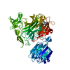
| ||||||||
|---|---|---|---|---|---|---|---|---|---|
| 1 |
| ||||||||
| Unit cell |
|
-Components
| #1: Protein | Mass: 77780.883 Da / Num. of mol.: 1 Source method: isolated from a genetically manipulated source Source: (gene. exp.) STREPTOCOCCUS PNEUMONIAE (bacteria) / Strain: TIGR / Production host: ESCHERICHIA COLI (E. coli) / Strain (production host): BL21(DE3) / References: UniProt: Q54727, exo-alpha-sialidase | ||||
|---|---|---|---|---|---|
| #2: Chemical | CHES (buffer)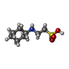  Mass: 207.290 Da / Num. of mol.: 2 / Source method: obtained synthetically / Formula: C8H17NO3S / Comment: pH buffer*YM Mass: 207.290 Da / Num. of mol.: 2 / Source method: obtained synthetically / Formula: C8H17NO3S / Comment: pH buffer*YM#3: Chemical | ChemComp-GOL / | Glycerol  Mass: 92.094 Da / Num. of mol.: 1 / Source method: obtained synthetically / Formula: C3H8O3 Mass: 92.094 Da / Num. of mol.: 1 / Source method: obtained synthetically / Formula: C3H8O3#4: Water | ChemComp-HOH / | Water Mass: 18.015 Da / Num. of mol.: 797 / Source method: isolated from a natural source / Formula: H2O Mass: 18.015 Da / Num. of mol.: 797 / Source method: isolated from a natural source / Formula: H2O |
-Experimental details
-Experiment
| Experiment | Method: X-RAY DIFFRACTION / Number of used crystals: 1 |
|---|
- Sample preparation
| Crystal | Density Matthews: 2.19 Å3/Da / Density % sol: 43.51 % / Description: NONE |
|---|---|
| Crystal grow | pH: 9.5 / Details: 10% PEG 8000, 0.15M NACL, 0.1M CHES PH 9.5 |
-Data collection
| Diffraction | Mean temperature: 100 K |
|---|---|
| Diffraction source | Source: ROTATING ANODE / Type: RIGAKU MICROMAX-007 HF / Wavelength: 1.5418 |
| Detector | Type: RIGAKU SATURN 944 / Detector: CCD |
| Radiation | Protocol: SINGLE WAVELENGTH / Monochromatic (M) / Laue (L): M / Scattering type: x-ray |
| Radiation wavelength | Wavelength: 1.5418 Å / Relative weight: 1 |
| Reflection | Resolution: 1.7→30 Å / Num. obs: 75667 / % possible obs: 92.1 % / Observed criterion σ(I): 0 / Redundancy: 3 % / Rmerge(I) obs: 0.08 / Net I/σ(I): 8.9 |
| Reflection shell | Resolution: 1.7→1.76 Å / Redundancy: 3 % / Rmerge(I) obs: 0.36 / Mean I/σ(I) obs: 2.2 / % possible all: 63 |
- Processing
| Software |
| ||||||||||||||||||||||||||||||||||||||||||||||||||||||||||||||||||||||||||||||||||||||||||||||||||||||||||||||||||||||||||||||||||||||||||||||||||||||||||||||||||||||||||||||||||||||
|---|---|---|---|---|---|---|---|---|---|---|---|---|---|---|---|---|---|---|---|---|---|---|---|---|---|---|---|---|---|---|---|---|---|---|---|---|---|---|---|---|---|---|---|---|---|---|---|---|---|---|---|---|---|---|---|---|---|---|---|---|---|---|---|---|---|---|---|---|---|---|---|---|---|---|---|---|---|---|---|---|---|---|---|---|---|---|---|---|---|---|---|---|---|---|---|---|---|---|---|---|---|---|---|---|---|---|---|---|---|---|---|---|---|---|---|---|---|---|---|---|---|---|---|---|---|---|---|---|---|---|---|---|---|---|---|---|---|---|---|---|---|---|---|---|---|---|---|---|---|---|---|---|---|---|---|---|---|---|---|---|---|---|---|---|---|---|---|---|---|---|---|---|---|---|---|---|---|---|---|---|---|---|---|
| Refinement | Method to determine structure: MOLECULAR REPLACEMENT Starting model: PDB ENTRY 1SLI Resolution: 1.7→67.42 Å / Cor.coef. Fo:Fc: 0.95 / Cor.coef. Fo:Fc free: 0.932 / SU B: 2.719 / SU ML: 0.087 / Cross valid method: THROUGHOUT / ESU R: 0.123 / ESU R Free: 0.118 / Stereochemistry target values: MAXIMUM LIKELIHOOD / Details: HYDROGENS HAVE BEEN ADDED IN THE RIDING POSITIONS.
| ||||||||||||||||||||||||||||||||||||||||||||||||||||||||||||||||||||||||||||||||||||||||||||||||||||||||||||||||||||||||||||||||||||||||||||||||||||||||||||||||||||||||||||||||||||||
| Solvent computation | Ion probe radii: 0.8 Å / Shrinkage radii: 0.8 Å / VDW probe radii: 1.4 Å / Solvent model: MASK | ||||||||||||||||||||||||||||||||||||||||||||||||||||||||||||||||||||||||||||||||||||||||||||||||||||||||||||||||||||||||||||||||||||||||||||||||||||||||||||||||||||||||||||||||||||||
| Displacement parameters | Biso mean: 18.51 Å2
| ||||||||||||||||||||||||||||||||||||||||||||||||||||||||||||||||||||||||||||||||||||||||||||||||||||||||||||||||||||||||||||||||||||||||||||||||||||||||||||||||||||||||||||||||||||||
| Refinement step | Cycle: LAST / Resolution: 1.7→67.42 Å
| ||||||||||||||||||||||||||||||||||||||||||||||||||||||||||||||||||||||||||||||||||||||||||||||||||||||||||||||||||||||||||||||||||||||||||||||||||||||||||||||||||||||||||||||||||||||
| Refine LS restraints |
|