 Movie
Movie Controller


+ Open data

- Basic information
| Entry | Database: PDB / ID: 3hy5 | ||||||
|---|---|---|---|---|---|---|---|
| Title | Crystal structure of CRALBP | ||||||
 Components Components | Retinaldehyde-binding protein 1 | ||||||
 Keywords Keywords | TRANSPORT PROTEIN / lipid transfer protein / 11-cis-retinal / bothnia dystrophy / Acetylation / Cytoplasm / Disease mutation / Retinitis pigmentosa / Retinol-binding / Sensory transduction / Transport / Vision | ||||||
| Function / homology |  Function and homology information Function and homology informationThe retinoid cycle in cones (daylight vision) / 11-cis retinal binding / vitamin A metabolic process / response to stimulus / retinol binding / phosphatidylinositol bisphosphate binding / Retinoid cycle disease events / The canonical retinoid cycle in rods (twilight vision) / visual perception / cell body ...The retinoid cycle in cones (daylight vision) / 11-cis retinal binding / vitamin A metabolic process / response to stimulus / retinol binding / phosphatidylinositol bisphosphate binding / Retinoid cycle disease events / The canonical retinoid cycle in rods (twilight vision) / visual perception / cell body / centrosome / nucleoplasm / cytosolSimilarity search - Function | ||||||
| Biological species |  Homo sapiens (human) Homo sapiens (human) | ||||||
| Method | X-RAY DIFFRACTION / SYNCHROTRON / MOLECULAR REPLACEMENT / molecular replacement / Resolution: 3.04 Å | ||||||
 Authors Authors | Stocker, A. / He, X. / Lobsiger, J. | ||||||
 Citation Citation | Journal: Proc.Natl.Acad.Sci.USA / Year: 2009 Title: Bothnia dystrophy is caused by domino-like rearrangements in cellular retinaldehyde-binding protein mutant R234W. Authors: He, X. / Lobsiger, J. / Stocker, A. | ||||||
| History |
|
- Structure visualization
| Structure viewer | Molecule: MolmilJmol/JSmol |
|---|
- Downloads & links
-Download
| PDBx/mmCIF format | 3hy5.cif.gz | 135.5 KB | Display | PDBx/mmCIF format |
|---|---|---|---|---|
| PDB format | pdb3hy5.ent.gz | 107.5 KB | Display | PDB format |
| PDBx/mmJSON format | 3hy5.json.gz | Tree view | PDBx/mmJSON format | |
| Others |  Other downloads Other downloads |
-Validation report
| Arichive directory | https://data.pdbj.org/pub/pdb/validation_reports/hy/3hy5ftp://data.pdbj.org/pub/pdb/validation_reports/hy/3hy5 | HTTPS FTP |
|---|
-Related structure data
| Related structure data | 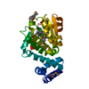 3hx3SC S: Starting model for refinement C: citing same article ( |
|---|---|
| Similar structure data |


-Links
 PDBj
PDBj


- Assembly
| Deposited unit | 
| ||||||||
|---|---|---|---|---|---|---|---|---|---|
| 1 |
| ||||||||
| Unit cell |
|
-Components
| #1: Protein | / Cellular retinaldehyde-binding protein Mass: 36382.957 Da / Num. of mol.: 1 Source method: isolated from a genetically manipulated source Source: (gene. exp.) Homo sapiens (human) / Gene: CRALBP, RLBP1 / Plasmid: pET28a / Production host:  Escherichia coli (E. coli) / Strain (production host): BL21(DE3) / References: UniProt: P12271 Escherichia coli (E. coli) / Strain (production host): BL21(DE3) / References: UniProt: P12271 |
|---|---|
| #2: Chemical | ChemComp-RET / Retinal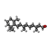  Mass: 284.436 Da / Num. of mol.: 1 / Source method: obtained synthetically / Formula: C20H28O Mass: 284.436 Da / Num. of mol.: 1 / Source method: obtained synthetically / Formula: C20H28O |
| #3: Chemical | ChemComp-TLA / Tartaric acid  Mass: 150.087 Da / Num. of mol.: 1 / Source method: obtained synthetically / Formula: C4H6O6 Mass: 150.087 Da / Num. of mol.: 1 / Source method: obtained synthetically / Formula: C4H6O6 |
-Experimental details
-Experiment
| Experiment | Method: X-RAY DIFFRACTION / Number of used crystals: 1 |
|---|
- Sample preparation
| Crystal | Density Matthews: 3.11 Å3/Da / Density % sol: 60.47 % |
|---|---|
| Crystal grow | Temperature: 298 K / Method: vapor diffusion / pH: 7 Details: 1M Sodium-potassium tartrate, 0.2M Lithium sulfate, pH 7.0, VAPOR DIFFUSION, temperature 298K |
-Data collection
| Diffraction | Mean temperature: 100 K |
|---|---|
| Diffraction source | Source: SYNCHROTRON / Site: ESRF  / Beamline: ID29 / Wavelength: 0.97618 Å / Beamline: ID29 / Wavelength: 0.97618 Å |
| Detector | Type: ADSC QUANTUM 315r / Detector: CCD / Date: Nov 6, 2007 |
| Radiation | Monochromator: Si(111) / Protocol: SINGLE WAVELENGTH / Scattering type: x-ray |
| Radiation wavelength | Wavelength: 0.97618 Å / Relative weight: 1 |
| Reflection | Resolution: 3.04→50 Å / Num. all: 9744 / Num. obs: 9692 / % possible obs: 99.5 % / Observed criterion σ(I): -3 / Redundancy: 10.6 % / Biso Wilson estimate: 89.676 Å2 / Rmerge(I) obs: 0.059 / Net I/σ(I): 22.8 |
| Reflection shell | Resolution: 3.04→3.22 Å / Redundancy: 9.6 % / Rmerge(I) obs: 0.534 / Mean I/σ(I) obs: 4.1 / Num. unique all: 1505 / Num. unique obs: 1499 / % possible all: 99.6 |
-Phasing
| Phasing | Method: molecular replacement | |||||||||
|---|---|---|---|---|---|---|---|---|---|---|
| Phasing MR | Rfactor: 57.68
|
- Processing
| Software |
| ||||||||||||||||||||||||||||||||||||||||||||||||||||||||||||||||||||||||||||||||||||||||||||||||||||||||||||||||||||||||||||||||||||||||||||||||||||||||||||||||||||||||||||||||||||||||||||||||||||||||||||||||||||||||||||||||||||||||||||||||||||||||||
|---|---|---|---|---|---|---|---|---|---|---|---|---|---|---|---|---|---|---|---|---|---|---|---|---|---|---|---|---|---|---|---|---|---|---|---|---|---|---|---|---|---|---|---|---|---|---|---|---|---|---|---|---|---|---|---|---|---|---|---|---|---|---|---|---|---|---|---|---|---|---|---|---|---|---|---|---|---|---|---|---|---|---|---|---|---|---|---|---|---|---|---|---|---|---|---|---|---|---|---|---|---|---|---|---|---|---|---|---|---|---|---|---|---|---|---|---|---|---|---|---|---|---|---|---|---|---|---|---|---|---|---|---|---|---|---|---|---|---|---|---|---|---|---|---|---|---|---|---|---|---|---|---|---|---|---|---|---|---|---|---|---|---|---|---|---|---|---|---|---|---|---|---|---|---|---|---|---|---|---|---|---|---|---|---|---|---|---|---|---|---|---|---|---|---|---|---|---|---|---|---|---|---|---|---|---|---|---|---|---|---|---|---|---|---|---|---|---|---|---|---|---|---|---|---|---|---|---|---|---|---|---|---|---|---|---|---|---|---|---|---|---|---|---|---|---|---|---|---|---|---|---|
| Refinement | Method to determine structure: MOLECULAR REPLACEMENT Starting model: PDB entry 3HX3 Resolution: 3.04→48.13 Å / Occupancy max: 1 / Occupancy min: 1 / SU ML: 0.35 / Stereochemistry target values: ML
| ||||||||||||||||||||||||||||||||||||||||||||||||||||||||||||||||||||||||||||||||||||||||||||||||||||||||||||||||||||||||||||||||||||||||||||||||||||||||||||||||||||||||||||||||||||||||||||||||||||||||||||||||||||||||||||||||||||||||||||||||||||||||||
| Solvent computation | Shrinkage radii: 0.9 Å / VDW probe radii: 1.11 Å / Solvent model: FLAT BULK SOLVENT MODEL / Bsol: 103.559 Å2 / ksol: 0.339 e/Å3 | ||||||||||||||||||||||||||||||||||||||||||||||||||||||||||||||||||||||||||||||||||||||||||||||||||||||||||||||||||||||||||||||||||||||||||||||||||||||||||||||||||||||||||||||||||||||||||||||||||||||||||||||||||||||||||||||||||||||||||||||||||||||||||
| Displacement parameters | Biso max: 282.79 Å2 / Biso mean: 132.072 Å2 / Biso min: 46.32 Å2
| ||||||||||||||||||||||||||||||||||||||||||||||||||||||||||||||||||||||||||||||||||||||||||||||||||||||||||||||||||||||||||||||||||||||||||||||||||||||||||||||||||||||||||||||||||||||||||||||||||||||||||||||||||||||||||||||||||||||||||||||||||||||||||
| Refinement step | Cycle: LAST / Resolution: 3.04→48.13 Å
| ||||||||||||||||||||||||||||||||||||||||||||||||||||||||||||||||||||||||||||||||||||||||||||||||||||||||||||||||||||||||||||||||||||||||||||||||||||||||||||||||||||||||||||||||||||||||||||||||||||||||||||||||||||||||||||||||||||||||||||||||||||||||||
| Refine LS restraints |
| ||||||||||||||||||||||||||||||||||||||||||||||||||||||||||||||||||||||||||||||||||||||||||||||||||||||||||||||||||||||||||||||||||||||||||||||||||||||||||||||||||||||||||||||||||||||||||||||||||||||||||||||||||||||||||||||||||||||||||||||||||||||||||
| LS refinement shell | Refine-ID: X-RAY DIFFRACTION / Total num. of bins used: 7
| ||||||||||||||||||||||||||||||||||||||||||||||||||||||||||||||||||||||||||||||||||||||||||||||||||||||||||||||||||||||||||||||||||||||||||||||||||||||||||||||||||||||||||||||||||||||||||||||||||||||||||||||||||||||||||||||||||||||||||||||||||||||||||
| Refinement TLS params. | Method: refined / Refine-ID: X-RAY DIFFRACTION
| ||||||||||||||||||||||||||||||||||||||||||||||||||||||||||||||||||||||||||||||||||||||||||||||||||||||||||||||||||||||||||||||||||||||||||||||||||||||||||||||||||||||||||||||||||||||||||||||||||||||||||||||||||||||||||||||||||||||||||||||||||||||||||
| Refinement TLS group |
|
