 Movie
Movie Controller


+ Open data

- Basic information
| Entry | Database: PDB / ID: 3bwq | ||||||
|---|---|---|---|---|---|---|---|
| Title | Structure of free SV40 VP1 pentamer | ||||||
 Components Components | Capsid protein VP1 | ||||||
 Keywords Keywords | VIRAL PROTEIN / SV40 / polyomavirus / ganglioside / GM1 / viral receptor / virus attachment / Capsid protein / Late protein / Nucleus / Virion | ||||||
| Function / homology |  Function and homology informationcapsomere / caveolin-mediated endocytosis of virus by host cell / viral capsid assembly / T=7 icosahedral viral capsid / host cell endoplasmic reticulum / molecular adaptor activity / host cell nucleus / virion attachment to host cell / structural molecule activity / identical protein binding Function and homology informationcapsomere / caveolin-mediated endocytosis of virus by host cell / viral capsid assembly / T=7 icosahedral viral capsid / host cell endoplasmic reticulum / molecular adaptor activity / host cell nucleus / virion attachment to host cell / structural molecule activity / identical protein bindingSimilarity search - Function | ||||||
| Biological species |  Simian virus 40 Simian virus 40 | ||||||
| Method | X-RAY DIFFRACTION / SYNCHROTRON / MOLECULAR REPLACEMENT / Resolution: 2.3 Å | ||||||
 Authors Authors | Neu, U. / Stehle, T. | ||||||
 Citation Citation | Journal: Proc.Natl.Acad.Sci.Usa / Year: 2008 Title: Structural basis of GM1 ganglioside recognition by simian virus 40. Authors: Neu, U. / Woellner, K. / Gauglitz, G. / Stehle, T. | ||||||
| History |
|
- Structure visualization
| Structure viewer | Molecule: MolmilJmol/JSmol |
|---|
- Downloads & links
-Download
| PDBx/mmCIF format | 3bwq.cif.gz | 270.8 KB | Display | PDBx/mmCIF format |
|---|---|---|---|---|
| PDB format | pdb3bwq.ent.gz | 219.2 KB | Display | PDB format |
| PDBx/mmJSON format | 3bwq.json.gz | Tree view | PDBx/mmJSON format | |
| Others |  Other downloads Other downloads |
-Validation report
| Arichive directory | https://data.pdbj.org/pub/pdb/validation_reports/bw/3bwqftp://data.pdbj.org/pub/pdb/validation_reports/bw/3bwq | HTTPS FTP |
|---|
-Related structure data
| Related structure data | 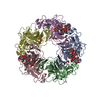 3bwrC  1svaS S: Starting model for refinement C: citing same article ( |
|---|---|
| Similar structure data |


-Links
 PDBj
PDBj


- Assembly
| Deposited unit | 
| ||||||||
|---|---|---|---|---|---|---|---|---|---|
| 1 |
| ||||||||
| Unit cell |
|
-Components
| #1: Protein | Mass: 29734.420 Da / Num. of mol.: 5 Source method: isolated from a genetically manipulated source Source: (gene. exp.) Simian virus 40 / Genus: PolyomavirusPolyomaviridae / Gene: VP1 / Production host:  Escherichia coli (E. coli) / References: UniProt: P03087 Escherichia coli (E. coli) / References: UniProt: P03087#2: Water | ChemComp-HOH / | Water Mass: 18.015 Da / Num. of mol.: 676 / Source method: isolated from a natural source / Formula: H2O Mass: 18.015 Da / Num. of mol.: 676 / Source method: isolated from a natural source / Formula: H2O |
|---|
-Experimental details
-Experiment
| Experiment | Method: X-RAY DIFFRACTION / Number of used crystals: 1 |
|---|
- Sample preparation
| Crystal | Density Matthews: 2.82 Å3/Da / Density % sol: 56.44 % |
|---|---|
| Crystal grow | Temperature: 273 K / Method: vapor diffusion, hanging drop Details: RESERVOIR: 0.1 M TRIS, PH 8.5, 24 % PEG3350. DROP: RESERVOIR MIXED 4:1 WITH 30 % ETHYLENE GLYCOL. THIS WAS MIXED 1:1 WITH VP1 (11 MG/ML), VAPOR DIFFUSION, HANGING DROP, temperature 273K |
-Data collection
| Diffraction | Mean temperature: 100 K |
|---|---|
| Diffraction source | Source: SYNCHROTRON / Site: SLS  / Beamline: X06SA / Wavelength: 1 Å / Beamline: X06SA / Wavelength: 1 Å |
| Detector | Type: MARMOSAIC 225 mm CCD / Detector: CCD / Date: Sep 29, 2006 |
| Radiation | Monochromator: Si (111) / Protocol: SINGLE WAVELENGTH / Monochromatic (M) / Laue (L): M / Scattering type: x-ray |
| Radiation wavelength | Wavelength: 1 Å / Relative weight: 1 |
| Reflection | Resolution: 2.3→50 Å / Num. all: 73715 / Num. obs: 63985 / % possible obs: 86.8 % / Redundancy: 4.8 % / Rmerge(I) obs: 0.099 / Net I/σ(I): 6.7 |
| Reflection shell | Resolution: 2.3→2.38 Å / Redundancy: 3.5 % / Rmerge(I) obs: 0.26 / Mean I/σ(I) obs: 1.4 / % possible all: 52.2 |
- Processing
| Software |
| ||||||||||||||||||||||||||||||||||||||||||||||||||||||||||||||||||||||||||||||||||||||||||||||||||||||||||||||||||||||||||||||||||||||||||||||||||||||||||||||||||||||||||
|---|---|---|---|---|---|---|---|---|---|---|---|---|---|---|---|---|---|---|---|---|---|---|---|---|---|---|---|---|---|---|---|---|---|---|---|---|---|---|---|---|---|---|---|---|---|---|---|---|---|---|---|---|---|---|---|---|---|---|---|---|---|---|---|---|---|---|---|---|---|---|---|---|---|---|---|---|---|---|---|---|---|---|---|---|---|---|---|---|---|---|---|---|---|---|---|---|---|---|---|---|---|---|---|---|---|---|---|---|---|---|---|---|---|---|---|---|---|---|---|---|---|---|---|---|---|---|---|---|---|---|---|---|---|---|---|---|---|---|---|---|---|---|---|---|---|---|---|---|---|---|---|---|---|---|---|---|---|---|---|---|---|---|---|---|---|---|---|---|---|---|---|
| Refinement | Method to determine structure: MOLECULAR REPLACEMENT Starting model: PDB ENTRY 1SVA Resolution: 2.3→39.78 Å / Cor.coef. Fo:Fc: 0.939 / Cor.coef. Fo:Fc free: 0.912 / SU B: 8.224 / SU ML: 0.196 / Cross valid method: THROUGHOUT / ESU R: 0.453 / ESU R Free: 0.275 / Stereochemistry target values: MAXIMUM LIKELIHOOD Details: HYDROGENS HAVE BEEN ADDED IN THE RIDING POSITIONS. some of the residues from Gly30 to Asp42 were disordered in several chains and were not modeled. The side chain of Asp42 of chain E was ...Details: HYDROGENS HAVE BEEN ADDED IN THE RIDING POSITIONS. some of the residues from Gly30 to Asp42 were disordered in several chains and were not modeled. The side chain of Asp42 of chain E was modeled as Ala due to missing density.
| ||||||||||||||||||||||||||||||||||||||||||||||||||||||||||||||||||||||||||||||||||||||||||||||||||||||||||||||||||||||||||||||||||||||||||||||||||||||||||||||||||||||||||
| Solvent computation | Ion probe radii: 0.8 Å / Shrinkage radii: 0.8 Å / VDW probe radii: 1.4 Å / Solvent model: MASK | ||||||||||||||||||||||||||||||||||||||||||||||||||||||||||||||||||||||||||||||||||||||||||||||||||||||||||||||||||||||||||||||||||||||||||||||||||||||||||||||||||||||||||
| Displacement parameters | Biso mean: 39.406 Å2
| ||||||||||||||||||||||||||||||||||||||||||||||||||||||||||||||||||||||||||||||||||||||||||||||||||||||||||||||||||||||||||||||||||||||||||||||||||||||||||||||||||||||||||
| Refinement step | Cycle: LAST / Resolution: 2.3→39.78 Å
| ||||||||||||||||||||||||||||||||||||||||||||||||||||||||||||||||||||||||||||||||||||||||||||||||||||||||||||||||||||||||||||||||||||||||||||||||||||||||||||||||||||||||||
| Refine LS restraints |
| ||||||||||||||||||||||||||||||||||||||||||||||||||||||||||||||||||||||||||||||||||||||||||||||||||||||||||||||||||||||||||||||||||||||||||||||||||||||||||||||||||||||||||
| LS refinement shell | Resolution: 2.3→2.36 Å / Total num. of bins used: 20
|
