 Movie
Movie Controller


+ Open data

- Basic information
| Entry | Database: PDB / ID: 2klj | ||||||
|---|---|---|---|---|---|---|---|
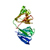
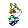
| Title | Solution Structure of gammaD-Crystallin with RDC and SAXS | ||||||
 Components Components | Gamma-crystallin D | ||||||
 Keywords Keywords |  STRUCTURAL PROTEIN / gammaD-Crystallin / 3D structure / RDC / SAXS / Cataract / Disease mutation / Eye lens protein / Oxidation / Polymorphism / Sensory transduction / Vision STRUCTURAL PROTEIN / gammaD-Crystallin / 3D structure / RDC / SAXS / Cataract / Disease mutation / Eye lens protein / Oxidation / Polymorphism / Sensory transduction / Vision | ||||||
| Function / homology |  Function and homology information Function and homology informationlens fiber cell differentiation / structural constituent of eye lens / lens development in camera-type eye / visual perception / cellular response to reactive oxygen species / nucleus / cytoplasmSimilarity search - Function | ||||||
| Biological species |  Homo sapiens (human) Homo sapiens (human) | ||||||
| Method | SOLUTION SCATTERING / SOLUTION NMR / simulated annealing | ||||||
| Model details | lowest energy, model 1 | ||||||
 Authors Authors | Wang, J. / Zuo, X. / Yu, P. / Byeon, I. / Jung, J. / Gronenborn, A.M. / Wang, Y. | ||||||
 Citation Citation | Journal: J.Am.Chem.Soc. / Year: 2009 Title: Determination of multicomponent protein structures in solution using global orientation and shape restraints. Authors: Wang, J. / Zuo, X. / Yu, P. / Byeon, I.J. / Jung, J. / Wang, X. / Dyba, M. / Seifert, S. / Schwieters, C.D. / Qin, J. / Gronenborn, A.M. / Wang, Y.X. #1: Journal: Biochemistry / Year: 2009Title: The Structure of the Cataract-Causing P23T Mutant of HgD Crystallin Exhibits Distinctive Local Conformational and Dynamic Changes Authors: Jung, J. / Byeon, I.L. / Wang, Y. / King, J. / Gronenborn, A.M. | ||||||
| History |
|
- Structure visualization
| Structure viewer | Molecule: MolmilJmol/JSmol |
|---|
- Downloads & links
-Download
| PDBx/mmCIF format | 2klj.cif.gz | 553 KB | Display | PDBx/mmCIF format |
|---|---|---|---|---|
| PDB format | pdb2klj.ent.gz | 477.8 KB | Display | PDB format |
| PDBx/mmJSON format | 2klj.json.gz | Tree view | PDBx/mmJSON format | |
| Others |  Other downloads Other downloads |
-Validation report
| Arichive directory | https://data.pdbj.org/pub/pdb/validation_reports/kl/2kljftp://data.pdbj.org/pub/pdb/validation_reports/kl/2klj | HTTPS FTP |
|---|
-Related structure data


-Links
 PDBj
PDBj
- Assembly
| Deposited unit | 
| |||||||||
|---|---|---|---|---|---|---|---|---|---|---|
| 1 |
| |||||||||
| NMR ensembles |
|
-Components
| #1: Protein | Mass: 20767.088 Da / Num. of mol.: 1 Source method: isolated from a genetically manipulated source Source: (gene. exp.) Homo sapiens (human) / Gene: CRYGD, CRYG4 / Production host:  Escherichia coli (E. coli) / References: UniProt: P07320 Escherichia coli (E. coli) / References: UniProt: P07320 |
|---|
-Experimental details
-Experiment
| Experiment |
| |||
|---|---|---|---|---|
| NMR experiment | Type: 2D 1H-15N HSQC |
- Sample preparation
| Details |
| ||||||||||||
|---|---|---|---|---|---|---|---|---|---|---|---|---|---|
| Sample |
| ||||||||||||
| Sample conditions | Ionic strength: 0.02 / pH: 6.2 / Pressure: ambient / Temperature: 298 K |
-Data collection
| NMR spectrometer | Type: Bruker Avance / Manufacturer: Bruker / Model: AVANCE / Field strength: 900 MHz |
|---|---|
| Soln scatter | Type: x-ray / Buffer name: 20 MM NACL 20 MM MES 5 MM DTT / Conc. range: 1.0-3.6 / Data analysis software list: GNOM Data reduction software list: MARDETECTOR, HOME- WRITTEN PROGRAM Detector specific: HOME-MADE / Detector type: CCD CAMERA / Mean guiner radius: 1.72 nm / Mean guiner radius esd: 0.03 nm / Num. of time frames: 20 / Protein length: 0.5 / Sample pH: 6.2 / Source beamline: 12-ID / Source class: Y / Source type: APS ARGONNE / Temperature: 298 K |
- Processing
| NMR software | Name: X-PLOR NIH / Version: 2.22 / Developer: Schwieters, Kuszewski, Tjandra and Clore / Classification: refinement |
|---|---|
| Refinement | Method: simulated annealing / Software ordinal: 1 |
| NMR representative | Selection criteria: lowest energy |
| NMR ensemble | Conformer selection criteria: structures with the lowest energy Conformers calculated total number: 100 / Conformers submitted total number: 10 |
| Soln scatter model | Conformer selection criteria: STRUCTURES WITH THE LOWEST ENERGY Num. of conformers calculated: 100 / Num. of conformers submitted: 10 / Representative conformer: 1 / Software list: GNOM |
