Mass: 18.015 Da / Num. of mol.: 4 / Source method: isolated from a natural source / Formula: H2O
-
Experimental details
-
Experiment
Experiment
Method: X-RAY DIFFRACTION / Number of used crystals: 1
-
Sample preparation
Crystal
Density Matthews: 2.74 Å3/Da / Density % sol: 55.05 %
Crystal grow
Temperature: 293 K / Method: vapor diffusion, sitting drop / pH: 7.8 Details: 0.45 mM EF-Tu in 50 mM Tris, pH 7.8, 10 mM GDP, 10 mM magnesium chloride, 0.5% PEG 3350, 5.5 mM ammonium acetate, 2.7 mM ammonium citrate, 1.34 mM ChemDiv compound 1013-0135, VAPOR ...Details: 0.45 mM EF-Tu in 50 mM Tris, pH 7.8, 10 mM GDP, 10 mM magnesium chloride, 0.5% PEG 3350, 5.5 mM ammonium acetate, 2.7 mM ammonium citrate, 1.34 mM ChemDiv compound 1013-0135, VAPOR DIFFUSION, SITTING DROP, temperature 293K
-
Data collection
Diffraction
Mean temperature: 95 K
Diffraction source
Source: SYNCHROTRON / Site: ALS / Beamline: 5.0.2 / Wavelength: 1.033 Å
Resolution: 3.4→60.232 Å / Isotropic thermal model: isotropic / Cross valid method: THROUGHOUT / σ(F): 0 / Stereochemistry target values: Engh & Huber Details: Used CNS specialized task files for refinement with hemihedrally twinned data. Twin operator was (-h,-k,l), and twin fraction was 0.294. R-factors reported are the twinned R-factors from CNS. ...Details: Used CNS specialized task files for refinement with hemihedrally twinned data. Twin operator was (-h,-k,l), and twin fraction was 0.294. R-factors reported are the twinned R-factors from CNS. (R-factors after detwinning: R-work=0.2867, R-free=0.3267)
Rfactor
Num. reflection
% reflection
Selection details
Rfree
0.2619
586
8.4 %
Used CNS task file make_cv_twin.inp to select cross-validation set such that pairs of twin-related reflections were designated to be in the same set, either the working set or the test set.
Rwork
0.197
-
-
-
all
-
6987
-
-
obs
-
6083
87.1 %
-
Solvent computation
Bsol: 73.771 Å2
Displacement parameters
Biso mean: 75.7 Å2
Baniso -1
Baniso -2
Baniso -3
1-
8.819 Å2
2.011 Å2
0 Å2
2-
-
8.819 Å2
0 Å2
3-
-
-
-17.639 Å2
Refinement step
Cycle: LAST / Resolution: 3.4→60.232 Å
Protein
Nucleic acid
Ligand
Solvent
Total
Num. atoms
2981
0
29
4
3014
Refine LS restraints
Refine-ID
Type
Dev ideal
X-RAY DIFFRACTION
c_bond_d
0.0114
X-RAY DIFFRACTION
c_angle_deg
1.73
X-RAY DIFFRACTION
c_dihedral_angle_d
24.688
X-RAY DIFFRACTION
c_improper_angle_d
1.184
Xplor file
Refine-ID
Serial no
Param file
X-RAY DIFFRACTION
1
CNS_TOPPAR:protein_rep.param
X-RAY DIFFRACTION
2
MYTOPPAR:gdp_cns4.param
X-RAY DIFFRACTION
3
CNS_TOPPAR:ion.param
X-RAY DIFFRACTION
4
CNS_TOPPAR:water_rep.param
+
About Yorodumi
-
News
-
Feb 9, 2022. New format data for meta-information of EMDB entries
New format data for meta-information of EMDB entries
Version 3 of the EMDB header file is now the official format.
The previous official version 1.9 will be removed from the archive.
In the structure databanks used in Yorodumi, some data are registered as the other names, "COVID-19 virus" and "2019-nCoV". Here are the details of the virus and the list of structure data.
Jan 31, 2019. EMDB accession codes are about to change! (news from PDBe EMDB page)
EMDB accession codes are about to change! (news from PDBe EMDB page)
The allocation of 4 digits for EMDB accession codes will soon come to an end. Whilst these codes will remain in use, new EMDB accession codes will include an additional digit and will expand incrementally as the available range of codes is exhausted. The current 4-digit format prefixed with “EMD-” (i.e. EMD-XXXX) will advance to a 5-digit format (i.e. EMD-XXXXX), and so on. It is currently estimated that the 4-digit codes will be depleted around Spring 2019, at which point the 5-digit format will come into force.
The EM Navigator/Yorodumi systems omit the EMD- prefix.
Related info.:Q: What is EMD? / ID/Accession-code notation in Yorodumi/EM Navigator
Yorodumi is a browser for structure data from EMDB, PDB, SASBDB, etc.
This page is also the successor to EM Navigator detail page, and also detail information page/front-end page for Omokage search.
The word "yorodu" (or yorozu) is an old Japanese word meaning "ten thousand". "mi" (miru) is to see.
Related info.:EMDB / PDB / SASBDB / Comparison of 3 databanks / Yorodumi Search / Aug 31, 2016. New EM Navigator & Yorodumi / Yorodumi Papers / Jmol/JSmol / Function and homology information / Changes in new EM Navigator and Yorodumi
 Movie
Movie Controller

 Yorodumi
 Open data
 Basic information
 Components
 EF-Tu
EF-Tu  Keywords
 Function and homology information
 Authors
 Citation
 Structure visualization
 Downloads & links
 Other downloads
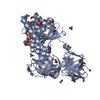


 PDBj
PDBj


 Assembly

 Mass: 24.305 Da / Num. of mol.: 1 / Source method: obtained synthetically / Formula: Mg
 Type: RNA linking / Mass: 443.201 Da / Num. of mol.: 1 / Source method: obtained synthetically / Formula: C10H15N5O11P2 / Comment: GDP, energy-carrying molecule*YM
 Mass: 18.015 Da / Num. of mol.: 4 / Source method: isolated from a natural source / Formula: H2O
 Sample preparation
 / Beamline: 5.0.2 / Wavelength: 1.033 Å
 Processing