 Movie
Movie Controller


+ Open data

- Basic information
| Entry | Database: PDB / ID: 1y56 | ||||||
|---|---|---|---|---|---|---|---|
| Title | Crystal structure of L-proline dehydrogenase from P.horikoshii | ||||||
 Components Components |
| ||||||
 Keywords Keywords |  OXIDOREDUCTASE / dehydrogenase / protein-protein complex OXIDOREDUCTASE / dehydrogenase / protein-protein complex | ||||||
| Function / homology |  Function and homology information Function and homology information | ||||||
| Biological species |   Pyrococcus horikoshii (archaea) Pyrococcus horikoshii (archaea) | ||||||
| Method | X-RAY DIFFRACTION / SYNCHROTRON / MIR / Resolution: 2.86 Å | ||||||
 Authors Authors | Tsuge, H. / Kawakami, R. / Sakuraba, H. / Ago, H. / Miyano, M. / Aki, K. / Katunuma, N. / Ohshima, T. | ||||||
 Citation Citation | Journal: J.Biol.Chem. / Year: 2005 Title: Crystal structure of a novel FAD-, FMN-, and ATP-containing L-proline dehydrogenase complex from Pyrococcus horikoshii Authors: Tsuge, H. / Kawakami, R. / Sakuraba, H. / Ago, H. / Miyano, M. / Aki, K. / Katunuma, N. / Ohshima, T. | ||||||
| History |
|
- Structure visualization
| Structure viewer | Molecule: MolmilJmol/JSmol |
|---|
- Downloads & links
-Download
| PDBx/mmCIF format | 1y56.cif.gz | 185.7 KB | Display | PDBx/mmCIF format |
|---|---|---|---|---|
| PDB format | pdb1y56.ent.gz | 152.5 KB | Display | PDB format |
| PDBx/mmJSON format | 1y56.json.gz | Tree view | PDBx/mmJSON format | |
| Others |  Other downloads Other downloads |
-Validation report
| Arichive directory | https://data.pdbj.org/pub/pdb/validation_reports/y5/1y56ftp://data.pdbj.org/pub/pdb/validation_reports/y5/1y56 | HTTPS FTP |
|---|
-Related structure data
| Similar structure data |
|---|


-Links
 PDBj
PDBj


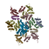
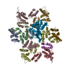
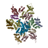
- Assembly
| Deposited unit | 
| ||||||||
|---|---|---|---|---|---|---|---|---|---|
| 1 | 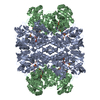
| ||||||||
| 2 |
| ||||||||
| 3 | 
| ||||||||
| 4 | 
| ||||||||
| Unit cell |
| ||||||||
| Details | The biological assembly is a octamer generated by tetramer of hetero-dimer |
-Components
-Protein , 2 types, 2 molecules AB
| #1: Protein | Hypothesis / L-proline dehydrogenase alpha subunit Mass: 55639.180 Da / Num. of mol.: 1 Source method: isolated from a genetically manipulated source Source: (gene. exp.) Pyrococcus horikoshii (archaea) / Strain: OT3 / Plasmid: pET11a / Production host:  Escherichia coli (E. coli) / Strain (production host): BL21 CodonPlus RIL(DE3) / References: UniProt: O59088, EC: 1.5.99.8 Escherichia coli (E. coli) / Strain (production host): BL21 CodonPlus RIL(DE3) / References: UniProt: O59088, EC: 1.5.99.8 |
|---|---|
| #2: Protein | / L-proline dehydrogenase beta subunit Mass: 42737.098 Da / Num. of mol.: 1 Source method: isolated from a genetically manipulated source Source: (gene. exp.) Pyrococcus horikoshii (archaea) / Strain: OT3 / Plasmid: pET11a / Production host: Escherichia coli (E. coli) / Strain (production host): BL21 CodonPlus RIL(DE3)References: UniProt: Q5R1N3, UniProt: O59089*PLUS, EC: 1.5.99.8 |
-Non-polymers , 8 types, 213 molecules 


| #3: Chemical | ChemComp-FE / Iron Mass: 55.845 Da / Num. of mol.: 1 / Source method: obtained synthetically / Formula: Fe Mass: 55.845 Da / Num. of mol.: 1 / Source method: obtained synthetically / Formula: Fe |
|---|---|
| #4: Chemical | ChemComp-SO4 / Sulfate Mass: 96.063 Da / Num. of mol.: 1 / Source method: obtained synthetically / Formula: SO4 Mass: 96.063 Da / Num. of mol.: 1 / Source method: obtained synthetically / Formula: SO4 |
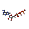
| #5: Chemical | ChemComp-FMN / Flavin mononucleotide Mass: 456.344 Da / Num. of mol.: 1 / Source method: obtained synthetically / Formula: C17H21N4O9P Mass: 456.344 Da / Num. of mol.: 1 / Source method: obtained synthetically / Formula: C17H21N4O9P |
| #6: Chemical | ChemComp-ATP / Adenosine triphosphate Mass: 507.181 Da / Num. of mol.: 1 / Source method: obtained synthetically / Formula: C10H16N5O13P3 / Comment: ATP, energy-carrying molecule*YM Mass: 507.181 Da / Num. of mol.: 1 / Source method: obtained synthetically / Formula: C10H16N5O13P3 / Comment: ATP, energy-carrying molecule*YM |
| #7: Chemical | ChemComp-CXS / CAPS (buffer) Mass: 221.317 Da / Num. of mol.: 1 / Source method: obtained synthetically / Formula: C9H19NO3S / Comment: pH buffer*YM Mass: 221.317 Da / Num. of mol.: 1 / Source method: obtained synthetically / Formula: C9H19NO3S / Comment: pH buffer*YM |
| #8: Chemical | ChemComp-CL / Chloride Mass: 35.453 Da / Num. of mol.: 1 / Source method: obtained synthetically / Formula: Cl Mass: 35.453 Da / Num. of mol.: 1 / Source method: obtained synthetically / Formula: Cl |
| #9: Chemical | ChemComp-FAD / Flavin adenine dinucleotide Mass: 785.550 Da / Num. of mol.: 1 / Source method: obtained synthetically / Formula: C27H33N9O15P2 / Comment: FAD*YM Mass: 785.550 Da / Num. of mol.: 1 / Source method: obtained synthetically / Formula: C27H33N9O15P2 / Comment: FAD*YM |
| #10: Water | ChemComp-HOH / WaterMass: 18.015 Da / Num. of mol.: 206 / Source method: isolated from a natural source / Formula: H2O |
-Experimental details
-Experiment
| Experiment | Method: X-RAY DIFFRACTION / Number of used crystals: 1 |
|---|
- Sample preparation
| Crystal | Density Matthews: 3.8 Å3/Da / Density % sol: 67 % |
|---|---|
| Crystal grow | Temperature: 298 K / Method: evaporation / pH: 10.5 Details: CAPS, NaH2PO4, K2HPO4, Li2SO4, pH 10.5, EVAPORATION, temperature 298K |
-Data collection
| Diffraction | Mean temperature: 100 K |
|---|---|
| Diffraction source | Source: SYNCHROTRON / Site: Photon Factory  / Beamline: AR-NW12A / Wavelength: 1 Å / Beamline: AR-NW12A / Wavelength: 1 Å |
| Detector | Type: ADSC QUANTUM 210 / Detector: CCD / Date: Nov 4, 2003 |
| Radiation | Monochromator: mirror / Protocol: SINGLE WAVELENGTH / Monochromatic (M) / Laue (L): M / Scattering type: x-ray |
| Radiation wavelength | Wavelength: 1 Å / Relative weight: 1 |
| Reflection | Resolution: 2.8→48.41 Å / Num. all: 38014 / Num. obs: 37913 / % possible obs: 98.7 % / Observed criterion σ(F): 1 / Observed criterion σ(I): 1 |
| Reflection shell | Resolution: 2.8→2.96 Å / % possible all: 93.8 |
- Processing
| Software |
| ||||||||||||||||||||||||||||||||||||||||||||||||||||||||||||||||||||||||||||||||||||||||||||||||||||||||||||||||||||||||||||||||||||||||||||||||||||||||||||||||||||||||||
|---|---|---|---|---|---|---|---|---|---|---|---|---|---|---|---|---|---|---|---|---|---|---|---|---|---|---|---|---|---|---|---|---|---|---|---|---|---|---|---|---|---|---|---|---|---|---|---|---|---|---|---|---|---|---|---|---|---|---|---|---|---|---|---|---|---|---|---|---|---|---|---|---|---|---|---|---|---|---|---|---|---|---|---|---|---|---|---|---|---|---|---|---|---|---|---|---|---|---|---|---|---|---|---|---|---|---|---|---|---|---|---|---|---|---|---|---|---|---|---|---|---|---|---|---|---|---|---|---|---|---|---|---|---|---|---|---|---|---|---|---|---|---|---|---|---|---|---|---|---|---|---|---|---|---|---|---|---|---|---|---|---|---|---|---|---|---|---|---|---|---|---|
| Refinement | Method to determine structure: MIR / Resolution: 2.86→19.85 Å / Cor.coef. Fo:Fc: 0.935 / Cor.coef. Fo:Fc free: 0.906 / SU B: 11.745 / SU ML: 0.222 / Cross valid method: THROUGHOUT / σ(F): 1 / ESU R: 0.908 / ESU R Free: 0.315 / Stereochemistry target values: MAXIMUM LIKELIHOOD / Details: HYDROGENS HAVE BEEN ADDED IN THE RIDING POSITIONS
| ||||||||||||||||||||||||||||||||||||||||||||||||||||||||||||||||||||||||||||||||||||||||||||||||||||||||||||||||||||||||||||||||||||||||||||||||||||||||||||||||||||||||||
| Solvent computation | Ion probe radii: 0.8 Å / Shrinkage radii: 0.8 Å / VDW probe radii: 1.2 Å / Solvent model: MASK | ||||||||||||||||||||||||||||||||||||||||||||||||||||||||||||||||||||||||||||||||||||||||||||||||||||||||||||||||||||||||||||||||||||||||||||||||||||||||||||||||||||||||||
| Displacement parameters | Biso mean: 35.551 Å2
| ||||||||||||||||||||||||||||||||||||||||||||||||||||||||||||||||||||||||||||||||||||||||||||||||||||||||||||||||||||||||||||||||||||||||||||||||||||||||||||||||||||||||||
| Refinement step | Cycle: LAST / Resolution: 2.86→19.85 Å
| ||||||||||||||||||||||||||||||||||||||||||||||||||||||||||||||||||||||||||||||||||||||||||||||||||||||||||||||||||||||||||||||||||||||||||||||||||||||||||||||||||||||||||
| Refine LS restraints |
| ||||||||||||||||||||||||||||||||||||||||||||||||||||||||||||||||||||||||||||||||||||||||||||||||||||||||||||||||||||||||||||||||||||||||||||||||||||||||||||||||||||||||||
| LS refinement shell | Resolution: 2.864→2.936 Å / Total num. of bins used: 20
|
