Movie
Movie Controller

[English] 日本語
 Yorodumi
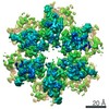
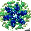
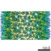
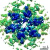
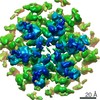
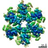
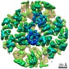
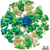
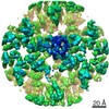
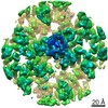
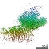
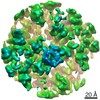
- PDB-6y9y: Structure of the native full-length HIV-1 capsid protein in compl... -
+ Open data

- Basic information
| Entry | Database: PDB / ID: 6y9y | |||||||||
|---|---|---|---|---|---|---|---|---|---|---|
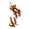
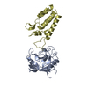
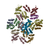
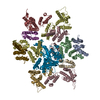
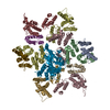
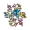
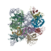
| Title | Structure of the native full-length HIV-1 capsid protein in complex with Cyclophilin A from helical assembly (-7,13) | |||||||||
 Components Components |
| |||||||||
 Keywords Keywords |  VIRAL PROTEIN / HIV / capsid / hexamer / helical assembly / curvature VIRAL PROTEIN / HIV / capsid / hexamer / helical assembly / curvature | |||||||||
| Function / homology |  Function and homology information Function and homology informationhost cellular component / Synthesis And Processing Of GAG, GAGPOL Polyproteins / host cell nuclear membrane / negative regulation of protein K48-linked ubiquitination / negative regulation of viral life cycle / regulation of apoptotic signaling pathway / cell adhesion molecule production / lipid droplet organization / heparan sulfate binding / regulation of viral genome replication ...host cellular component / Synthesis And Processing Of GAG, GAGPOL Polyproteins / host cell nuclear membrane / negative regulation of protein K48-linked ubiquitination / negative regulation of viral life cycle / regulation of apoptotic signaling pathway / cell adhesion molecule production / lipid droplet organization / heparan sulfate binding / regulation of viral genome replication / leukocyte chemotaxis / negative regulation of stress-activated MAPK cascade / endothelial cell activation / virion binding / Basigin interactions / Integration of viral DNA into host genomic DNA / Autointegration results in viral DNA circles / cyclosporin A binding / Minus-strand DNA synthesis / Plus-strand DNA synthesis / Uncoating of the HIV Virion / 2-LTR circle formation / viral budding via host ESCRT complex / Early Phase of HIV Life Cycle / Vpr-mediated nuclear import of PICs / Integration of provirus / APOBEC3G mediated resistance to HIV-1 infection / Calcineurin activates NFAT / viral release from host cell / Binding and entry of HIV virion / positive regulation of viral genome replication / protein peptidyl-prolyl isomerization / negative regulation of oxidative stress-induced intrinsic apoptotic signaling pathway / positive regulation of protein dephosphorylation / Membrane binding and targetting of GAG proteins / Gene and protein expression by JAK-STAT signaling after Interleukin-12 stimulation / activation of protein kinase B activity / neutrophil chemotaxis / negative regulation of protein phosphorylation / peptidylprolyl isomerase / peptidyl-prolyl cis-trans isomerase activity / positive regulation of protein secretion / negative regulation of protein kinase activity / Assembly Of The HIV Virion / HIV-1 retropepsin / : / retroviral ribonuclease H / Budding and maturation of HIV virion / exoribonuclease H / : / exoribonuclease H activity / neuron differentiation / platelet aggregation / platelet activation / host multivesicular body / DNA integration / RNA-directed DNA polymerase / viral genome integration into host DNA / viral penetration into host nucleus / establishment of integrated proviral latency / SARS-CoV-1 activates/modulates innate immune responses / RNA-directed DNA polymerase activity / Transferases; Transferring phosphorus-containing groups; Nucleotidyltransferases / unfolded protein binding / RNA-DNA hybrid ribonuclease activity / integrin binding / protein folding / Platelet degranulation / cellular response to oxidative stress / positive regulation of NF-kappaB transcription factor activity / viral nucleocapsid / secretory granule lumen / DNA recombination / vesicle / ficolin-1-rich granule lumen / positive regulation of MAPK cascade / Hydrolases; Acting on ester bonds / DNA-directed DNA polymerase / aspartic-type endopeptidase activity / DNA-directed DNA polymerase activity / positive regulation of protein phosphorylation / symbiont entry into host cell / symbiont-mediated suppression of host gene expression / focal adhesion / apoptotic process / lipid binding / host cell nucleus / Neutrophil degranulation / host cell plasma membrane / virion membrane / structural molecule activity / protein-containing complex / proteolysis / DNA binding / extracellular space / RNA binding / extracellular exosome / zinc ion binding / extracellular region / membraneSimilarity search - Function | |||||||||
| Biological species |  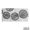 Human immunodeficiency virus 1 Human immunodeficiency virus 1 Homo sapiens (human) Homo sapiens (human) | |||||||||
| Method | ELECTRON MICROSCOPY / single particle reconstruction / cryo EM / Resolution: 6.1 Å | |||||||||
 Authors Authors | Ni, T. / Gerard, S. / Zhao, G. / Ning, J. / Zhang, P. | |||||||||
| Funding support |  United Kingdom, United Kingdom,  United States, 2items United States, 2items
| |||||||||
 Citation Citation | Journal: Nat Struct Mol Biol / Year: 2020 Title: Intrinsic curvature of the HIV-1 CA hexamer underlies capsid topology and interaction with cyclophilin A. Authors: Tao Ni / Samuel Gerard / Gongpu Zhao / Kyle Dent / Jiying Ning / Jing Zhou / Jiong Shi / Jordan Anderson-Daniels / Wen Li / Sooin Jang / Alan N Engelman / Christopher Aiken / Peijun Zhang / Abstract: The mature retrovirus capsid consists of a variably curved lattice of capsid protein (CA) hexamers and pentamers. High-resolution structures of the curved assembly, or in complex with host factors, ...The mature retrovirus capsid consists of a variably curved lattice of capsid protein (CA) hexamers and pentamers. High-resolution structures of the curved assembly, or in complex with host factors, have not been available. By devising cryo-EM methodologies for exceedingly flexible and pleomorphic assemblies, we have determined cryo-EM structures of apo-CA hexamers and in complex with cyclophilin A (CypA) at near-atomic resolutions. The CA hexamers are intrinsically curved, flexible and asymmetric, revealing the capsomere and not the previously touted dimer or trimer interfaces as the key contributor to capsid curvature. CypA recognizes specific geometries of the curved lattice, simultaneously interacting with three CA protomers from adjacent hexamers via two noncanonical interfaces, thus stabilizing the capsid. By determining multiple structures from various helical symmetries, we further revealed the essential plasticity of the CA molecule, which allows formation of continuously curved conical capsids and the mechanism of capsid pattern sensing by CypA. | |||||||||
| History |
|
- Structure visualization
| Movie |
Movie viewer |
|---|---|
| Structure viewer | Molecule: MolmilJmol/JSmol |
- Downloads & links
-Download
| PDBx/mmCIF format | 6y9y.cif.gz | 400.3 KB | Display | PDBx/mmCIF format |
|---|---|---|---|---|
| PDB format | pdb6y9y.ent.gz | 340.9 KB | Display | PDB format |
| PDBx/mmJSON format | 6y9y.json.gz | Tree view | PDBx/mmJSON format | |
| Others |  Other downloads Other downloads |
-Validation report
| Arichive directory | https://data.pdbj.org/pub/pdb/validation_reports/y9/6y9yftp://data.pdbj.org/pub/pdb/validation_reports/y9/6y9y | HTTPS FTP |
|---|
-Related structure data
| Related structure data | 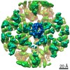 10741MC 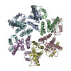 6skkC 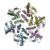 6skmC  6sknC 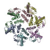 6slqC  6sluC  6smuC  6y9vC  6y9wC 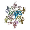 6y9xC 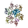 6y9zC 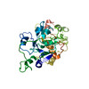 6yj5C 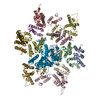 6zdjC M: map data used to model this data C: citing same article ( |
|---|---|
| Similar structure data |


-Links
 PDBj
PDBj


- Assembly
| Deposited unit | 
|
|---|---|
| 1 |
|
-Components
| #1: Protein | Mass: 24531.094 Da / Num. of mol.: 12 Source method: isolated from a genetically manipulated source Source: (gene. exp.) Human immunodeficiency virus 1 / Gene: gag-pol / Production host:  Escherichia coli (E. coli) Escherichia coli (E. coli)References: UniProt: P0C6F2, UniProt: P04591*PLUS, HIV-1 retropepsin, RNA-directed DNA polymerase, DNA-directed DNA polymerase, retroviral ribonuclease H, exoribonuclease H, Transferases; ...References: UniProt: P0C6F2, UniProt: P04591*PLUS, HIV-1 retropepsin, RNA-directed DNA polymerase, DNA-directed DNA polymerase, retroviral ribonuclease H, exoribonuclease H, Transferases; Transferring phosphorus-containing groups; Nucleotidyltransferases, Hydrolases; Acting on ester bonds#2: Protein | | Mass: 17905.307 Da / Num. of mol.: 1 Source method: isolated from a genetically manipulated source Source: (gene. exp.) Homo sapiens (human) / Gene: PPIA, CYPA / Production host: Escherichia coli (E. coli) / References: UniProt: P62937, peptidylprolyl isomerase |
|---|
-Experimental details
-Experiment
| Experiment | Method: ELECTRON MICROSCOPY |
|---|---|
| EM experiment | Aggregation state: HELICAL ARRAY / 3D reconstruction method: single particle reconstruction |
- Sample preparation
| Component |
| ||||||||||||||||||||||||
|---|---|---|---|---|---|---|---|---|---|---|---|---|---|---|---|---|---|---|---|---|---|---|---|---|---|
| Molecular weight | Experimental value: NO | ||||||||||||||||||||||||
| Source (natural) |
| ||||||||||||||||||||||||
| Source (recombinant) |
| ||||||||||||||||||||||||
| Buffer solution | pH: 8 | ||||||||||||||||||||||||
| Specimen | Conc.: 2 mg/ml / Embedding applied: NO / Shadowing applied: NO / Staining applied: NO / Vitrification applied: YES Details: Purified capsid protein were assembled in the presence of Cyclophilin A. | ||||||||||||||||||||||||
| Specimen support | Grid type: Quantifoil R2/1 | ||||||||||||||||||||||||
| Vitrification | Cryogen name: ETHANE |
- Electron microscopy imaging
| Experimental equipment |  Model: Titan Krios / Image courtesy: FEI Company |
|---|---|
| Microscopy | Model: FEI TITAN KRIOS |
| Electron gun | Electron source: FIELD EMISSION GUN / Accelerating voltage: 300 kV / Illumination mode: FLOOD BEAM |
| Electron lens | Mode: BRIGHT FIELDBright-field microscopy / Cs: 2.7 mm / C2 aperture diameter: 100 µm / Alignment procedure: COMA FREE |
| Specimen holder | Cryogen: NITROGEN / Specimen holder model: FEI TITAN KRIOS AUTOGRID HOLDER |
| Image recording | Electron dose: 40 e/Å2 / Detector mode: COUNTING / Film or detector model: GATAN K2 SUMMIT (4k x 4k) / Num. of real images: 6500 |
- Processing
| Software | Name: PHENIX / Version: dev_3699: / Classification: refinement | ||||||||||||||||||||||||||||||||||||||||
|---|---|---|---|---|---|---|---|---|---|---|---|---|---|---|---|---|---|---|---|---|---|---|---|---|---|---|---|---|---|---|---|---|---|---|---|---|---|---|---|---|---|
| EM software |
| ||||||||||||||||||||||||||||||||||||||||
| CTF correction | Type: PHASE FLIPPING AND AMPLITUDE CORRECTION | ||||||||||||||||||||||||||||||||||||||||
| 3D reconstruction | Resolution: 6.1 Å / Resolution method: FSC 0.143 CUT-OFF / Num. of particles: 91864 / Symmetry type: POINT | ||||||||||||||||||||||||||||||||||||||||
| Atomic model building | Protocol: RIGID BODY FIT / Space: REAL / Target criteria: Correlation coefficient | ||||||||||||||||||||||||||||||||||||||||
| Atomic model building |
| ||||||||||||||||||||||||||||||||||||||||
| Refine LS restraints |
|