 Movie
Movie Controller

[English] 日本語
 Yorodumi
- PDB-1r6w: Crystal structure of the K133R mutant of o-Succinylbenzoate synth... -
+ Open data

- Basic information
| Entry | Database: PDB / ID: 1r6w | ||||||
|---|---|---|---|---|---|---|---|
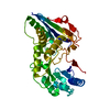
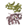
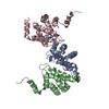
| Title | Crystal structure of the K133R mutant of o-Succinylbenzoate synthase (OSBS) from Escherichia coli. Complex with SHCHC | ||||||
 Components Components | o-Succinylbenzoate Synthase | ||||||
 Keywords Keywords | LYASE / Enolase superfamily / TIM barrel / capping alpha+beta domain | ||||||
| Function / homology |  Function and homology informationo-succinylbenzoate synthase / O-succinylbenzoate synthase activity / menaquinone biosynthetic process / hydro-lyase activity / magnesium ion binding Function and homology informationo-succinylbenzoate synthase / O-succinylbenzoate synthase activity / menaquinone biosynthetic process / hydro-lyase activity / magnesium ion bindingSimilarity search - Function | ||||||
| Biological species |  Escherichia coli (E. coli) Escherichia coli (E. coli) | ||||||
| Method | X-RAY DIFFRACTION / SYNCHROTRON / MOLECULAR REPLACEMENT / Resolution: 1.62 Å | ||||||
 Authors Authors | Klenchin, V.A. / Taylor Ringia, E.A. / Gerlt, J.A. / Rayment, I. | ||||||
 Citation Citation | Journal: Biochemistry / Year: 2003 Title: Evolution of Enzymatic Activity in the Enolase Superfamily: Structural and Mutagenic Studies of the Mechanism of the Reaction Catalyzed by o-Succinylbenzoate Synthase from Escherichia coli Authors: Klenchin, V.A. / Taylor Ringia, E.A. / Gerlt, J.A. / Rayment, I. | ||||||
| History |
|
- Structure visualization
| Structure viewer | Molecule: MolmilJmol/JSmol |
|---|
- Downloads & links
-Download
| PDBx/mmCIF format | 1r6w.cif.gz | 85.3 KB | Display | PDBx/mmCIF format |
|---|---|---|---|---|
| PDB format | pdb1r6w.ent.gz | 62.1 KB | Display | PDB format |
| PDBx/mmJSON format | 1r6w.json.gz | Tree view | PDBx/mmJSON format | |
| Others |  Other downloads Other downloads |
-Validation report
| Arichive directory | https://data.pdbj.org/pub/pdb/validation_reports/r6/1r6wftp://data.pdbj.org/pub/pdb/validation_reports/r6/1r6w | HTTPS FTP |
|---|
-Related structure data
| Related structure data | 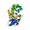 1fhvS S: Starting model for refinement |
|---|---|
| Similar structure data |


-Links
 PDBj
PDBj
- Assembly
| Deposited unit | 
| ||||||||||
|---|---|---|---|---|---|---|---|---|---|---|---|
| 1 | 
| ||||||||||
| Unit cell |
|
-Components
| #1: Protein | / E.C.4.2.1.- / OSB synthase / OSBS / 4-(2'-carboxyphenyl)-4-oxybutyric acid synthase / O-succinylbenzoic acid synthase Mass: 35765.734 Da / Num. of mol.: 1 / Mutation: K133R Source method: isolated from a genetically manipulated source Source: (gene. exp.) Escherichia coli (E. coli) / Production host: Escherichia coli (E. coli)References: UniProt: P29208, Lyases; Carbon-oxygen lyases; Hydro-lyases |
|---|---|
| #2: Chemical | ChemComp-MG /   Mass: 24.305 Da / Num. of mol.: 1 / Source method: obtained synthetically / Formula: Mg Mass: 24.305 Da / Num. of mol.: 1 / Source method: obtained synthetically / Formula: Mg |
| #3: Chemical | ChemComp-164 /   Mass: 240.209 Da / Num. of mol.: 1 / Source method: obtained synthetically / Formula: C11H12O6 Mass: 240.209 Da / Num. of mol.: 1 / Source method: obtained synthetically / Formula: C11H12O6 |
| #4: Water | ChemComp-HOH / Water Mass: 18.015 Da / Num. of mol.: 344 / Source method: isolated from a natural source / Formula: H2O Mass: 18.015 Da / Num. of mol.: 344 / Source method: isolated from a natural source / Formula: H2O |
-Experimental details
-Experiment
| Experiment | Method: X-RAY DIFFRACTION / Number of used crystals: 1 |
|---|
- Sample preparation
| Crystal | Density Matthews: 2.11 Å3/Da / Density % sol: 41.2 % | |||||||||||||||||||||||||||||||||||||||||||||||||
|---|---|---|---|---|---|---|---|---|---|---|---|---|---|---|---|---|---|---|---|---|---|---|---|---|---|---|---|---|---|---|---|---|---|---|---|---|---|---|---|---|---|---|---|---|---|---|---|---|---|---|
| Crystal grow | Temperature: 293 K / Method: microbatch / pH: 5.5 Details: The K133R mutant of OSBS was concentrated to 15 mg/ml, dialyzed against 5 mM Tris pH 8.3 containing 2 mM MgCl2, drop frozen as small pellets in liquid nitrogen and stored at -80 C. Crystals ...Details: The K133R mutant of OSBS was concentrated to 15 mg/ml, dialyzed against 5 mM Tris pH 8.3 containing 2 mM MgCl2, drop frozen as small pellets in liquid nitrogen and stored at -80 C. Crystals were grown at 20 C by small-scale batch experiments by combining 15 ml of protein solution and 15 ml of a solution containing 12-13% MePEG 5000, 100 mM sodium acetate, 60 mM MgCl2, at pH 5.5. SHCHC was included in the crystallization at a final concentration of approximately 2.5 mM., microbatch, temperature 293K | |||||||||||||||||||||||||||||||||||||||||||||||||
| Crystal grow | *PLUS Temperature: 20 ℃ / Method: batch method | |||||||||||||||||||||||||||||||||||||||||||||||||
| Components of the solutions | *PLUS
|
-Data collection
| Diffraction | Mean temperature: 150 K |
|---|---|
| Diffraction source | Source: SYNCHROTRON / Site: APS  / Beamline: 14-BM-D / Wavelength: 0.979 Å / Beamline: 14-BM-D / Wavelength: 0.979 Å |
| Radiation | Protocol: SINGLE WAVELENGTH / Monochromatic (M) / Laue (L): M / Scattering type: x-ray |
| Radiation wavelength | Wavelength: 0.979 Å / Relative weight: 1 |
| Reflection | Resolution: 1.62→31 Å / Num. all: 43694 / Num. obs: 43694 / % possible obs: 99.9 % / Observed criterion σ(F): 0 / Observed criterion σ(I): 0 / Redundancy: 7.1 % / Biso Wilson estimate: 25.9 Å2 / Rmerge(I) obs: 0.046 / Net I/σ(I): 41 |
| Reflection shell | Resolution: 1.62→1.68 Å / Rmerge(I) obs: 0.293 / Mean I/σ(I) obs: 17.1 / % possible all: 98.9 |
| Reflection | *PLUS Lowest resolution: 31.3 Å / % possible obs: 99.8 % / Redundancy: 7.05 % / Num. measured all: 307914 |
| Reflection shell | *PLUS % possible obs: 99.9 % |
- Processing
| Software |
| ||||||||||||||||||||||||||||||||||||||||||||||||||||||||||||||||||||||||||||||||||||||||||||||||||||
|---|---|---|---|---|---|---|---|---|---|---|---|---|---|---|---|---|---|---|---|---|---|---|---|---|---|---|---|---|---|---|---|---|---|---|---|---|---|---|---|---|---|---|---|---|---|---|---|---|---|---|---|---|---|---|---|---|---|---|---|---|---|---|---|---|---|---|---|---|---|---|---|---|---|---|---|---|---|---|---|---|---|---|---|---|---|---|---|---|---|---|---|---|---|---|---|---|---|---|---|---|---|
| Refinement | Method to determine structure: MOLECULAR REPLACEMENT Starting model: 1fhv Resolution: 1.62→30 Å / Cor.coef. Fo:Fc: 0.965 / Cor.coef. Fo:Fc free: 0.951 / SU B: 1.489 / SU ML: 0.052 / Cross valid method: THROUGHOUT / σ(F): 0 / σ(I): 0 / ESU R: 0.085 / ESU R Free: 0.086 / Details: HYDROGENS HAVE BEEN ADDED IN THE RIDING POSITIONS
| ||||||||||||||||||||||||||||||||||||||||||||||||||||||||||||||||||||||||||||||||||||||||||||||||||||
| Solvent computation | Ion probe radii: 0.8 Å / Shrinkage radii: 0.8 Å / VDW probe radii: 1.4 Å / Solvent model: BABINET MODEL WITH MASK | ||||||||||||||||||||||||||||||||||||||||||||||||||||||||||||||||||||||||||||||||||||||||||||||||||||
| Displacement parameters | Biso mean: 17.89 Å2
| ||||||||||||||||||||||||||||||||||||||||||||||||||||||||||||||||||||||||||||||||||||||||||||||||||||
| Refinement step | Cycle: LAST / Resolution: 1.62→30 Å
| ||||||||||||||||||||||||||||||||||||||||||||||||||||||||||||||||||||||||||||||||||||||||||||||||||||
| Refine LS restraints |
| ||||||||||||||||||||||||||||||||||||||||||||||||||||||||||||||||||||||||||||||||||||||||||||||||||||
| LS refinement shell | Resolution: 1.62→1.66 Å / Total num. of bins used: 20 /
| ||||||||||||||||||||||||||||||||||||||||||||||||||||||||||||||||||||||||||||||||||||||||||||||||||||
| Refinement | *PLUS Rfactor Rfree: 0.2 / Rfactor Rwork: 0.167 | ||||||||||||||||||||||||||||||||||||||||||||||||||||||||||||||||||||||||||||||||||||||||||||||||||||
| Solvent computation | *PLUS | ||||||||||||||||||||||||||||||||||||||||||||||||||||||||||||||||||||||||||||||||||||||||||||||||||||
| Displacement parameters | *PLUS | ||||||||||||||||||||||||||||||||||||||||||||||||||||||||||||||||||||||||||||||||||||||||||||||||||||
| Refine LS restraints | *PLUS
|
