 Movie
Movie Controller


+ Open data

- Basic information
| Entry | Database: PDB / ID: 3rgf | ||||||
|---|---|---|---|---|---|---|---|
| Title | Crystal Structure of human CDK8/CycC | ||||||
 Components Components |
| ||||||
 Keywords Keywords |  TRANSFERASE / TRANSCRIPTION / protein kinase complex TRANSFERASE / TRANSCRIPTION / protein kinase complex | ||||||
| Function / homology |  Function and homology information Function and homology informationCKM complex / G0 to G1 transition / negative regulation of triglyceride metabolic process / mediator complex / Generic Transcription Pathway / [RNA-polymerase]-subunit kinase / cyclin-dependent protein serine/threonine kinase regulator activity / negative regulation of Notch signaling pathway / cyclin-dependent protein kinase holoenzyme complex / cyclin-dependent kinase ...CKM complex / G0 to G1 transition / negative regulation of triglyceride metabolic process / mediator complex / Generic Transcription Pathway / [RNA-polymerase]-subunit kinase / cyclin-dependent protein serine/threonine kinase regulator activity / negative regulation of Notch signaling pathway / cyclin-dependent protein kinase holoenzyme complex / cyclin-dependent kinase / cyclin-dependent protein serine/threonine kinase activity / ubiquitin ligase complex / RNA polymerase II CTD heptapeptide repeat kinase activity / SMAD2/SMAD3:SMAD4 heterotrimer regulates transcription / PPARA activates gene expression / NOTCH1 Intracellular Domain Regulates Transcription / Constitutive Signaling by NOTCH1 PEST Domain Mutants / Constitutive Signaling by NOTCH1 HD+PEST Domain Mutants / Transcriptional regulation of white adipocyte differentiation / ubiquitin protein ligase activity / protein ubiquitination / protein kinase activity / phosphorylation / protein serine kinase activity / protein serine/threonine kinase activity / nucleolus / positive regulation of transcription by RNA polymerase II / protein-containing complex / nucleoplasm / ATP binding / identical protein binding / nucleusSimilarity search - Function | ||||||
| Biological species |  Homo sapiens (human) Homo sapiens (human) | ||||||
| Method | X-RAY DIFFRACTION / SYNCHROTRON / MOLECULAR REPLACEMENT / Resolution: 2.2 Å | ||||||
 Authors Authors | Schneider, E.V. / Boettcher, J. / Blaesse, M. / Huber, R. / Maskos, K. | ||||||
 Citation Citation | Journal: J.Mol.Biol. / Year: 2011 Title: The Structure of CDK8/CycC Implicates Specificity in the CDK/Cyclin Family and Reveals Interaction with a Deep Pocket Binder. Authors: Schneider, E.V. / Bottcher, J. / Blaesse, M. / Neumann, L. / Huber, R. / Maskos, K. | ||||||
| History |
|
- Structure visualization
| Structure viewer | Molecule: MolmilJmol/JSmol |
|---|
- Downloads & links
-Download
| PDBx/mmCIF format | 3rgf.cif.gz | 263.7 KB | Display | PDBx/mmCIF format |
|---|---|---|---|---|
| PDB format | pdb3rgf.ent.gz | 209.8 KB | Display | PDB format |
| PDBx/mmJSON format | 3rgf.json.gz | Tree view | PDBx/mmJSON format | |
| Others |  Other downloads Other downloads |
-Validation report
| Arichive directory | https://data.pdbj.org/pub/pdb/validation_reports/rg/3rgfftp://data.pdbj.org/pub/pdb/validation_reports/rg/3rgf | HTTPS FTP |
|---|
-Related structure data
| Related structure data |  1zp2S  2i53S  2pk2S  2r3nS  2r3qS  2rk3S 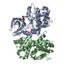 2uzeS 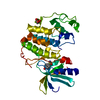 2vthS  2wh2 S: Starting model for refinement |
|---|---|
| Similar structure data |


-Links
 PDBj
PDBj


- Assembly
| Deposited unit | 
| ||||||||
|---|---|---|---|---|---|---|---|---|---|
| 1 |
| ||||||||
| Unit cell |
|
-Components
-Protein , 2 types, 2 molecules AB
| #1: Protein | / CDK8 / Cell division protein kinase 8 / Mediator complex subunit CDK8 / Mediator of RNA polymerase ...CDK8 / Cell division protein kinase 8 / Mediator complex subunit CDK8 / Mediator of RNA polymerase II transcription subunit CDK8 / Protein kinase K35 Mass: 46990.801 Da / Num. of mol.: 1 / Fragment: UNP residues 1-403 Source method: isolated from a genetically manipulated source Source: (gene. exp.) Homo sapiens (human) / Gene: CDK8 / Production host:   Spodoptera frugiperda (fall armyworm) Spodoptera frugiperda (fall armyworm)References: UniProt: P49336, cyclin-dependent kinase, [RNA-polymerase]-subunit kinase |
|---|---|
| #2: Protein | Mass: 33479.953 Da / Num. of mol.: 1 Source method: isolated from a genetically manipulated source Source: (gene. exp.) Homo sapiens (human) / Gene: CCNC / Production host: Spodoptera frugiperda (fall armyworm) / References: UniProt: P24863 |
-Non-polymers , 4 types, 188 molecules 


| #3: Chemical | ChemComp-BAX / Sorafenib Mass: 464.825 Da / Num. of mol.: 1 / Source method: obtained synthetically / Formula: C21H16ClF3N4O3 / Comment: inhibitor*YM Mass: 464.825 Da / Num. of mol.: 1 / Source method: obtained synthetically / Formula: C21H16ClF3N4O3 / Comment: inhibitor*YM | ||||
|---|---|---|---|---|---|
| #4: Chemical | ChemComp-EDO / Ethylene glycol Mass: 62.068 Da / Num. of mol.: 11 / Source method: obtained synthetically / Formula: C2H6O2 Mass: 62.068 Da / Num. of mol.: 11 / Source method: obtained synthetically / Formula: C2H6O2#5: Chemical | ChemComp-GOL / | Glycerol Mass: 92.094 Da / Num. of mol.: 1 / Source method: obtained synthetically / Formula: C3H8O3 Mass: 92.094 Da / Num. of mol.: 1 / Source method: obtained synthetically / Formula: C3H8O3#6: Water | ChemComp-HOH / | WaterMass: 18.015 Da / Num. of mol.: 175 / Source method: isolated from a natural source / Formula: H2O |
-Experimental details
-Experiment
| Experiment | Method: X-RAY DIFFRACTION / Number of used crystals: 1 |
|---|
- Sample preparation
| Crystal | Density Matthews: 2.15 Å3/Da / Density % sol: 42.87 % |
|---|---|
| Crystal grow | Temperature: 293 K / Method: vapor diffusion, hanging drop Details: 20% PEG3350, 0.2M lithium chloride, VAPOR DIFFUSION, HANGING DROP, temperature 293K |
-Data collection
| Diffraction | Mean temperature: 100 K |
|---|---|
| Diffraction source | Source: SYNCHROTRON / Site: SLS  / Beamline: X06SA / Wavelength: 1 / Beamline: X06SA / Wavelength: 1 |
| Detector | Type: PSI PILATUS 6M / Detector: PIXEL / Date: May 10, 2010 |
| Radiation | Protocol: SINGLE WAVELENGTH / Monochromatic (M) / Laue (L): M / Scattering type: x-ray |
| Radiation wavelength | Wavelength: 1 Å / Relative weight: 1 |
| Reflection | Resolution: 2.2→37.567 Å / Num. obs: 37725 / % possible obs: 100 % / Redundancy: 4.6 % / Rmerge(I) obs: 0.089 / Rsym value: 0.089 / Net I/σ(I): 10.7 |
| Reflection shell | Resolution: 2.2→2.32 Å / Redundancy: 4.6 % / Rmerge(I) obs: 0.542 / Mean I/σ(I) obs: 2.6 / Rsym value: 0.542 / % possible all: 99.9 |
- Processing
| Software |
| ||||||||||||||||||||||||||||||||||||||||||||||||||||||||||||||||||||||||||||||||||||||||||||||||||||
|---|---|---|---|---|---|---|---|---|---|---|---|---|---|---|---|---|---|---|---|---|---|---|---|---|---|---|---|---|---|---|---|---|---|---|---|---|---|---|---|---|---|---|---|---|---|---|---|---|---|---|---|---|---|---|---|---|---|---|---|---|---|---|---|---|---|---|---|---|---|---|---|---|---|---|---|---|---|---|---|---|---|---|---|---|---|---|---|---|---|---|---|---|---|---|---|---|---|---|---|---|---|
| Refinement | Method to determine structure: MOLECULAR REPLACEMENT Starting model: PDB ENTRIES 1ZP2, 2I53, 2PK2, 2WH2, 2PK2, 2RK3, 2UZE, 2R3Q, 2R3N, 2VTH Resolution: 2.2→37.567 Å / Cor.coef. Fo:Fc: 0.95 / Cor.coef. Fo:Fc free: 0.922 / Occupancy max: 1 / Occupancy min: 0 / SU B: 10.809 / SU ML: 0.128 / Cross valid method: THROUGHOUT / σ(F): 0 / ESU R: 0.228 / ESU R Free: 0.185 / Stereochemistry target values: MAXIMUM LIKELIHOOD Details: HYDROGENS HAVE BEEN ADDED IN THE RIDING POSITIONS U VALUES : RESIDUAL ONLY
| ||||||||||||||||||||||||||||||||||||||||||||||||||||||||||||||||||||||||||||||||||||||||||||||||||||
| Solvent computation | Ion probe radii: 0.8 Å / Shrinkage radii: 0.8 Å / VDW probe radii: 1.4 Å / Solvent model: BABINET MODEL WITH MASK | ||||||||||||||||||||||||||||||||||||||||||||||||||||||||||||||||||||||||||||||||||||||||||||||||||||
| Displacement parameters | Biso max: 105.28 Å2 / Biso mean: 44.536 Å2 / Biso min: 10.4 Å2
| ||||||||||||||||||||||||||||||||||||||||||||||||||||||||||||||||||||||||||||||||||||||||||||||||||||
| Refinement step | Cycle: LAST / Resolution: 2.2→37.567 Å
| ||||||||||||||||||||||||||||||||||||||||||||||||||||||||||||||||||||||||||||||||||||||||||||||||||||
| Refine LS restraints |
| ||||||||||||||||||||||||||||||||||||||||||||||||||||||||||||||||||||||||||||||||||||||||||||||||||||
| LS refinement shell | Resolution: 2.2→2.257 Å / Total num. of bins used: 20
| ||||||||||||||||||||||||||||||||||||||||||||||||||||||||||||||||||||||||||||||||||||||||||||||||||||
| Refinement TLS params. | Method: refined / Refine-ID: X-RAY DIFFRACTION
| ||||||||||||||||||||||||||||||||||||||||||||||||||||||||||||||||||||||||||||||||||||||||||||||||||||
| Refinement TLS group |
|
