[English] 日本語
Yorodumi
- EMDB-43892: Composite cryo-EM map of the Chlamydomonas reinhardtii mastigoneme -

+
Open data


ID or keywords:

Loading...

-
Basic information

Entry
Database: EMDB / ID: EMD-43892
TitleComposite cryo-EM map of the Chlamydomonas reinhardtii mastigoneme
Map dataComposite cryo-EM map of the C. reinhardtii mastigoneme
Sample
  • Complex: Native mastigonemes
KeywordsMastigoneme / cilia / STRUCTURAL PROTEIN
Function / homology
Function and homology information


Tyrosine-protein kinase ephrin type A/B receptor-like / Tyrosine-protein kinase ephrin type A/B receptor-like / Putative ephrin-receptor like / Lectin C-type domain / C-type lectin domain profile. / C-type lectin-like / C-type lectin (CTL) or carbohydrate-recognition domain (CRD) / C-type lectin-like/link domain superfamily / C-type lectin fold / Growth factor receptor cysteine-rich domain superfamily / Immunoglobulin-like fold
Similarity search - Domain/homology
C-type lectin domain-containing protein / Tyrosine-protein kinase ephrin type A/B receptor-like domain-containing protein
Similarity search - Component
Biological speciesChlamydomonas reinhardtii (plant)
Methodsingle particle reconstruction / cryo EM / Resolution: 3.1 Å
AuthorsDai J / Ma M / Zhang R / Brown A
Funding support United States, 6 items
OrganizationGrant numberCountry
National Institutes of Health/National Institute of General Medical Sciences (NIH/NIGMS)1R01GM141109 United States
National Institutes of Health/National Institute of General Medical Sciences (NIH/NIGMS)R01GM143183 United States
National Institutes of Health/National Institute of General Medical Sciences (NIH/NIGMS)1R01GM138854 United States
National Institutes of Health/National Institute of General Medical Sciences (NIH/NIGMS)R01GM139856 United States
National Institutes of Health/National Institute of General Medical Sciences (NIH/NIGMS)R35GM131909 United States
National Institutes of Health/National Heart, Lung, and Blood Institute (NIH/NHLBI)R01HL128370 United States
Citation
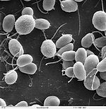
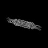
Journal: Cell / Year: 2024
Title: Mastigoneme structure reveals insights into the O-linked glycosylation code of native hydroxyproline-rich helices.
Authors: Jin Dai / Meisheng Ma / Qingwei Niu / Robyn J Eisert / Xiangli Wang / Poulomi Das / Karl F Lechtreck / Susan K Dutcher / Rui Zhang / Alan Brown /
Abstract: Hydroxyproline-rich glycoproteins (HRGPs) are a ubiquitous class of protein in the extracellular matrices and cell walls of plants and algae, yet little is known of their native structures or ...Hydroxyproline-rich glycoproteins (HRGPs) are a ubiquitous class of protein in the extracellular matrices and cell walls of plants and algae, yet little is known of their native structures or interactions. Here, we used electron cryomicroscopy (cryo-EM) to determine the structure of the hydroxyproline-rich mastigoneme, an extracellular filament isolated from the cilia of the alga Chlamydomonas reinhardtii. The structure demonstrates that mastigonemes are formed from two HRGPs (a filament of MST1 wrapped around a single copy of MST3) that both have hyperglycosylated poly(hydroxyproline) helices. Within the helices, O-linked glycosylation of the hydroxyproline residues and O-galactosylation of interspersed serine residues create a carbohydrate casing. Analysis of the associated glycans reveals how the pattern of hydroxyproline repetition determines the type and extent of glycosylation. MST3 possesses a PKD2-like transmembrane domain that forms a heteromeric polycystin-like cation channel with PKD2 and SIP, explaining how mastigonemes are tethered to ciliary membranes.
History
DepositionFeb 29, 2024-
Header (metadata) releaseApr 10, 2024-
Map releaseApr 10, 2024-
UpdateApr 24, 2024-
Current statusApr 24, 2024Processing site: RCSB / Status: Released

-
Structure visualization
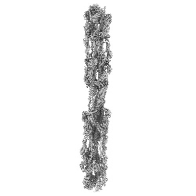
Supplemental images

Downloads & links

-
Map

FileDownload / File: emd_43892.map.gz / Format: CCP4 / Size: 512 MB / Type: IMAGE STORED AS FLOATING POINT NUMBER (4 BYTES)
AnnotationComposite cryo-EM map of the C. reinhardtii mastigoneme
Projections & slices

Image control

Size
Brightness
Contrast
Others
AxesZ (Sec.)Y (Row.)X (Col.)
1.39 Å/pix.
x 512 pix.
= 711.68 Å
1.39 Å/pix.
x 512 pix.
= 711.68 Å
1.39 Å/pix.
x 512 pix.
= 711.68 Å

Surface

Projections

Slices (1/3)

Slices (1/2)

Slices (2/3)

Images are generated by Spider.

Voxel sizeX=Y=Z: 1.39 Å
Density
Contour LevelBy AUTHOR: 0.5
Minimum - Maximum0.0 - 2.7712102
Average (Standard dev.)0.005317868 (±0.05399376)
SymmetrySpace group: 1
Details

EMDB XML:

Map geometry
Axis orderXYZ
Origin000
Dimensions512512512
Spacing512512512
CellA=B=C: 711.68 Å
α=β=γ: 90.0 °

-
Supplemental data

-
Sample components

-
Entire : Native mastigonemes

EntireName: Native mastigonemes
Components
  • Complex: Native mastigonemes

-
Supramolecule #1: Native mastigonemes

SupramoleculeName: Native mastigonemes / type: complex / ID: 1 / Parent: 0 / Macromolecule list: #1
Source (natural)Organism: Chlamydomonas reinhardtii (plant)

-
Experimental details

-
Structure determination

Methodcryo EM
Processingsingle particle reconstruction
Aggregation statefilament

-
Sample preparation

Concentration0.01 mg/mL
BufferpH: 7.4
Details: HMDEKP buffer (30 mM HEPES, 5 mM MgSO4, 1 mM DTT, 0.5 mM EGTA, 25 mM KCl, pH 7.4) containing 1x ProteaseArrest protease inhibitors (G-Biosciences)
GridModel: C-flat-1.2/1.3 / Material: COPPER / Mesh: 400 / Pretreatment - Type: GLOW DISCHARGE / Pretreatment - Time: 15 sec. / Pretreatment - Atmosphere: AIR
VitrificationCryogen name: ETHANE / Chamber humidity: 95 % / Chamber temperature: 277.15 K / Instrument: FEI VITROBOT MARK IV

-
Electron microscopy

MicroscopeFEI TITAN KRIOS
Electron beamAcceleration voltage: 300 kV / Electron source: FIELD EMISSION GUN
Electron opticsIllumination mode: FLOOD BEAM / Imaging mode: BRIGHT FIELDBright-field microscopy / Cs: 0.01 mm / Nominal defocus max: 3.5 µm / Nominal defocus min: 1.0 µm / Nominal magnification: 81000
Sample stageSpecimen holder model: FEI TITAN KRIOS AUTOGRID HOLDER / Cooling holder cryogen: NITROGEN
Image recordingFilm or detector model: GATAN K2 SUMMIT (4k x 4k) / Detector mode: COUNTING / Digitization - Dimensions - Width: 3838 pixel / Digitization - Dimensions - Height: 3710 pixel / Number real images: 19601 / Average exposure time: 9.0 sec. / Average electron dose: 38.9 e/Å2
Experimental equipment
Model: Titan Krios / Image courtesy: FEI Company

-
Image processing

Startup modelType of model: OTHER / Details: ab initio by cryoSPARC
Initial angle assignmentType: PROJECTION MATCHING / Software - Name: cryoSPARC
Final 3D classificationNumber classes: 2 / Software - Name: cryoSPARC
Final angle assignmentType: PROJECTION MATCHING / Software - Name: cryoSPARC (ver. 3.4)
Final reconstructionNumber classes used: 1 / Applied symmetry - Point group: C1 (asymmetric) / Algorithm: FOURIER SPACE / Resolution.type: BY AUTHOR / Resolution: 3.1 Å / Resolution method: FSC 0.143 CUT-OFF / Software - Name: cryoSPARC / Number images used: 687452

-
Atomic model buiding 1

RefinementProtocol: AB INITIO MODEL
Output model

PDB-9b4h:
Chlamydomonas reinhardtii mastigoneme filament

+
About Yorodumi

-
News

-
Feb 9, 2022. New format data for meta-information of EMDB entries

New format data for meta-information of EMDB entries

  • Version 3 of the EMDB header file is now the official format.
  • The previous official version 1.9 will be removed from the archive.

Related info.:EMDB header

External links:wwPDB to switch to version 3 of the EMDB data model

-
Aug 12, 2020. Covid-19 info

Covid-19 info

URL: https://pdbj.org/emnavi/covid19.php

New page: Covid-19 featured information page in EM Navigator.

Related info.:Covid-19 info / Mar 5, 2020. Novel coronavirus structure data

+
Mar 5, 2020. Novel coronavirus structure data

Novel coronavirus structure data

Related info.:Yorodumi Speices / Aug 12, 2020. Covid-19 info

External links:COVID-19 featured content - PDBj / Molecule of the Month (242):Coronavirus Proteases

+
Jan 31, 2019. EMDB accession codes are about to change! (news from PDBe EMDB page)

EMDB accession codes are about to change! (news from PDBe EMDB page)

  • The allocation of 4 digits for EMDB accession codes will soon come to an end. Whilst these codes will remain in use, new EMDB accession codes will include an additional digit and will expand incrementally as the available range of codes is exhausted. The current 4-digit format prefixed with “EMD-” (i.e. EMD-XXXX) will advance to a 5-digit format (i.e. EMD-XXXXX), and so on. It is currently estimated that the 4-digit codes will be depleted around Spring 2019, at which point the 5-digit format will come into force.
  • The EM Navigator/Yorodumi systems omit the EMD- prefix.

Related info.:Q: What is EMD? / ID/Accession-code notation in Yorodumi/EM Navigator

External links:EMDB Accession Codes are Changing Soon! / Contact to PDBj

+
Jul 12, 2017. Major update of PDB

Major update of PDB

  • wwPDB released updated PDB data conforming to the new PDBx/mmCIF dictionary.
  • This is a major update changing the version number from 4 to 5, and with Remediation, in which all the entries are updated.
  • In this update, many items about electron microscopy experimental information are reorganized (e.g. em_software).
  • Now, EM Navigator and Yorodumi are based on the updated data.

External links:wwPDB Remediation / Enriched Model Files Conforming to OneDep Data Standards Now Available in the PDB FTP Archive

-
Yorodumi

Thousand views of thousand structures

  • Yorodumi is a browser for structure data from EMDB, PDB, SASBDB, etc.
  • This page is also the successor to EM Navigator detail page, and also detail information page/front-end page for Omokage search.
  • The word "yorodu" (or yorozu) is an old Japanese word meaning "ten thousand". "mi" (miru) is to see.

Related info.:EMDB / PDB / SASBDB / Comparison of 3 databanks / Yorodumi Search / Aug 31, 2016. New EM Navigator & Yorodumi / Yorodumi Papers / Jmol/JSmol / Function and homology information / Changes in new EM Navigator and Yorodumi

Read more