Movie
Movie Controller


+ Open data

- Basic information
| Entry |  | |||||||||
|---|---|---|---|---|---|---|---|---|---|---|
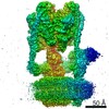
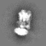
| Title | State 2 Yeast V-ATPase without Oxr1p bound | |||||||||
 Map data Map data | Yeast V-ATPase in state 2 | |||||||||
 Sample Sample |
| |||||||||
 Keywords Keywords | Vacuolar ATPase / Proton pump / Oxr1p / MEMBRANE PROTEIN | |||||||||
| Biological species |  Saccharomyces cerevisiae (brewer's yeast) Saccharomyces cerevisiae (brewer's yeast) | |||||||||
| Method | single particle reconstruction / cryo EM / Resolution: 8.1 Å | |||||||||
 Authors Authors | Khan MM / Wilkens S | |||||||||
| Funding support |  United States, 1 items United States, 1 items
| |||||||||
 Citation Citation | Journal: EMBO Rep / Year: 2024 Title: Molecular mechanism of Oxr1p mediated disassembly of yeast V-ATPase. Authors: Md Murad Khan / Stephan Wilkens / Abstract: The eukaryotic vacuolar H-ATPase (V-ATPase) is regulated by reversible disassembly into autoinhibited V-ATPase and V proton channel subcomplexes. We recently reported that the TLDc protein Oxr1p ...The eukaryotic vacuolar H-ATPase (V-ATPase) is regulated by reversible disassembly into autoinhibited V-ATPase and V proton channel subcomplexes. We recently reported that the TLDc protein Oxr1p induces V-ATPase disassembly in vitro. Whether and how Oxr1p is involved in enzyme disassembly in vivo, however, is not known. Here, using yeast genetics and fluorescence microscopy, we show that Oxr1p is essential for efficient V-ATPase disassembly in the cell. Supporting biochemical and biophysical in vitro experiments show that whereas Oxr1p-driven holoenzyme disassembly can occur in the absence of nucleotides, the presence of ATP greatly accelerates the process. ATP hydrolysis is needed, however, for subsequent release of Oxr1p so that the free V can adopt the autoinhibited conformation. Overall, our study unravels the molecular mechanism of Oxr1p-induced disassembly that occurs in vivo as part of the canonical V-ATPase regulation by reversible disassembly. | |||||||||
| History |
|
- Structure visualization
| Supplemental images |
|---|

- Downloads & links
-EMDB archive
| Map data | emd_42881.map.gz | 11.9 MB |  EMDB map data format EMDB map data format | |
|---|---|---|---|---|
| Header (meta data) | emd-42881-v30.xmlemd-42881.xml | 12.8 KB 12.8 KB | Display Display | EMDB header |
| FSC (resolution estimation) | emd_42881_fsc.xml | 5.8 KB | Display | FSC data file |
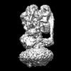
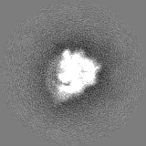
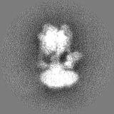
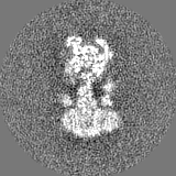
| Images | 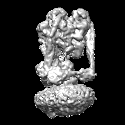 emd_42881.png emd_42881.png | 73.1 KB | ||
| Masks | emd_42881_msk_1.map | 15.6 MB | Mask map | |
| Filedesc metadata | emd-42881.cif.gz | 3.9 KB | ||
| Others | emd_42881_half_map_1.map.gzemd_42881_half_map_2.map.gz | 11.9 MB 11.9 MB | ||
| Archive directory |  http://ftp.pdbj.org/pub/emdb/structures/EMD-42881ftp://ftp.pdbj.org/pub/emdb/structures/EMD-42881 http://ftp.pdbj.org/pub/emdb/structures/EMD-42881ftp://ftp.pdbj.org/pub/emdb/structures/EMD-42881 | HTTPS FTP |
-Related structure data
-Links
| EMDB pages | EMDB (EBI/PDBe) / EMDataResource |
|---|
-Map
| File | Download / File: emd_42881.map.gz / Format: CCP4 / Size: 15.6 MB / Type: IMAGE STORED AS FLOATING POINT NUMBER (4 BYTES) | ||||||||||||||||||||
|---|---|---|---|---|---|---|---|---|---|---|---|---|---|---|---|---|---|---|---|---|---|
| Annotation | Yeast V-ATPase in state 2 | ||||||||||||||||||||
| Voxel size | X=Y=Z: 2.85 Å | ||||||||||||||||||||
| Density |
| ||||||||||||||||||||
| Symmetry | Space group: 1 | ||||||||||||||||||||
| Details | EMDB XML:
|
-Supplemental data
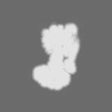
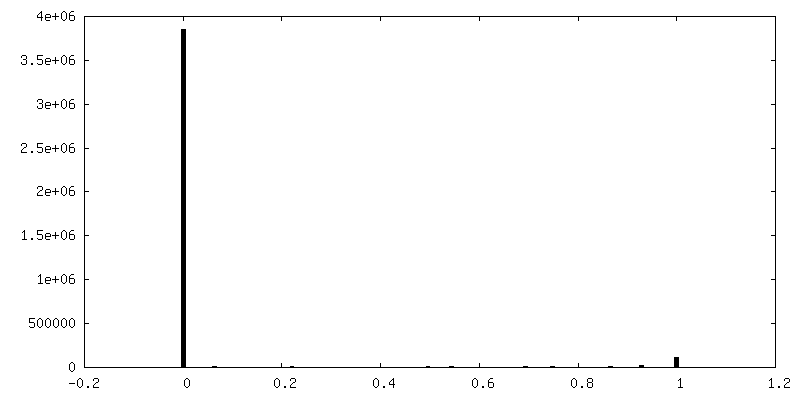
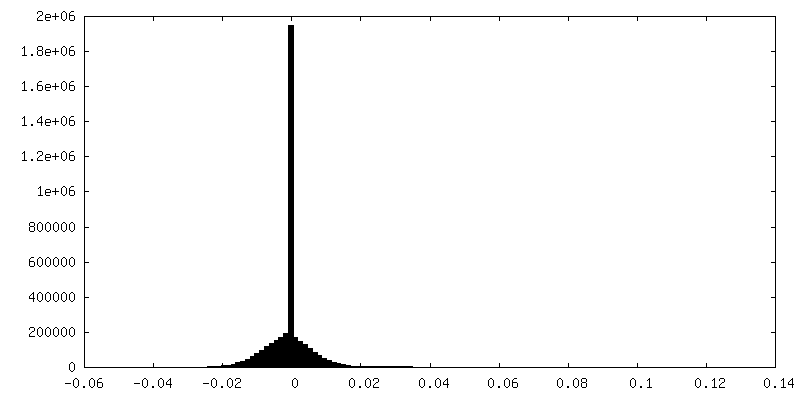
-Mask #1
| File | emd_42881_msk_1.map | ||||||||||||
|---|---|---|---|---|---|---|---|---|---|---|---|---|---|
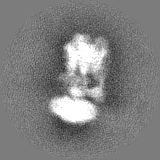
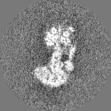
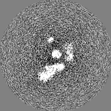
| Projections & Slices |
| ||||||||||||
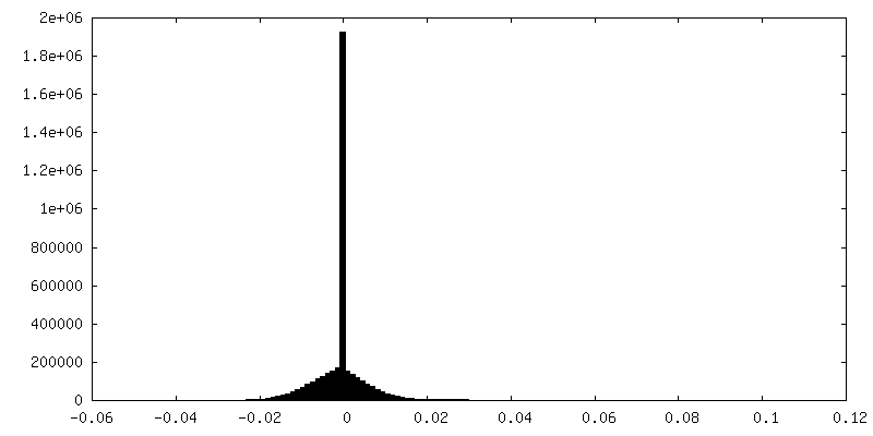
| Density Histograms |
 Z
Z Y
Y X
X


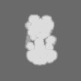
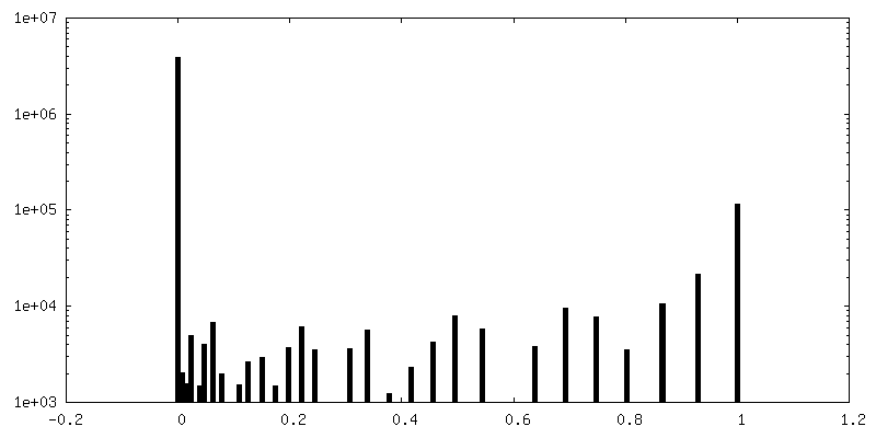
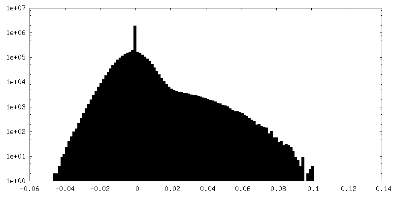
-Half map: #1
| File | emd_42881_half_map_1.map | ||||||||||||
|---|---|---|---|---|---|---|---|---|---|---|---|---|---|
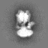
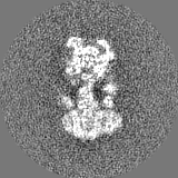
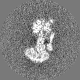
| Projections & Slices |
| ||||||||||||
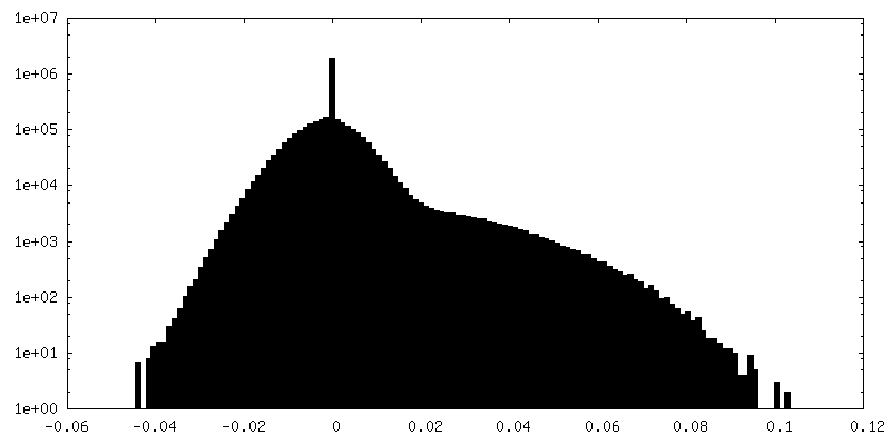
| Density Histograms |
-Half map: #2
| File | emd_42881_half_map_2.map | ||||||||||||
|---|---|---|---|---|---|---|---|---|---|---|---|---|---|
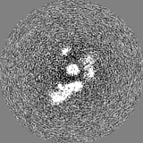
| Projections & Slices |
| ||||||||||||
| Density Histograms |

- Sample components
-Entire : Yeast Vacuolar ATPase vitrified in presence of Oxr1p
| Entire | Name: Yeast Vacuolar ATPase vitrified in presence of Oxr1p |
|---|---|
| Components |
|
-Supramolecule #1: Yeast Vacuolar ATPase vitrified in presence of Oxr1p
| Supramolecule | Name: Yeast Vacuolar ATPase vitrified in presence of Oxr1p / type: complex / ID: 1 / Parent: 0 |
|---|---|
| Source (natural) | Organism: Saccharomyces cerevisiae (brewer's yeast) / Organelle: Vacuole |
| Molecular weight | Theoretical: 1 MDa |
-Experimental details
-Structure determination
| Method | cryo EM |
|---|---|
 Processing Processing | single particle reconstruction |
| Aggregation state | particle |
-Sample preparation
| Concentration | 2 mg/mL |
|---|---|
| Buffer | pH: 7.4 / Details: TBS |
| Vitrification | Cryogen name: ETHANE / Chamber humidity: 85 % / Chamber temperature: 279 K / Instrument: LEICA EM GP |
- Electron microscopy
| Microscope | JEOL 2100F |
|---|---|
| Electron beam | Acceleration voltage: 200 kV / Electron source: FIELD EMISSION GUN |
| Electron optics | Illumination mode: FLOOD BEAM / Imaging mode: BRIGHT FIELDBright-field microscopy / Nominal defocus max: 3.5 µm / Nominal defocus min: 1.2 µm |
| Sample stage | Specimen holder model: GATAN 914 HIGH TILT LIQUID NITROGEN CRYO TRANSFER TOMOGRAPHY HOLDER Cooling holder cryogen: NITROGEN |
| Image recording | Film or detector model: GATAN K2 SUMMIT (4k x 4k) / Detector mode: COUNTING / Average electron dose: 50.0 e/Å2 |
-Image processing
| Startup model | Type of model: EMDB MAP EMDB ID: |
|---|---|
| Initial angle assignment | Type: PROJECTION MATCHING |
| Final angle assignment | Type: PROJECTION MATCHING / Software - Name: RELION (ver. 4) |
| Final reconstruction | Resolution.type: BY AUTHOR / Resolution: 8.1 Å / Resolution method: FSC 0.143 CUT-OFF / Software - Name: RELION (ver. 4) / Number images used: 13950 |
| FSC plot (resolution estimation) | 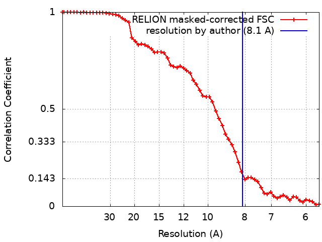 |