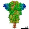
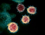
Journal: Proc Natl Acad Sci U S A / Year: 2023 Title: In situ architecture and membrane fusion of SARS-CoV-2 Delta variant. Authors: Yutong Song / Hangping Yao / Nanping Wu / Jialu Xu / Zheyuan Zhang / Cheng Peng / Shibo Li / Weizheng Kong / Yong Chen / Miaojin Zhu / Jiaqi Wang / Danrong Shi / Chongchong Zhao / Xiangyun ...Authors: Yutong Song / Hangping Yao / Nanping Wu / Jialu Xu / Zheyuan Zhang / Cheng Peng / Shibo Li / Weizheng Kong / Yong Chen / Miaojin Zhu / Jiaqi Wang / Danrong Shi / Chongchong Zhao / Xiangyun Lu / Martín Echavarría Galindo / Sai Li / Abstract: Among the current five Variants of Concern, infections caused by SARS-CoV-2 B.1.617.2 (Delta) variant are often associated with the greatest severity. Despite recent advances on the molecular basis ...Among the current five Variants of Concern, infections caused by SARS-CoV-2 B.1.617.2 (Delta) variant are often associated with the greatest severity. Despite recent advances on the molecular basis of elevated pathogenicity using recombinant proteins, the architecture of intact Delta virions remains veiled. Moreover, pieces of molecular evidence for the detailed mechanism of S-mediated membrane fusion are missing. Here, we showed the pleomorphic nature of Delta virions from electron beam inactivated samples and reported the in situ structure and distribution of S on the authentic Delta variant. We also captured the virus-virus fusion events, which provided pieces of structural evidence for Delta's attenuated dependency on cellular factors for fusion activation, and proposed a model of S-mediated membrane fusion. Besides, site-specific glycan analysis revealed increased oligomannose-type glycosylation of native Delta S than that of the WT S. Together, these results disclose distinctive factors of Delta being the most virulent SARS-CoV-2 variant.
In the structure databanks used in Yorodumi, some data are registered as the other names, "COVID-19 virus" and "2019-nCoV". Here are the details of the virus and the list of structure data.
Jan 31, 2019. EMDB accession codes are about to change! (news from PDBe EMDB page)
EMDB accession codes are about to change! (news from PDBe EMDB page)
The allocation of 4 digits for EMDB accession codes will soon come to an end. Whilst these codes will remain in use, new EMDB accession codes will include an additional digit and will expand incrementally as the available range of codes is exhausted. The current 4-digit format prefixed with “EMD-” (i.e. EMD-XXXX) will advance to a 5-digit format (i.e. EMD-XXXXX), and so on. It is currently estimated that the 4-digit codes will be depleted around Spring 2019, at which point the 5-digit format will come into force.
The EM Navigator/Yorodumi systems omit the EMD- prefix.
Related info.:Q: What is EMD? / ID/Accession-code notation in Yorodumi/EM Navigator
Yorodumi is a browser for structure data from EMDB, PDB, SASBDB, etc.
This page is also the successor to EM Navigator detail page, and also detail information page/front-end page for Omokage search.
The word "yorodu" (or yorozu) is an old Japanese word meaning "ten thousand". "mi" (miru) is to see.
Related info.:EMDB / PDB / SASBDB / Comparison of 3 databanks / Yorodumi Search / Aug 31, 2016. New EM Navigator & Yorodumi / Yorodumi Papers / Jmol/JSmol / Function and homology information / Changes in new EM Navigator and Yorodumi
 Movie
Movie Controller

 Yorodumi
 Open data
 Basic information
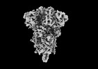
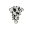
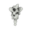
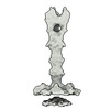
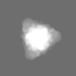
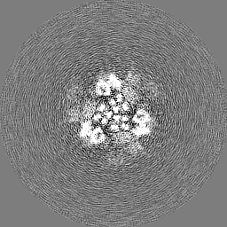
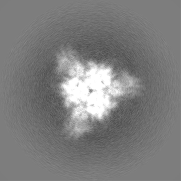
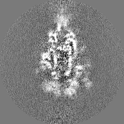
 Map data
 Sample
 Function and homology information
 membrane fusion / positive regulation of viral entry into host cell / receptor-mediated virion attachment to host cell /
 Authors
 China, 1 items
  Citation
 Structure visualization
 Downloads & links
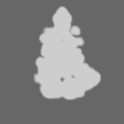
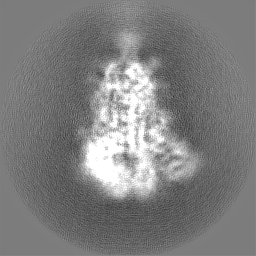
 emd_33205.png
 http://ftp.pdbj.org/pub/emdb/structures/EMD-33205
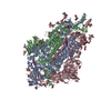


 Z
Z Y
Y X
X


 Sample components
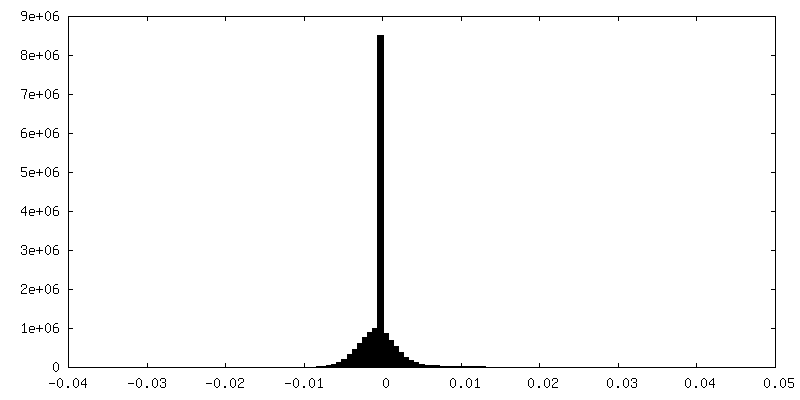
 Processing
 Electron microscopy