Movie
Movie Controller


+ Open data

- Basic information
| Entry | 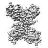 | |||||||||
|---|---|---|---|---|---|---|---|---|---|---|
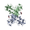
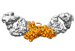
| Title | CryoEM structure of a dimer of loose sNS1 tetramer | |||||||||
 Map data Map data | ||||||||||
 Sample Sample |
| |||||||||
| Function / homology |  Function and homology information Function and homology informationsymbiont-mediated suppression of host JAK-STAT cascade via inhibition of host TYK2 activity / host cell mitochondrion / symbiont-mediated suppression of host JAK-STAT cascade via inhibition of STAT2 activity / symbiont-mediated suppression of host cytoplasmic pattern recognition receptor signaling pathway via inhibition of MAVS activity / ribonucleoside triphosphate phosphatase activity / : /  viral capsid / double-stranded RNA binding / protein complex oligomerization / monoatomic ion channel activity ...symbiont-mediated suppression of host JAK-STAT cascade via inhibition of host TYK2 activity / host cell mitochondrion / symbiont-mediated suppression of host JAK-STAT cascade via inhibition of STAT2 activity / symbiont-mediated suppression of host cytoplasmic pattern recognition receptor signaling pathway via inhibition of MAVS activity / ribonucleoside triphosphate phosphatase activity / : / viral capsid / double-stranded RNA binding / protein complex oligomerization / monoatomic ion channel activity / clathrin-dependent endocytosis of virus by host cell / mRNA (nucleoside-2'-O-)-methyltransferase activity / mRNA 5'-cap (guanine-N7-)-methyltransferase activity / RNA helicase activity / host cell endoplasmic reticulum membrane / protein dimerization activity / induction by virus of host autophagy / viral RNA genome replication / RNA-dependent RNA polymerase activity / serine-type endopeptidase activity / fusion of virus membrane with host endosome membrane / viral envelope / symbiont-mediated suppression of host type I interferon-mediated signaling pathway / host cell nucleus / virion attachment to host cell / virion membrane / structural molecule activity / proteolysis / extracellular region / ATP binding / membrane / metal ion binding viral capsid / double-stranded RNA binding / protein complex oligomerization / monoatomic ion channel activity ...symbiont-mediated suppression of host JAK-STAT cascade via inhibition of host TYK2 activity / host cell mitochondrion / symbiont-mediated suppression of host JAK-STAT cascade via inhibition of STAT2 activity / symbiont-mediated suppression of host cytoplasmic pattern recognition receptor signaling pathway via inhibition of MAVS activity / ribonucleoside triphosphate phosphatase activity / : / viral capsid / double-stranded RNA binding / protein complex oligomerization / monoatomic ion channel activity / clathrin-dependent endocytosis of virus by host cell / mRNA (nucleoside-2'-O-)-methyltransferase activity / mRNA 5'-cap (guanine-N7-)-methyltransferase activity / RNA helicase activity / host cell endoplasmic reticulum membrane / protein dimerization activity / induction by virus of host autophagy / viral RNA genome replication / RNA-dependent RNA polymerase activity / serine-type endopeptidase activity / fusion of virus membrane with host endosome membrane / viral envelope / symbiont-mediated suppression of host type I interferon-mediated signaling pathway / host cell nucleus / virion attachment to host cell / virion membrane / structural molecule activity / proteolysis / extracellular region / ATP binding / membrane / metal ion bindingSimilarity search - Function | |||||||||
| Biological species |  Dengue virus 2 Dengue virus 2 | |||||||||
| Method | single particle reconstruction / cryo EM / Resolution: 3.4 Å | |||||||||
 Authors Authors | Shu B / Lok SM | |||||||||
| Funding support |  Singapore, 1 items Singapore, 1 items
| |||||||||
 Citation Citation | Journal: Nat Commun / Year: 2022 Title: CryoEM structures of the multimeric secreted NS1, a major factor for dengue hemorrhagic fever. Authors: Bo Shu / Justin S G Ooi / Aaron W K Tan / Thiam-Seng Ng / Wanwisa Dejnirattisai / Juthathip Mongkolsapaya / Guntur Fibriansah / Jian Shi / Victor A Kostyuchenko / Gavin R Screaton / Shee-Mei Lok /  Abstract: Dengue virus infection can cause dengue hemorrhagic fever (DHF). Dengue NS1 is multifunctional. The intracellular dimeric NS1 (iNS1) forms part of the viral replication complex. Previous studies ...Dengue virus infection can cause dengue hemorrhagic fever (DHF). Dengue NS1 is multifunctional. The intracellular dimeric NS1 (iNS1) forms part of the viral replication complex. Previous studies suggest the extracellular secreted NS1 (sNS1), which is a major factor contributing to DHF, exists as hexamers. The structure of the iNS1 is well-characterised but not that of sNS1. Here we show by cryoEM that the recombinant sNS1 exists in multiple oligomeric states: the tetrameric (stable and loose conformation) and hexameric structures. Stability of the stable and loose tetramers is determined by the conformation of their N-terminal domain - elongated β-sheet or β-roll. Binding of an anti-NS1 Fab breaks the loose tetrameric and hexameric sNS1 into dimers, whereas the stable tetramer remains largely unbound. Our results show detailed quaternary organization of different oligomeric states of sNS1 and will contribute towards the design of dengue therapeutics. | |||||||||
| History |
|
- Structure visualization
| Supplemental images |
|---|

- Downloads & links
-EMDB archive
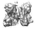
| Map data | emd_32840.map.gz | 14.5 MB | EMDB map data format | |
|---|---|---|---|---|
| Header (meta data) | emd-32840-v30.xmlemd-32840.xml | 10.1 KB 10.1 KB | Display Display | EMDB header |
| Images |  emd_32840.png emd_32840.png | 109.9 KB | ||
| Archive directory |  http://ftp.pdbj.org/pub/emdb/structures/EMD-32840ftp://ftp.pdbj.org/pub/emdb/structures/EMD-32840 http://ftp.pdbj.org/pub/emdb/structures/EMD-32840ftp://ftp.pdbj.org/pub/emdb/structures/EMD-32840 | HTTPS FTP |
-Related structure data
| Related structure data | 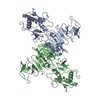 7wusMC  7wurC  7wutC 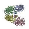 7wuuC  7wuvC M: atomic model generated by this map C: citing same article ( |
|---|---|
| Similar structure data |


-Links
| EMDB pages | EMDB (EBI/PDBe) / EMDataResource |
|---|---|
| Related items in Molecule of the Month |


-Map
| File | Download / File: emd_32840.map.gz / Format: CCP4 / Size: 15.6 MB / Type: IMAGE STORED AS FLOATING POINT NUMBER (4 BYTES) | ||||||||||||||||||||
|---|---|---|---|---|---|---|---|---|---|---|---|---|---|---|---|---|---|---|---|---|---|
| Voxel size | X=Y=Z: 1.32 Å | ||||||||||||||||||||
| Density |
| ||||||||||||||||||||
| Symmetry | Space group: 1 | ||||||||||||||||||||
| Details | EMDB XML:
|
-Supplemental data
- Sample components
-Entire : Dengue NS1 dimer of loose tetramer
| Entire | Name: Dengue NS1 dimer of loose tetramer |
|---|---|
| Components |
|
-Supramolecule #1: Dengue NS1 dimer of loose tetramer
| Supramolecule | Name: Dengue NS1 dimer of loose tetramer / type: organelle_or_cellular_component / ID: 1 / Parent: 0 / Macromolecule list: all |
|---|---|
| Source (natural) | Organism: Dengue virus 2 |
-Macromolecule #1: Core protein
| Macromolecule | Name: Core protein / type: protein_or_peptide / ID: 1 / Number of copies: 2 / Enantiomer: LEVO |
|---|---|
| Source (natural) | Organism: Dengue virus 2 |
| Molecular weight | Theoretical: 38.875215 KDa |
| Recombinant expression | Organism:  Homo sapiens (human) Homo sapiens (human) |
| Sequence | String: DSGCVVSWKN KELKCGSGIF ITDNVHTWTE QYKFQPESPS KLASAIQKAH EEGICGIRSV TRLENLMWKQ ITPELNHILT ENEVKLTIM TGDIKGIMQA GKRSLRPQPT ELKYSWKAWG KAKMLSTELH NHTFLIDGPE TAECPNTNRA WNSLEVEDYG F GVFTTNIW ...String: DSGCVVSWKN KELKCGSGIF ITDNVHTWTE QYKFQPESPS KLASAIQKAH EEGICGIRSV TRLENLMWKQ ITPELNHILT ENEVKLTIM TGDIKGIMQA GKRSLRPQPT ELKYSWKAWG KAKMLSTELH NHTFLIDGPE TAECPNTNRA WNSLEVEDYG F GVFTTNIW LKLKERQDVF CDSKLMSAAI KDNRAVHADM GYWIESALND TWKIEKASFI EVKSCHWPKS HTLWSNGVLE SE MIIPKNF AGPVSQHNYR PGYHTQTAGP WHLGRLEMDF DFCEGTTVVV TEDCGNRGPS LRTTTASGKL ITEWCCRSCT LPP LRYRGE DGCWYGMEIR PLKEK |
-Experimental details
-Structure determination
| Method | cryo EM |
|---|---|
 Processing Processing | single particle reconstruction |
| Aggregation state | particle |
-Sample preparation
| Concentration | 0.2 mg/mL |
|---|---|
| Buffer | pH: 8 |
| Vitrification | Cryogen name: ETHANE |
- Electron microscopy
| Microscope | FEI TITAN KRIOS |
|---|---|
| Electron beam | Acceleration voltage: 300 kV / Electron source: FIELD EMISSION GUN |
| Electron optics | Illumination mode: FLOOD BEAM / Imaging mode: BRIGHT FIELDBright-field microscopy / Nominal defocus max: 2.0 µm / Nominal defocus min: 0.8 µm |
| Image recording | Film or detector model: GATAN K3 (6k x 4k) / Average electron dose: 81.5 e/Å2 |
| Experimental equipment |  Model: Titan Krios / Image courtesy: FEI Company |
-Image processing
| Startup model | Type of model: PDB ENTRY PDB model - PDB ID: |
|---|---|
| Initial angle assignment | Type: MAXIMUM LIKELIHOOD |
| Final angle assignment | Type: MAXIMUM LIKELIHOOD |
| Final reconstruction | Resolution.type: BY AUTHOR / Resolution: 3.4 Å / Resolution method: FSC 0.143 CUT-OFF / Number images used: 181587 |