Entry Database : PDB / ID : 5gs6Title Full-length NS1 structure of Zika virus from 2015 Brazil strain NS1 of Zika virus from 2015 Brazil strain Keywords / / / / Function / homology Function Domain/homology Component
/ / / / / / / / / / / / / / / / / / / / / / / / / / / / / / / / / / / / / / / / / / / / / / / / / / / / / / / / / / / / / / / / / / / / / / / / / / / / / / / / / / / / / / / / / / / / / / / / / / / / / / Biological species Method / / / Resolution : 2.852 Å Authors Xu, X.Y. / Song, H. / Qi, J.X. / Shi, Y. / Gao, G.F. Journal : Embo J. / Year : 2016Title : Contribution of intertwined loop to membrane association revealed by Zika virus full-length NS1 structureAuthors : Xu, X. / Song, H. / Qi, J. / Liu, Y. / Wang, H. / Su, C. / Shi, Y. / Gao, G.F. History Deposition Aug 14, 2016 Deposition site / Processing site Revision 1.0 Oct 5, 2016 Provider / Type Revision 1.1 Nov 16, 2016 Group Revision 1.2 Jul 29, 2020 Group Data collection / Database references ... Data collection / Database references / Derived calculations / Structure summary Category chem_comp / citation ... chem_comp / citation / entity / pdbx_chem_comp_identifier / pdbx_entity_nonpoly / pdbx_struct_oper_list / struct_conn / struct_site / struct_site_gen Item _chem_comp.name / _chem_comp.type ... _chem_comp.name / _chem_comp.type / _citation.journal_id_CSD / _entity.pdbx_description / _pdbx_entity_nonpoly.name / _pdbx_struct_oper_list.symmetry_operation / _struct_conn.pdbx_role Description / Provider / Type Revision 1.3 Nov 8, 2023 Group Data collection / Database references ... Data collection / Database references / Refinement description / Structure summary Category chem_comp / chem_comp_atom ... chem_comp / chem_comp_atom / chem_comp_bond / database_2 / pdbx_initial_refinement_model Item / _database_2.pdbx_DOI / _database_2.pdbx_database_accessionRevision 1.4 Mar 20, 2024 Group / Category
Show all Show less
 Movie
Movie Controller


 Open data
 Basic information
 Components
 Keywords
 VIRAL PROTEIN /
  Function and homology information
 Authors
 Citation
 Structure visualization
 Downloads & links
 Other downloads
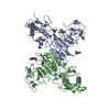


 PDBj
PDBj

 Assembly


 Type: D-saccharide, beta linking / Mass: 221.208 Da / Num. of mol.: 2
 Sample preparation
 / Beamline: BL17U / Wavelength: 0.97853 Å
 Processing