 Movie
Movie Controller


+ Open data

- Basic information
| Entry | 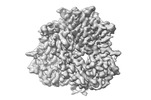 | |||||||||
|---|---|---|---|---|---|---|---|---|---|---|
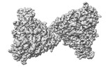
| Title | Prefusion-stabilized Nipah virus fusion protein | |||||||||
 Map data Map data | Prefusion-stabilized Nipah virus fusion protein | |||||||||
 Sample Sample |
| |||||||||
 Keywords Keywords | Nipah /  Nipah virus / NiV / fusion / F / antibody / neutralizing / conserved epitope / neutralizing antibody / prefusion / prefusion-stabilized / vaccine / vaccine design / antigen / antigen design / VIRAL PROTEIN Nipah virus / NiV / fusion / F / antibody / neutralizing / conserved epitope / neutralizing antibody / prefusion / prefusion-stabilized / vaccine / vaccine design / antigen / antigen design / VIRAL PROTEIN | |||||||||
| Function / homology |  Function and homology information Function and homology informationmembrane fusion involved in viral entry into host cell / symbiont entry into host cell / fusion of virus membrane with host plasma membrane / viral envelope / host cell plasma membrane / virion membrane / membraneSimilarity search - Function | |||||||||
| Biological species |  Nipah henipavirus Nipah henipavirus | |||||||||
| Method | single particle reconstruction / cryo EM / Resolution: 3.0 Å | |||||||||
 Authors Authors | Byrne PO / Blade EG / McLellan JS | |||||||||
| Funding support |  United States, 1 items United States, 1 items
| |||||||||
 Citation Citation | Journal: J Virol / Year: 2024 Title: Prefusion stabilization of the Hendra and Langya virus F proteins. Authors: Patrick O Byrne / Elizabeth G Blade / Brian E Fisher / David R Ambrozak / Ajit R Ramamohan / Barney S Graham / Rebecca J Loomis / Jason S McLellan / Abstract: Nipah virus (NiV) and Hendra virus (HeV) are pathogenic paramyxoviruses that cause mild-to-severe disease in humans. As members of the genus, NiV and HeV use an attachment (G) glycoprotein and a ...Nipah virus (NiV) and Hendra virus (HeV) are pathogenic paramyxoviruses that cause mild-to-severe disease in humans. As members of the genus, NiV and HeV use an attachment (G) glycoprotein and a class I fusion (F) glycoprotein to invade host cells. The F protein rearranges from a metastable prefusion form to an extended postfusion form to facilitate host cell entry. Prefusion NiV F elicits higher neutralizing antibody titers than postfusion NiV F, indicating that stabilization of prefusion F may aid vaccine development. A combination of amino acid substitutions (L104C/I114C, L172F, and S191P) is known to stabilize NiV F in its prefusion conformation, although the extent to which substitutions transfer to other henipavirus F proteins is not known. Here, we perform biophysical and structural studies to investigate the mechanism of prefusion stabilization in F proteins from three henipaviruses: NiV, HeV, and Langya virus (LayV). Three known stabilizing substitutions from NiV F transfer to HeV F and exert similar structural and functional effects. One engineered disulfide bond, located near the fusion peptide, is sufficient to stabilize the prefusion conformations of both HeV F and LayV F. Although LayV F shares low overall sequence identity with NiV F and HeV F, the region around the fusion peptide exhibits high sequence conservation across all henipaviruses. Our findings indicate that substitutions targeting this site of conformational change might be applicable to prefusion stabilization of other henipavirus F proteins and support the use of NiV as a prototypical pathogen for henipavirus vaccine antigen design.IMPORTANCEPathogenic henipaviruses such as Nipah virus (NiV) and Hendra virus (HeV) cause respiratory symptoms, with severe cases resulting in encephalitis, seizures, and coma. The work described here shows that the NiV and HeV fusion (F) proteins share common structural features with the F protein from an emerging henipavirus Langya virus (LayV). Sequence alignment alone was sufficient to predict which known prefusion-stabilizing amino acid substitutions from NiV F would stabilize the prefusion conformations of HeV F and LayV F. This work also reveals an unexpected oligomeric interface shared by prefusion HeV F and NiV F. Together, these advances lay a foundation for future antigen design targeting henipavirus F proteins. In this way, Nipah virus can serve as a prototypical pathogen for the development of protective vaccines and monoclonal antibodies to prepare for potential henipavirus outbreaks. | |||||||||
| History |
|
- Structure visualization
| Supplemental images |
|---|

- Downloads & links
-EMDB archive
| Map data | emd_27566.map.gz | 97.2 MB | EMDB map data format | |
|---|---|---|---|---|
| Header (meta data) | emd-27566-v30.xmlemd-27566.xml | 18.8 KB 18.8 KB | Display Display | EMDB header |
| FSC (resolution estimation) | emd_27566_fsc.xml | 10.8 KB | Display | FSC data file |
| Images |  emd_27566.png emd_27566.png | 66.2 KB | ||
| Masks | emd_27566_msk_1.map | 103 MB | Mask map | |
| Filedesc metadata | emd-27566.cif.gz | 5.7 KB | ||
| Others | emd_27566_additional_1.map.gzemd_27566_additional_2.map.gzemd_27566_half_map_1.map.gzemd_27566_half_map_2.map.gz | 51.2 MB 92 MB 95.5 MB 95.5 MB | ||
| Archive directory |  http://ftp.pdbj.org/pub/emdb/structures/EMD-27566ftp://ftp.pdbj.org/pub/emdb/structures/EMD-27566 http://ftp.pdbj.org/pub/emdb/structures/EMD-27566ftp://ftp.pdbj.org/pub/emdb/structures/EMD-27566 | HTTPS FTP |
-Related structure data
| Related structure data |  8dngMC  8dnrC  8do4C M: atomic model generated by this map C: citing same article ( |
|---|---|
| Similar structure data |


-Links
| EMDB pages | EMDB (EBI/PDBe) / EMDataResource |
|---|---|
| Related items in Molecule of the Month |


-Map
| File | Download / File: emd_27566.map.gz / Format: CCP4 / Size: 103 MB / Type: IMAGE STORED AS FLOATING POINT NUMBER (4 BYTES) | ||||||||||||||||||||
|---|---|---|---|---|---|---|---|---|---|---|---|---|---|---|---|---|---|---|---|---|---|
| Annotation | Prefusion-stabilized Nipah virus fusion protein | ||||||||||||||||||||
| Voxel size | X=Y=Z: 0.94 Å | ||||||||||||||||||||
| Density |
| ||||||||||||||||||||
| Symmetry | Space group: 1 | ||||||||||||||||||||
| Details | EMDB XML:
|
-Supplemental data
-Mask #1
| File | emd_27566_msk_1.map | ||||||||||||
|---|---|---|---|---|---|---|---|---|---|---|---|---|---|
| Projections & Slices |
| ||||||||||||
| Density Histograms |
 Z
Z Y
Y X
X


-Additional map: Additional Map 1
| File | emd_27566_additional_1.map | ||||||||||||
|---|---|---|---|---|---|---|---|---|---|---|---|---|---|
| Annotation | Additional Map 1 | ||||||||||||
| Projections & Slices |
| ||||||||||||
| Density Histograms |


-Additional map: Additional Map 2
| File | emd_27566_additional_2.map | ||||||||||||
|---|---|---|---|---|---|---|---|---|---|---|---|---|---|
| Annotation | Additional Map 2 | ||||||||||||
| Projections & Slices |
| ||||||||||||
| Density Histograms |


-Half map: Half Map 1
| File | emd_27566_half_map_1.map | ||||||||||||
|---|---|---|---|---|---|---|---|---|---|---|---|---|---|
| Annotation | Half Map 1 | ||||||||||||
| Projections & Slices |
| ||||||||||||
| Density Histograms |


-Half map: Half Map 2
| File | emd_27566_half_map_2.map | ||||||||||||
|---|---|---|---|---|---|---|---|---|---|---|---|---|---|
| Annotation | Half Map 2 | ||||||||||||
| Projections & Slices |
| ||||||||||||
| Density Histograms |


- Sample components
-Entire : Prefusion-stabilized Nipah virus fusion protein
| Entire | Name: Prefusion-stabilized Nipah virus fusion protein |
|---|---|
| Components |
|
-Supramolecule #1: Prefusion-stabilized Nipah virus fusion protein
| Supramolecule | Name: Prefusion-stabilized Nipah virus fusion protein / type: complex / ID: 1 / Parent: 0 / Macromolecule list: #1 |
|---|---|
| Source (natural) | Organism: Nipah henipavirus |
-Macromolecule #1: Fusion glycoprotein F0
| Macromolecule | Name: Fusion glycoprotein F0 / type: protein_or_peptide / ID: 1 / Number of copies: 3 / Enantiomer: LEVO |
|---|---|
| Source (natural) | Organism: Nipah henipavirus |
| Molecular weight | Theoretical: 48.503523 KDa |
| Recombinant expression | Organism:  Homo sapiens (human) Homo sapiens (human) |
| Sequence | String: ILHYEKLSKI GLVKGVTRKY KIKSNPLTKD IVIKMIPNVS NMSQCTGSVM ENYKTRLNGI LTPIKGALEI YKNNTHDCVG DVRLAGVCM AGVAIGIATA AQITAGVALY EAMKNADNIN KLKSSIESTN EAVVKLQETA EKTVYVFTAL QDYINTNLVP T IDKIPCKQ ...String: ILHYEKLSKI GLVKGVTRKY KIKSNPLTKD IVIKMIPNVS NMSQCTGSVM ENYKTRLNGI LTPIKGALEI YKNNTHDCVG DVRLAGVCM AGVAIGIATA AQITAGVALY EAMKNADNIN KLKSSIESTN EAVVKLQETA EKTVYVFTAL QDYINTNLVP T IDKIPCKQ TELSLDLALS KYLSDLLFVF GPNLQDPVSN SMTIQAISQA FGGNYETLLR TLGYATEDFD DLLESDSITG QI IYVDLSS YYIIVRVYFP ILTEIQQAYI QELLPVSFNN DNSEWISIVP NFILVRNTLI SNIEIGFCLI TKRSVICNQD YAT PMTNNM RECLTGSTEK CPRELVVSSH VPRFALSNGV LFANCISVTC QCQTTGRAIS QSGEQTLLMI DNTTCPTAVL GNVI ISLGK YLGSVNYNSE GIAIGPPVFT DKVDISSQIS SMNQSLQ UniProtKB: Fusion glycoprotein F0 |
-Macromolecule #2: 2-acetamido-2-deoxy-beta-D-glucopyranose
| Macromolecule | Name: 2-acetamido-2-deoxy-beta-D-glucopyranose / type: ligand / ID: 2 / Number of copies: 12 / Formula: NAG |
|---|---|
| Molecular weight | Theoretical: 221.208 Da |
| Chemical component information |  ChemComp-NAG: |
-Experimental details
-Structure determination
| Method | cryo EM |
|---|---|
 Processing Processing | single particle reconstruction |
| Aggregation state | particle |
-Sample preparation
| Buffer | pH: 8 |
|---|---|
| Vitrification | Cryogen name: ETHANE |
- Electron microscopy
| Microscope | FEI TITAN KRIOS |
|---|---|
| Electron beam | Acceleration voltage: 300 kV / Electron source: FIELD EMISSION GUN |
| Electron optics | Illumination mode: FLOOD BEAM / Imaging mode: BRIGHT FIELDBright-field microscopy / Nominal defocus max: 2.5 µm / Nominal defocus min: 1.5 µm |
| Image recording | Film or detector model: GATAN K3 (6k x 4k) / Average electron dose: 70.0 e/Å2 |
| Experimental equipment |  Model: Titan Krios / Image courtesy: FEI Company |
-Image processing
| Startup model | Type of model: NONE |
|---|---|
| Initial angle assignment | Type: MAXIMUM LIKELIHOOD |
| Final angle assignment | Type: MAXIMUM LIKELIHOOD |
| Final reconstruction | Applied symmetry - Point group: C3 (3 fold cyclic) / Resolution.type: BY AUTHOR / Resolution: 3.0 Å / Resolution method: FSC 0.143 CUT-OFF / Number images used: 447701 |
| FSC plot (resolution estimation) |  |
