 Movie
Movie Controller


+ Open data

- Basic information
| Entry | 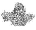 | |||||||||
|---|---|---|---|---|---|---|---|---|---|---|
| Title | Postfusion Nipah virus fusion protein in complex with Fab 1H1 | |||||||||
 Map data Map data | Postfusion Nipah virus fusion protein in complex with Fab 1H1 | |||||||||
 Sample Sample |
| |||||||||
 Keywords Keywords | Nipah /  Nipah virus / NiV / fusion / F / antibody / neutralizing / conserved epitope / neutralizing antibody / VIRAL PROTEIN / VIRAL PROTEIN-Immune System complex Nipah virus / NiV / fusion / F / antibody / neutralizing / conserved epitope / neutralizing antibody / VIRAL PROTEIN / VIRAL PROTEIN-Immune System complex | |||||||||
| Function / homology |  Function and homology information Function and homology informationmembrane fusion involved in viral entry into host cell / symbiont entry into host cell / fusion of virus membrane with host plasma membrane / viral envelope / host cell plasma membrane / virion membrane / membraneSimilarity search - Function | |||||||||
| Biological species |  Nipah henipavirus / Nipah henipavirus /  Mus musculus (house mouse) Mus musculus (house mouse) | |||||||||
| Method | single particle reconstruction / cryo EM / Resolution: 3.2 Å | |||||||||
 Authors Authors | Byrne PO / Blade EG / McLellan JS | |||||||||
| Funding support |  United States, 1 items United States, 1 items
| |||||||||
 Citation Citation | Journal: To Be Published Title: Postfusion Nipah virus fusion protein in complex with Fab 1H1 Authors: Byrne PO / Blade EG / McLellan JS | |||||||||
| History |
|
- Structure visualization
| Supplemental images |
|---|

- Downloads & links
-EMDB archive
| Map data | emd_27541.map.gz | 285.7 MB | EMDB map data format | |
|---|---|---|---|---|
| Header (meta data) | emd-27541-v30.xmlemd-27541.xml | 19.4 KB 19.4 KB | Display Display | EMDB header |
| FSC (resolution estimation) | emd_27541_fsc.xml | 16.1 KB | Display | FSC data file |
| Images |  emd_27541.png emd_27541.png | 87.9 KB | ||
| Masks | emd_27541_msk_1.map | 343 MB | Mask map | |
| Others | emd_27541_additional_1.map.gzemd_27541_additional_2.map.gzemd_27541_half_map_1.map.gzemd_27541_half_map_2.map.gz | 171.4 MB 323.6 MB 318.7 MB 318.7 MB | ||
| Archive directory |  http://ftp.pdbj.org/pub/emdb/structures/EMD-27541ftp://ftp.pdbj.org/pub/emdb/structures/EMD-27541 http://ftp.pdbj.org/pub/emdb/structures/EMD-27541ftp://ftp.pdbj.org/pub/emdb/structures/EMD-27541 | HTTPS FTP |
-Related structure data
| Related structure data |  8dmjMC M: atomic model generated by this map C: citing same article ( |
|---|---|
| Similar structure data |


-Links
| EMDB pages | EMDB (EBI/PDBe) / EMDataResource |
|---|---|
| Related items in Molecule of the Month |


-Map
| File | Download / File: emd_27541.map.gz / Format: CCP4 / Size: 343 MB / Type: IMAGE STORED AS FLOATING POINT NUMBER (4 BYTES) | ||||||||||||||||||||
|---|---|---|---|---|---|---|---|---|---|---|---|---|---|---|---|---|---|---|---|---|---|
| Annotation | Postfusion Nipah virus fusion protein in complex with Fab 1H1 | ||||||||||||||||||||
| Voxel size | X=Y=Z: 0.81 Å | ||||||||||||||||||||
| Density |
| ||||||||||||||||||||
| Symmetry | Space group: 1 | ||||||||||||||||||||
| Details | EMDB XML:
|
-Supplemental data
-Mask #1
| File | emd_27541_msk_1.map | ||||||||||||
|---|---|---|---|---|---|---|---|---|---|---|---|---|---|
| Projections & Slices |
| ||||||||||||
| Density Histograms |
 Z
Z Y
Y X
X


-Additional map: Additional Map 1
| File | emd_27541_additional_1.map | ||||||||||||
|---|---|---|---|---|---|---|---|---|---|---|---|---|---|
| Annotation | Additional Map 1 | ||||||||||||
| Projections & Slices |
| ||||||||||||
| Density Histograms |


-Additional map: Additional Map 2
| File | emd_27541_additional_2.map | ||||||||||||
|---|---|---|---|---|---|---|---|---|---|---|---|---|---|
| Annotation | Additional Map 2 | ||||||||||||
| Projections & Slices |
| ||||||||||||
| Density Histograms |


-Half map: Half Map 1
| File | emd_27541_half_map_1.map | ||||||||||||
|---|---|---|---|---|---|---|---|---|---|---|---|---|---|
| Annotation | Half Map 1 | ||||||||||||
| Projections & Slices |
| ||||||||||||
| Density Histograms |


-Half map: Half Map 2
| File | emd_27541_half_map_2.map | ||||||||||||
|---|---|---|---|---|---|---|---|---|---|---|---|---|---|
| Annotation | Half Map 2 | ||||||||||||
| Projections & Slices |
| ||||||||||||
| Density Histograms |


- Sample components
-Entire : Postfusion Nipah virus fusion protein in complex with Fab 1H1
| Entire | Name: Postfusion Nipah virus fusion protein in complex with Fab 1H1 |
|---|---|
| Components |
|
-Supramolecule #1: Postfusion Nipah virus fusion protein in complex with Fab 1H1
| Supramolecule | Name: Postfusion Nipah virus fusion protein in complex with Fab 1H1 type: complex / ID: 1 / Parent: 0 / Macromolecule list: all |
|---|---|
| Source (natural) | Organism: Nipah henipavirus |
-Macromolecule #1: Fusion glycoprotein F0,Fusion glycoprotein F1
| Macromolecule | Name: Fusion glycoprotein F0,Fusion glycoprotein F1 / type: protein_or_peptide / ID: 1 / Number of copies: 3 / Enantiomer: LEVO |
|---|---|
| Source (natural) | Organism: Nipah henipavirus |
| Molecular weight | Theoretical: 58.45198 KDa |
| Recombinant expression | Organism:  Homo sapiens (human) Homo sapiens (human) |
| Sequence | String: MYSMQLASCV TLTLVLLVNS QGILHYEKLS KIGLVKGVTR KYKIKSNPLT KDIVIKMIPN VSNMSQCTGS VMENYKTRLN GILTPIKGA LEIYKNGGSG VAIGIATAAQ ITAGVALYEA MKNADNINKL KSSIESTNEA VVKLQETAEK TVYVLTALQD Y INTNLVPT ...String: MYSMQLASCV TLTLVLLVNS QGILHYEKLS KIGLVKGVTR KYKIKSNPLT KDIVIKMIPN VSNMSQCTGS VMENYKTRLN GILTPIKGA LEIYKNGGSG VAIGIATAAQ ITAGVALYEA MKNADNINKL KSSIESTNEA VVKLQETAEK TVYVLTALQD Y INTNLVPT IDKISCKQTE LSLDLALSKY LSDLLFVFGP NLQDPVSNSM TIQAISQAFG GNYETLLRTL GYATEDFDDL LE SDSITGQ IIYVDLSSYY IIVRVYFPIL TEIQQAYIQE LLPVSFNNDN SEWISIVPNF ILVRNTLISN IEIGFCLITK RSV ICNQDY ATPMTNNMRE CLTGSTEKCP RELVVSSHVP RFALSNGVLF ANCISVTCQC QTTGRAISQS GEQTLLMIDN TTCP TAVLG NVIISLGKYL GSVNYNSEGI AIGPPVFTDK VDISSQISSM NQSLQQSKDY IKEAQRLLDT VNPSLKLMKQ IEDKI EEIL SKIYHIENEI ARIKKLIGEA PGGLVPRGSH HHHHHSAWSH PQFEK UniProtKB: Fusion glycoprotein F0, Fusion glycoprotein F0 |
-Macromolecule #2: antibody 1H1 heavy chain
| Macromolecule | Name: antibody 1H1 heavy chain / type: protein_or_peptide / ID: 2 / Number of copies: 2 / Enantiomer: LEVO |
|---|---|
| Source (natural) | Organism: Mus musculus (house mouse) |
| Molecular weight | Theoretical: 13.256589 KDa |
| Recombinant expression | Organism: Homo sapiens (human) |
| Sequence | String: AVQLQQSGAE LMRPGASMKI SCKATGYTFS SYWIDWVKQR PGHGLEWIGE ILPGSGDTNY NENFKGKAAF TADTSSNTAY MQLTSLTSE DSAVFYCARG GRYHGQGFFD YWGQGTTLTV SS |
-Macromolecule #3: antibody 1H1 light chain
| Macromolecule | Name: antibody 1H1 light chain / type: protein_or_peptide / ID: 3 / Number of copies: 2 / Enantiomer: LEVO |
|---|---|
| Source (natural) | Organism: Mus musculus (house mouse) |
| Molecular weight | Theoretical: 11.752168 KDa |
| Recombinant expression | Organism: Homo sapiens (human) |
| Sequence | String: AIQMTQSPAS LSASVGETVT ITCRPSENVH IYLAWYQQKQ GKSPQLLVYN AKTLADGVPS RFSGSASGTQ FSLKINSLQP EDFGSYYCQ HFWSIPYTFG GGTKLEIK |
-Experimental details
-Structure determination
| Method | cryo EM |
|---|---|
 Processing Processing | single particle reconstruction |
| Aggregation state | particle |
-Sample preparation
| Buffer | pH: 8 |
|---|---|
| Vitrification | Cryogen name: ETHANE |
- Electron microscopy
| Microscope | FEI TITAN KRIOS |
|---|---|
| Electron beam | Acceleration voltage: 300 kV / Electron source: FIELD EMISSION GUN |
| Electron optics | Illumination mode: FLOOD BEAM / Imaging mode: BRIGHT FIELDBright-field microscopy / Nominal defocus max: 2.5 µm / Nominal defocus min: 1.5 µm |
| Image recording | Film or detector model: GATAN K3 (6k x 4k) / Average electron dose: 70.0 e/Å2 |
| Experimental equipment |  Model: Titan Krios / Image courtesy: FEI Company |
-Image processing
| Startup model | Type of model: NONE |
|---|---|
| Initial angle assignment | Type: MAXIMUM LIKELIHOOD |
| Final angle assignment | Type: MAXIMUM LIKELIHOOD |
| Final reconstruction | Applied symmetry - Point group: C1 (asymmetric) / Resolution.type: BY AUTHOR / Resolution: 3.2 Å / Resolution method: FSC 0.143 CUT-OFF / Number images used: 312805 |
| FSC plot (resolution estimation) |  |
