National Institutes of Health/National Library of Medicine (NIH/NLM)
CA13202
United States
Citation
Journal: J Virol / Year: 2022 Title: Rotavirus VP4 Epitope of a Broadly Neutralizing Human Antibody Defined by Its Structure Bound with an Attenuated-Strain Virion. Authors: Simon Jenni / Zongli Li / Yuhuan Wang / Theresa Bessey / Eric N Salgado / Aaron G Schmidt / Harry B Greenberg / Baoming Jiang / Stephen C Harrison / Abstract: Rotavirus live-attenuated vaccines, both mono- and pentavalent, generate broadly heterotypic protection. B-cells isolated from adults encode neutralizing antibodies, some with affinity for VP5*, that ...Rotavirus live-attenuated vaccines, both mono- and pentavalent, generate broadly heterotypic protection. B-cells isolated from adults encode neutralizing antibodies, some with affinity for VP5*, that afford broad protection in mice. We have mapped the epitope of one such antibody by determining the high-resolution cryo-EM structure of its antigen-binding fragment (Fab) bound to the virion of a candidate vaccine strain, CDC-9. The Fab contacts both the distal end of a VP5* β-barrel domain and the two VP8* lectin-like domains at the tip of a projecting spike. Its interactions with VP8* do not impinge on the likely receptor-binding site, suggesting that the mechanism of neutralization is at a step subsequent to initial attachment. We also examined structures of CDC-9 virions from two different stages of serial passaging. Nearly all the VP4 (cleaved to VP8*/VP5*) spikes on particles from the earlier passage (wild-type isolate) had transitioned from the "upright" conformation present on fully infectious virions to the "reversed" conformation that is probably the end state of membrane insertion, unable to mediate penetration, consistent with the very low infectivity of the wild-type isolate. About half the VP4 spikes were upright on particles from the later passage, which had recovered substantial infectivity but had acquired an attenuated phenotype in neonatal rats. A mutation in VP4 that occurred during passaging appears to stabilize the interface at the apex of the spike and could account for the greater stability of the upright spikes on the late-passage, attenuated isolate. Rotavirus live-attenuated vaccines generate broadly heterotypic protection, and B-cells isolated from adults encode antibodies that are broadly protective in mice. Determining the structural and mechanistic basis of broad protection can contribute to understanding the current limitations of vaccine efficacy in developing countries. The structure of an attenuated human rotavirus isolate (CDC-9) bound with the Fab fragment of a broadly heterotypic protective antibody shows that protection is probably due to inhibition of the conformational transition in the viral spike protein (VP4) critical for viral penetration, rather than to inhibition of receptor binding. A comparison of structures of CDC-9 virus particles at two stages of serial passaging supports a proposed mechanism for initial steps in rotavirus membrane penetration.
In the structure databanks used in Yorodumi, some data are registered as the other names, "COVID-19 virus" and "2019-nCoV". Here are the details of the virus and the list of structure data.
Jan 31, 2019. EMDB accession codes are about to change! (news from PDBe EMDB page)
EMDB accession codes are about to change! (news from PDBe EMDB page)
The allocation of 4 digits for EMDB accession codes will soon come to an end. Whilst these codes will remain in use, new EMDB accession codes will include an additional digit and will expand incrementally as the available range of codes is exhausted. The current 4-digit format prefixed with “EMD-” (i.e. EMD-XXXX) will advance to a 5-digit format (i.e. EMD-XXXXX), and so on. It is currently estimated that the 4-digit codes will be depleted around Spring 2019, at which point the 5-digit format will come into force.
The EM Navigator/Yorodumi systems omit the EMD- prefix.
Related info.:Q: What is EMD? / ID/Accession-code notation in Yorodumi/EM Navigator
Yorodumi is a browser for structure data from EMDB, PDB, SASBDB, etc.
This page is also the successor to EM Navigator detail page, and also detail information page/front-end page for Omokage search.
The word "yorodu" (or yorozu) is an old Japanese word meaning "ten thousand". "mi" (miru) is to see.
Related info.:EMDB / PDB / SASBDB / Comparison of 3 databanks / Yorodumi Search / Aug 31, 2016. New EM Navigator & Yorodumi / Yorodumi Papers / Jmol/JSmol / Function and homology information / Changes in new EM Navigator and Yorodumi
 Movie
Movie Controller

 Yorodumi
 Open data
 Basic information
 Map data
 Sample
 Function and homology information
 viral envelope / virion attachment to host cell / host cell plasma membrane / structural molecule activity /
 Authors
 United States, 1 items
  Citation
 Structure visualization
 Downloads & links
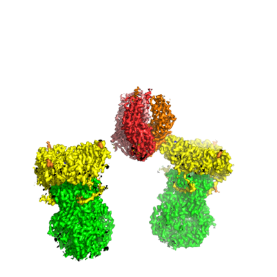 emd_26609.png
 http://ftp.pdbj.org/pub/emdb/structures/EMD-26609


 Z
Z Y
Y X
X


 Sample components

 Processing
 Electron microscopy