 Movie
Movie Controller

[English] 日本語
 Yorodumi
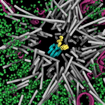
- EMDB-19781: Cryo-ET of a mitotic centrosome in an embryonic C. elegans cell -
+ Open data

- Basic information
| Entry |  | |||||||||
|---|---|---|---|---|---|---|---|---|---|---|
| Title | Cryo-ET of a mitotic centrosome in an embryonic C. elegans cell | |||||||||
 Map data Map data | Representative C. elegans centrosome tomogram | |||||||||
 Sample Sample |
| |||||||||
 Keywords Keywords | cryo-ET / cryo-FIB /  C. elegans / mitosis / centrosome / centriole / microtubules / CELL CYCLE C. elegans / mitosis / centrosome / centriole / microtubules / CELL CYCLE | |||||||||
| Biological species |  Caenorhabditis elegans (invertebrata) Caenorhabditis elegans (invertebrata) | |||||||||
| Method | electron tomography / cryo EM | |||||||||
 Authors Authors | Tollervey F / Rios MU / Zagoriy I / Woodruff JB / Mahamid J | |||||||||
| Funding support | European Union, 1 items
| |||||||||
 Citation Citation | Journal: bioRxiv / Year: 2024 Title: Native molecular architectures of centrosomes in embryos. Authors: Fergus Tollervey / Manolo U Rios / Evgenia Zagoriy / Jeffrey B Woodruff / Julia Mahamid /   Abstract: Centrosomes organize microtubules that are essential for mitotic divisions in animal cells. They consist of centrioles surrounded by Pericentriolar Material (PCM). Questions related to mechanisms of ...Centrosomes organize microtubules that are essential for mitotic divisions in animal cells. They consist of centrioles surrounded by Pericentriolar Material (PCM). Questions related to mechanisms of centriole assembly, PCM organization, and microtubule formation remain unanswered, in part due to limited availability of molecular-resolution structural analyses . Here, we use cryo-electron tomography to visualize centrosomes across the cell cycle in cells isolated from embryos. We describe a pseudo-timeline of centriole assembly and identify distinct structural features including a cartwheel in daughter centrioles, and incomplete microtubule doublets surrounded by a star-shaped density in mother centrioles. We find that centriole and PCM microtubules differ in protofilament number (13 versus 11) indicating distinct nucleation mechanisms. This difference could be explained by atypical γ-tubulin ring complexes with 11-fold symmetry identified at the minus ends of short PCM microtubules. We further characterize a porous and disordered network that forms the interconnected PCM. Thus, our work builds a three-dimensional structural atlas that helps explain how centrosomes assemble, grow, and achieve function. | |||||||||
| History |
|
- Structure visualization
| Supplemental images |
|---|
- Downloads & links
-EMDB archive
| Map data | emd_19781.map.gz | 1.5 GB |  EMDB map data format EMDB map data format | |
|---|---|---|---|---|
| Header (meta data) | emd-19781-v30.xmlemd-19781.xml | 17.7 KB 17.7 KB | Display Display | EMDB header |
| Images |  emd_19781.png emd_19781.png | 245.7 KB | ||
| Filedesc metadata | emd-19781.cif.gz | 4.3 KB | ||
| Others | emd_19781_additional_1.map.gzemd_19781_additional_2.map.gzemd_19781_additional_3.map.gzemd_19781_additional_4.map.gz | 447.8 KB 1.1 MB 882.8 KB 718.6 KB | ||
| Archive directory |  http://ftp.pdbj.org/pub/emdb/structures/EMD-19781ftp://ftp.pdbj.org/pub/emdb/structures/EMD-19781 http://ftp.pdbj.org/pub/emdb/structures/EMD-19781ftp://ftp.pdbj.org/pub/emdb/structures/EMD-19781 | HTTPS FTP |
-Related structure data
-Links
| EMDB pages | EMDB (EBI/PDBe) / EMDataResource |
|---|
-Map
| File | Download / File: emd_19781.map.gz / Format: CCP4 / Size: 1.7 GB / Type: IMAGE STORED AS FLOATING POINT NUMBER (4 BYTES) | ||||||||||||||||||||||||||||||||
|---|---|---|---|---|---|---|---|---|---|---|---|---|---|---|---|---|---|---|---|---|---|---|---|---|---|---|---|---|---|---|---|---|---|
| Annotation | Representative C. elegans centrosome tomogram | ||||||||||||||||||||||||||||||||
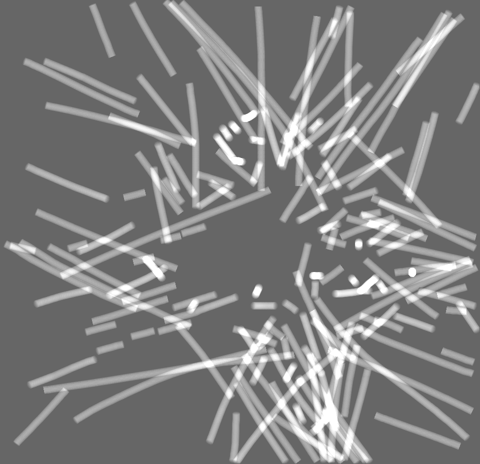
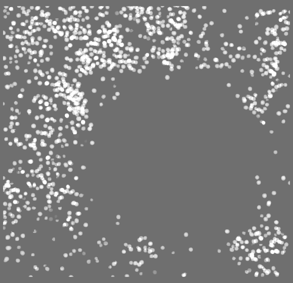
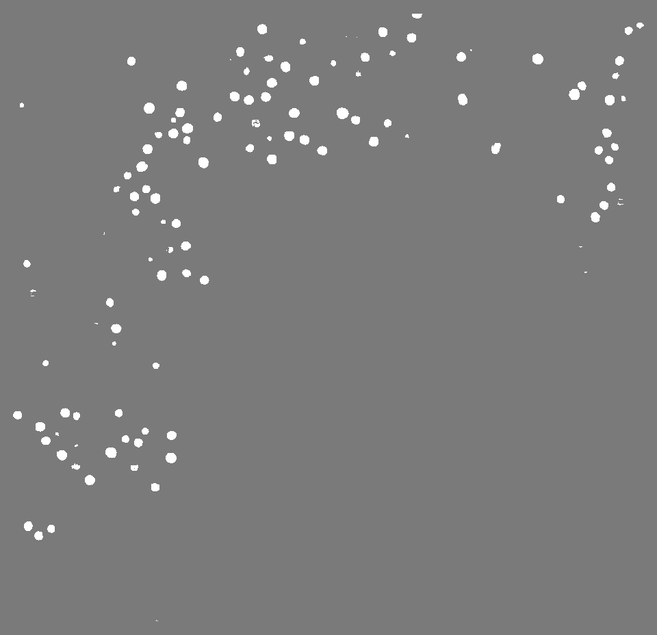
| Projections & slices | Image control
Images are generated by Spider. generated in cubic-lattice coordinate | ||||||||||||||||||||||||||||||||
| Voxel size | X=Y=Z: 13.4808 Å | ||||||||||||||||||||||||||||||||
| Density |
| ||||||||||||||||||||||||||||||||
| Symmetry | Space group: 1 | ||||||||||||||||||||||||||||||||
| Details | EMDB XML:
|
 Z (Sec.)
 Y (Row.)
 X (Col.)


-Supplemental data
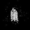
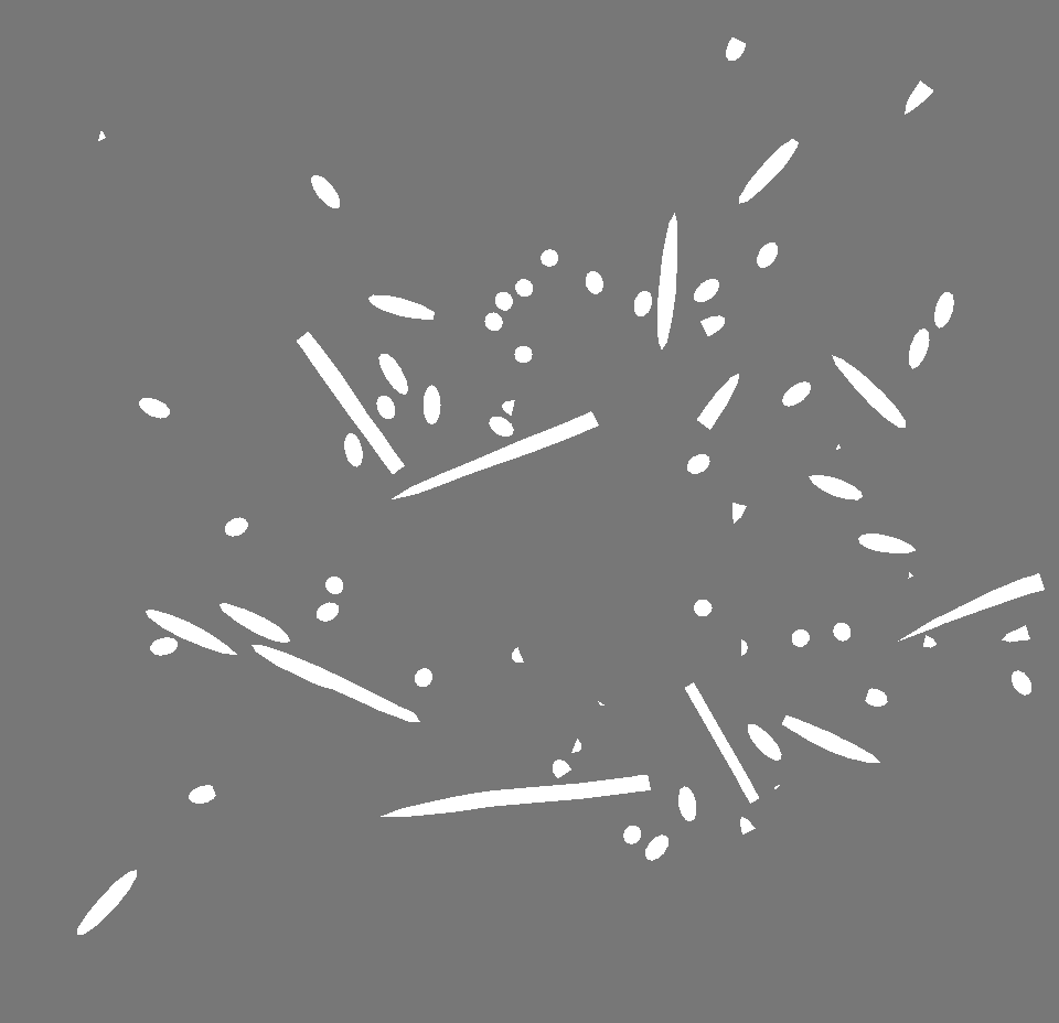
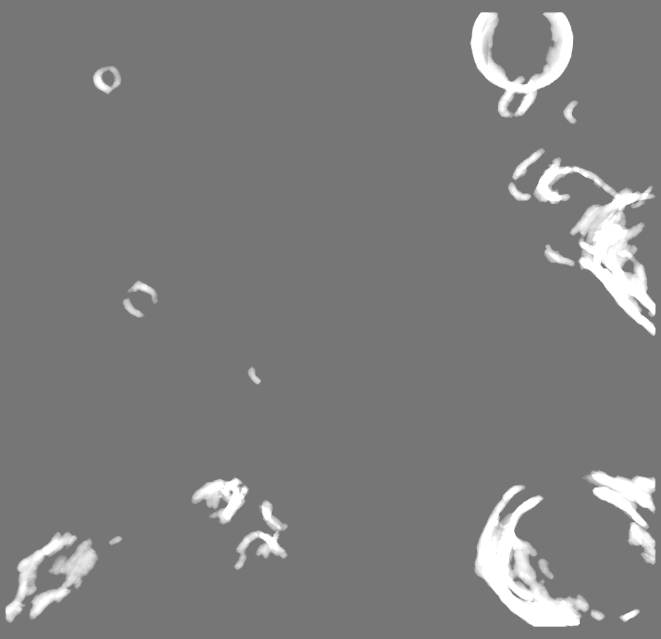
-Additional map: Binary segmentation of centrioles
| File | emd_19781_additional_1.map | ||||||||||||
|---|---|---|---|---|---|---|---|---|---|---|---|---|---|
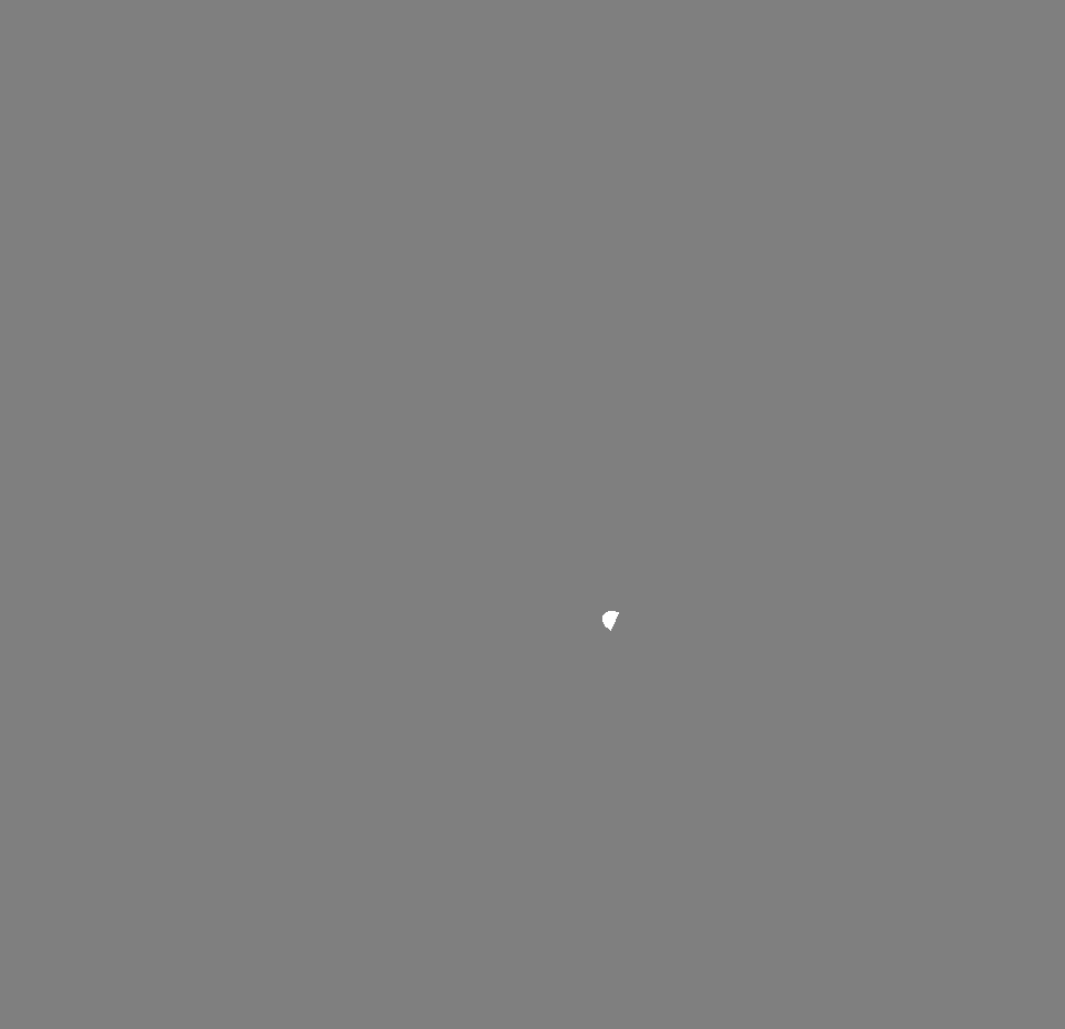
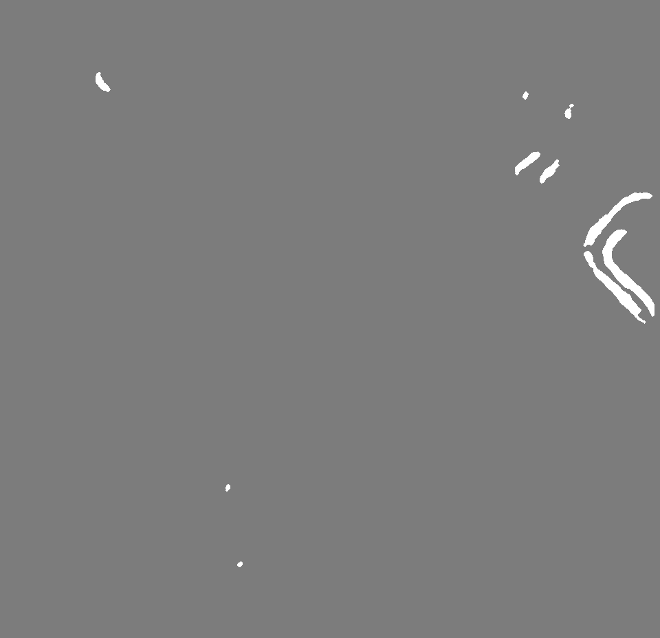
| Annotation | Binary segmentation of centrioles | ||||||||||||
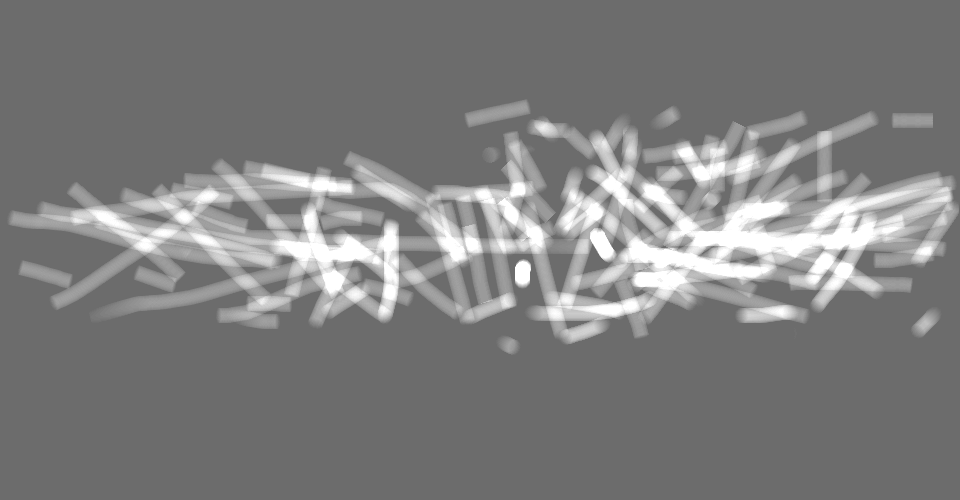
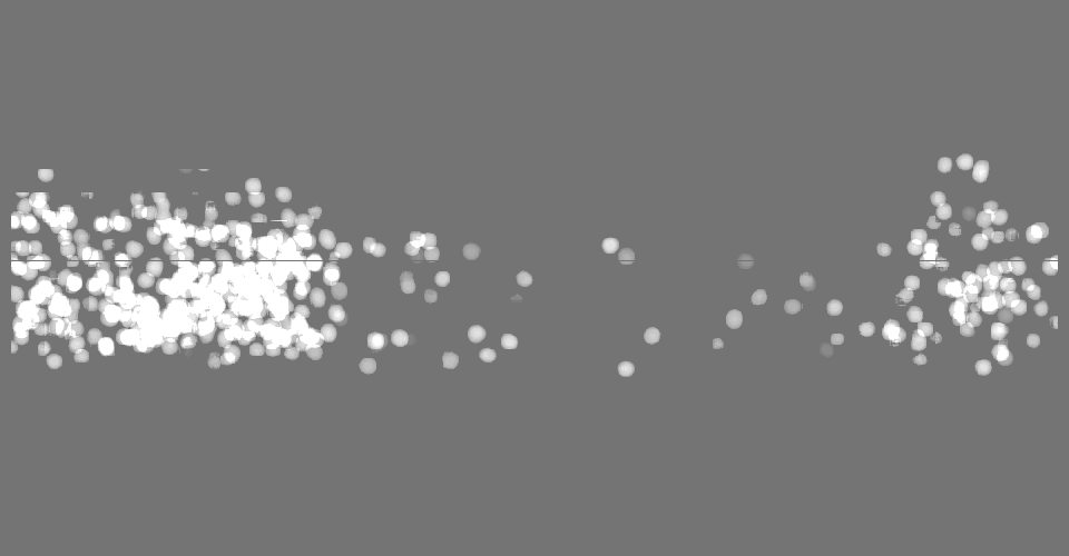
| Projections & Slices |
| ||||||||||||
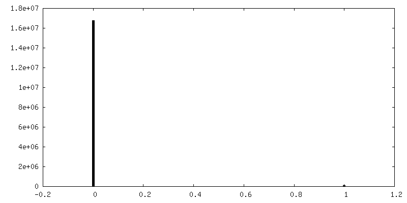
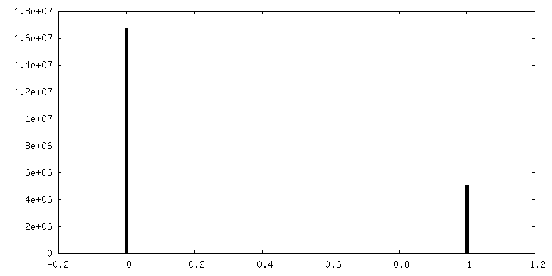
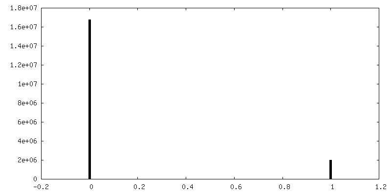
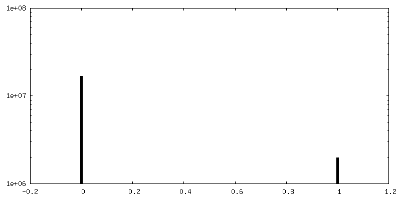
| Density Histograms |
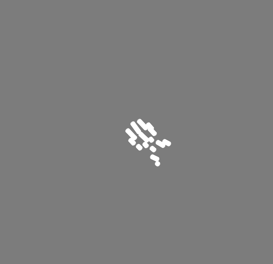
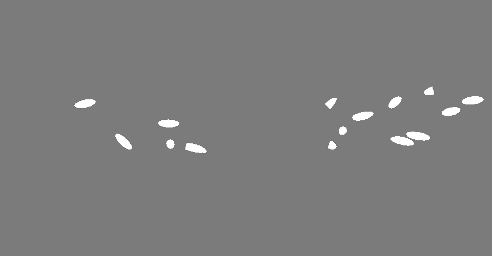
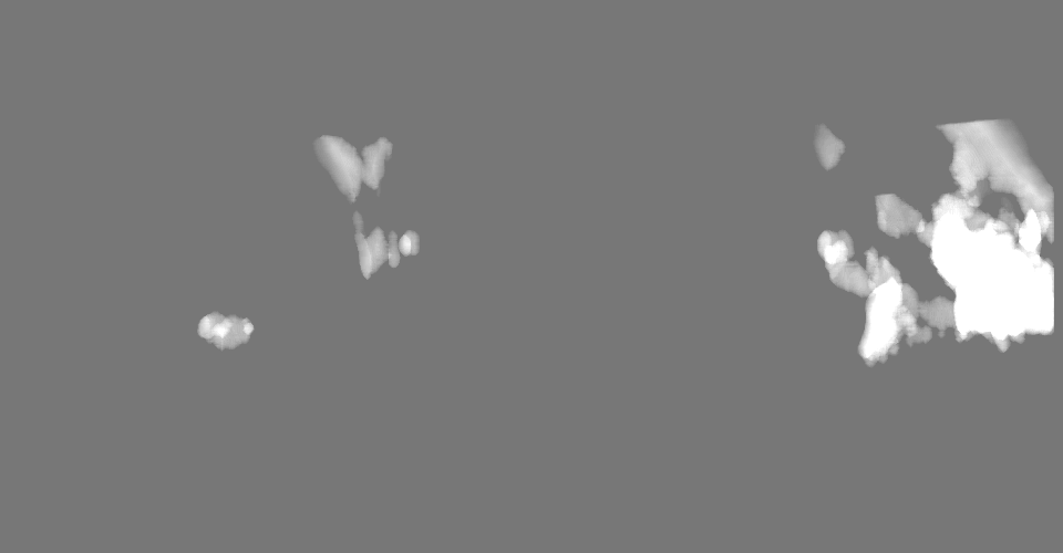
-Additional map: Binary segmentation of microtubules
| File | emd_19781_additional_2.map | ||||||||||||
|---|---|---|---|---|---|---|---|---|---|---|---|---|---|
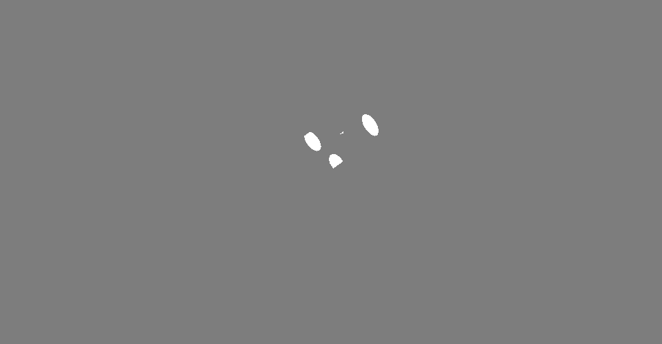
| Annotation | Binary segmentation of microtubules | ||||||||||||
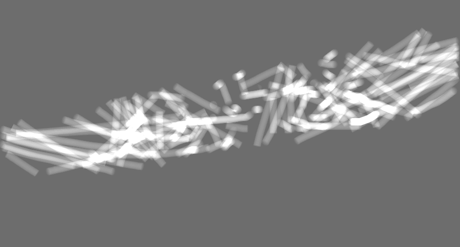
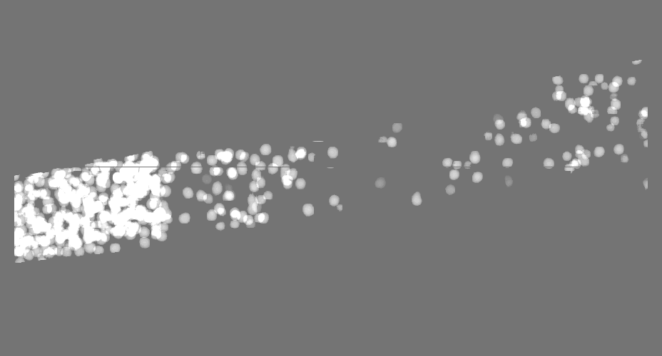
| Projections & Slices |
| ||||||||||||
| Density Histograms |
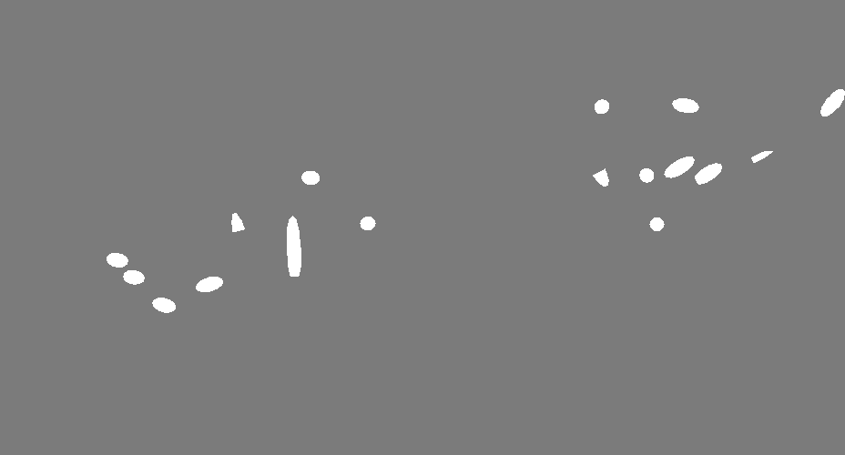
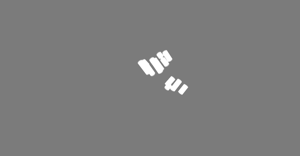
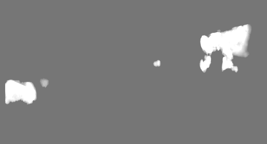
-Additional map: Binary segmentation of ribosomes
| File | emd_19781_additional_3.map | ||||||||||||
|---|---|---|---|---|---|---|---|---|---|---|---|---|---|
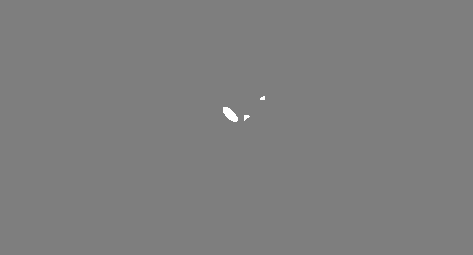
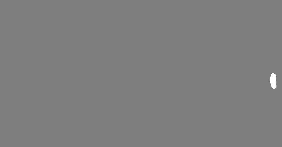
| Annotation | Binary segmentation of ribosomes | ||||||||||||
| Projections & Slices |
| ||||||||||||
| Density Histograms |
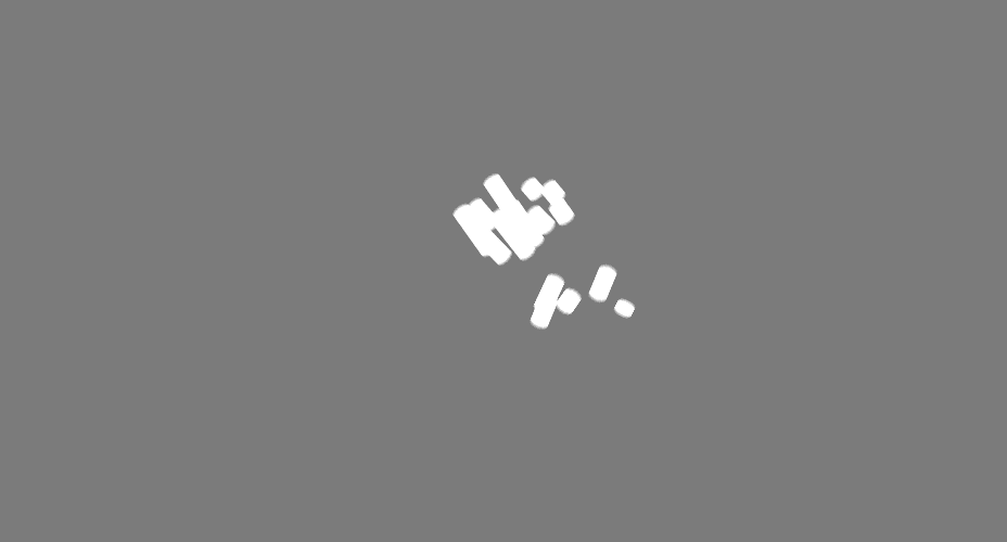
-Additional map: Binary segmentation of ribosomes
| File | emd_19781_additional_4.map | ||||||||||||
|---|---|---|---|---|---|---|---|---|---|---|---|---|---|
| Annotation | Binary segmentation of ribosomes | ||||||||||||
| Projections & Slices |
| ||||||||||||
| Density Histograms |


- Sample components
-Entire : Dissociated C. elegans embryonic cells
| Entire | Name: Dissociated C. elegans embryonic cells |
|---|---|
| Components |
|
-Supramolecule #1: Dissociated C. elegans embryonic cells
| Supramolecule | Name: Dissociated C. elegans embryonic cells / type: cell / ID: 1 / Parent: 0 |
|---|---|
| Source (natural) | Organism: Caenorhabditis elegans (invertebrata) / Tissue: Embryo |
-Experimental details
-Structure determination
| Method | cryo EM |
|---|---|
 Processing Processing | electron tomography |
| Aggregation state | cell |
-Sample preparation
| Buffer | pH: 7.4 |
|---|---|
| Grid | Model: Quantifoil / Material: GOLD / Mesh: 200 / Support film - topology: HOLEY / Pretreatment - Type: GLOW DISCHARGE / Pretreatment - Time: 45 sec. / Pretreatment - Atmosphere: AIR / Pretreatment - Pressure: 0.037 kPa |
| Vitrification | Cryogen name: ETHANE / Instrument: LEICA EM GP |
| Sectioning | Focused ion beam - Instrument: OTHER / Focused ion beam - Ion: OTHER / Focused ion beam - Voltage: 30 / Focused ion beam - Current: 0.5 / Focused ion beam - Duration: 600 / Focused ion beam - Temperature: 95 K / Focused ion beam - Initial thickness: 1000 / Focused ion beam - Final thickness: 200 Focused ion beam - Details: The value given for _em_focused_ion_beam.instrument is FEI Aquilos cryo-FIB. This is not in a list of allowed values {'OTHER', 'DB235'} so OTHER is written into the XML file. |
- Electron microscopy
| Microscope | FEI TITAN KRIOS |
|---|---|
| Electron beam | Acceleration voltage: 300 kV / Electron source: FIELD EMISSION GUN |
| Electron optics | Illumination mode: FLOOD BEAM / Imaging mode: BRIGHT FIELDBright-field microscopy / Cs: 2.7 mm / Nominal defocus max: 4.5 µm / Nominal defocus min: 2.5 µm |
| Specialist optics | Phase plate: VOLTA PHASE PLATE / Energy filter - Slit width: 20 eV |
| Sample stage | Specimen holder model: FEI TITAN KRIOS AUTOGRID HOLDER / Cooling holder cryogen: NITROGEN |
| Image recording | Film or detector model: GATAN K2 QUANTUM (4k x 4k) / Detector mode: COUNTING / Average electron dose: 120.0 e/Å2 |
| Experimental equipment |  Model: Titan Krios / Image courtesy: FEI Company |
-Image processing
| Final reconstruction | Software - Name: Warp (ver. 1.0.7) / Number images used: 61 |
|---|
