 ムービー
ムービー コントローラー


+ データを開く

- 基本情報
基本情報
| 登録情報 |  | |||||||||
|---|---|---|---|---|---|---|---|---|---|---|
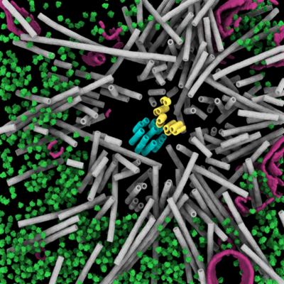
| タイトル | Cryo-ET of a mitotic centrosome in an embryonic C. elegans cell | |||||||||
 マップデータ マップデータ | Representative C. elegans centrosome tomogram | |||||||||
 試料 試料 |
| |||||||||
 キーワード キーワード | cryo-ET / cryo-FIB /  C. elegans (カエノラブディティス・エレガンス) / mitosis (有糸分裂) / centrosome (中心体) / centriole (中心小体) / microtubules (微小管) / CELL CYCLE (細胞周期) C. elegans (カエノラブディティス・エレガンス) / mitosis (有糸分裂) / centrosome (中心体) / centriole (中心小体) / microtubules (微小管) / CELL CYCLE (細胞周期) | |||||||||
| 生物種 |  Caenorhabditis elegans (センチュウ) Caenorhabditis elegans (センチュウ) | |||||||||
| 手法 | 電子線トモグラフィー法 / クライオ電子顕微鏡法 | |||||||||
 データ登録者 データ登録者 | Tollervey F / Rios MU / Zagoriy I / Woodruff JB / Mahamid J | |||||||||
| 資金援助 | European Union, 1件
| |||||||||
 引用 引用 | ジャーナル: bioRxiv / 年: 2024 タイトル: Native molecular architectures of centrosomes in embryos. 著者: Fergus Tollervey / Manolo U Rios / Evgenia Zagoriy / Jeffrey B Woodruff / Julia Mahamid /   要旨: Centrosomes organize microtubules that are essential for mitotic divisions in animal cells. They consist of centrioles surrounded by Pericentriolar Material (PCM). Questions related to mechanisms of ...Centrosomes organize microtubules that are essential for mitotic divisions in animal cells. They consist of centrioles surrounded by Pericentriolar Material (PCM). Questions related to mechanisms of centriole assembly, PCM organization, and microtubule formation remain unanswered, in part due to limited availability of molecular-resolution structural analyses . Here, we use cryo-electron tomography to visualize centrosomes across the cell cycle in cells isolated from embryos. We describe a pseudo-timeline of centriole assembly and identify distinct structural features including a cartwheel in daughter centrioles, and incomplete microtubule doublets surrounded by a star-shaped density in mother centrioles. We find that centriole and PCM microtubules differ in protofilament number (13 versus 11) indicating distinct nucleation mechanisms. This difference could be explained by atypical γ-tubulin ring complexes with 11-fold symmetry identified at the minus ends of short PCM microtubules. We further characterize a porous and disordered network that forms the interconnected PCM. Thus, our work builds a three-dimensional structural atlas that helps explain how centrosomes assemble, grow, and achieve function. | |||||||||
| 履歴 |
|
- 構造の表示
構造の表示
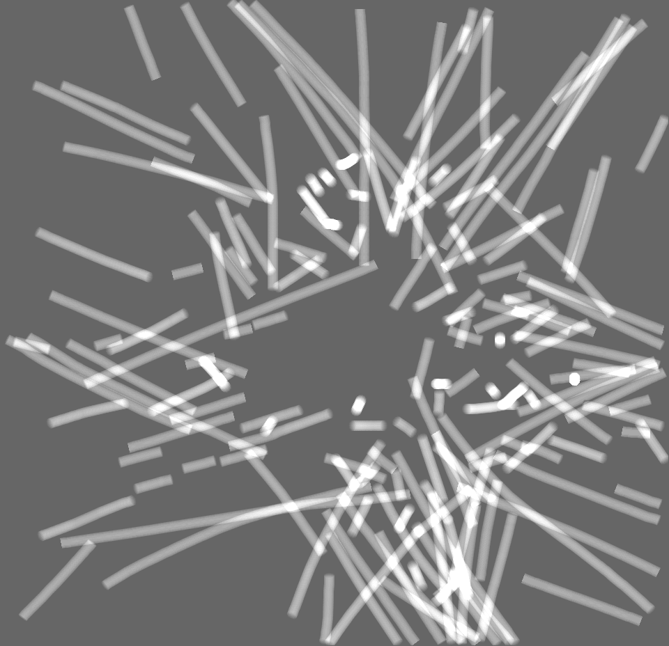
| 添付画像 |
|---|
- ダウンロードとリンク
-EMDBアーカイブ
| マップデータ | emd_19781.map.gz | 1.5 GB |  EMDBマップデータ形式 EMDBマップデータ形式 | |
|---|---|---|---|---|
| ヘッダ (付随情報) | emd-19781-v30.xmlemd-19781.xml | 17.7 KB 17.7 KB | 表示 表示 | EMDBヘッダ |
| 画像 |  emd_19781.png emd_19781.png | 245.7 KB | ||
| Filedesc metadata | emd-19781.cif.gz | 4.3 KB | ||
| その他 | emd_19781_additional_1.map.gzemd_19781_additional_2.map.gzemd_19781_additional_3.map.gzemd_19781_additional_4.map.gz | 447.8 KB 1.1 MB 882.8 KB 718.6 KB | ||
| アーカイブディレクトリ |  http://ftp.pdbj.org/pub/emdb/structures/EMD-19781ftp://ftp.pdbj.org/pub/emdb/structures/EMD-19781 http://ftp.pdbj.org/pub/emdb/structures/EMD-19781ftp://ftp.pdbj.org/pub/emdb/structures/EMD-19781 | HTTPS FTP |
-関連構造データ


-リンク
| EMDBのページ | EMDB (EBI/PDBe) / EMDataResource |
|---|
-マップ
| ファイル | ダウンロード / ファイル: emd_19781.map.gz / 形式: CCP4 / 大きさ: 1.7 GB / タイプ: IMAGE STORED AS FLOATING POINT NUMBER (4 BYTES) | ||||||||||||||||||||
|---|---|---|---|---|---|---|---|---|---|---|---|---|---|---|---|---|---|---|---|---|---|
| 注釈 | Representative C. elegans centrosome tomogram | ||||||||||||||||||||
| ボクセルのサイズ | X=Y=Z: 13.4808 Å | ||||||||||||||||||||
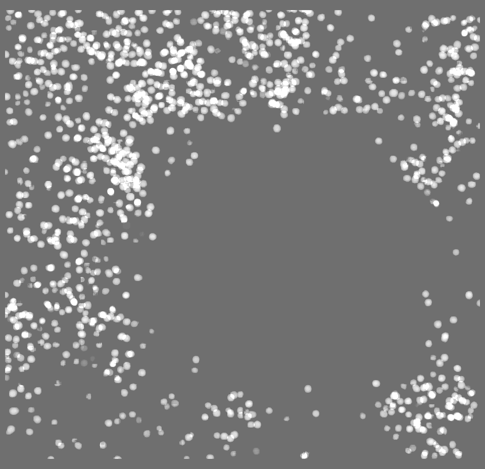
| 密度 |
| ||||||||||||||||||||
| 対称性 | 空間群: 1 | ||||||||||||||||||||
| 詳細 | EMDB XML:
|
-添付データ
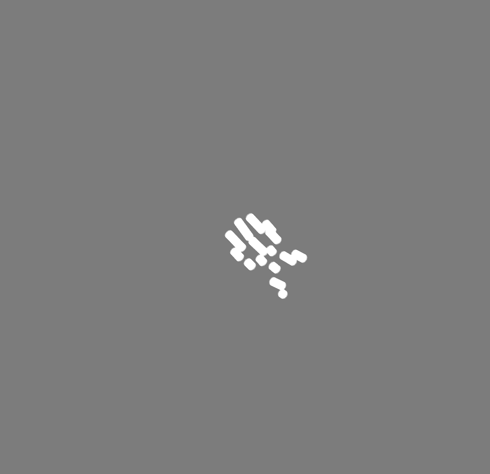
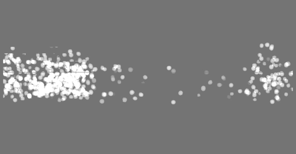
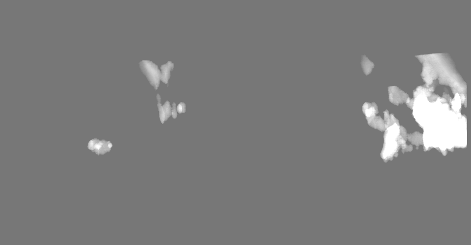
-追加マップ: Binary segmentation of centrioles
| ファイル | emd_19781_additional_1.map | ||||||||||||
|---|---|---|---|---|---|---|---|---|---|---|---|---|---|
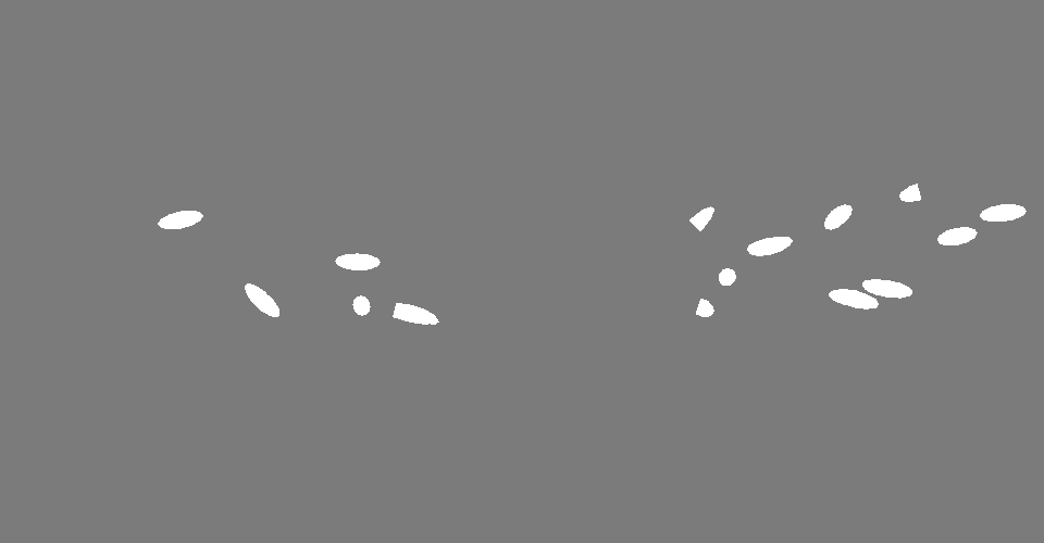
| 注釈 | Binary segmentation of centrioles | ||||||||||||
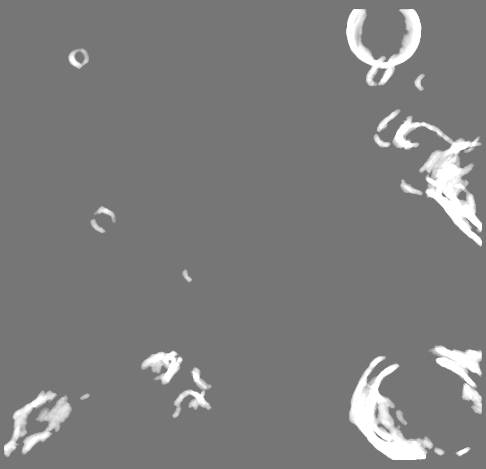
| 投影像・断面図 |
| ||||||||||||
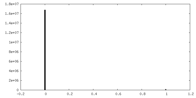
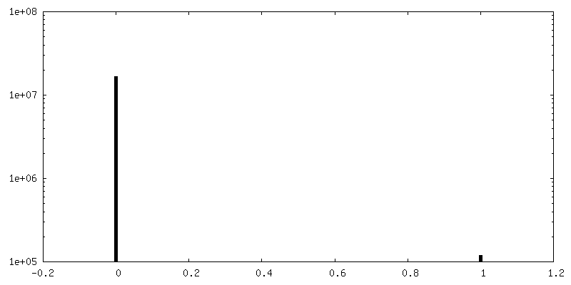
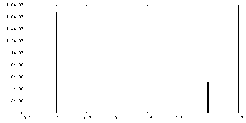
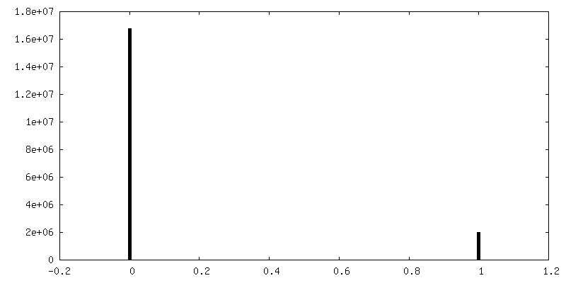
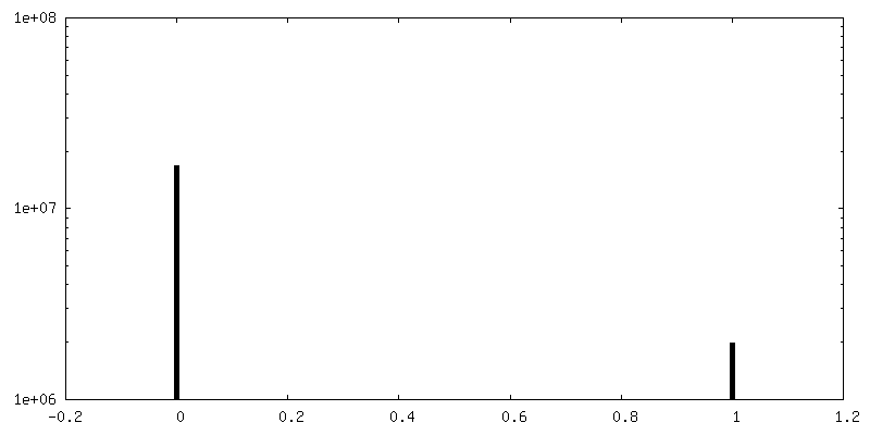
| 密度ヒストグラム |
 Z
Z Y
Y X
X


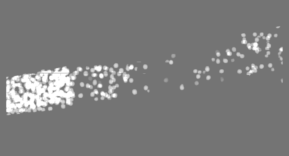
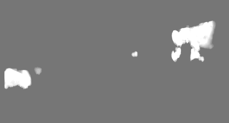
-追加マップ: Binary segmentation of microtubules
| ファイル | emd_19781_additional_2.map | ||||||||||||
|---|---|---|---|---|---|---|---|---|---|---|---|---|---|
| 注釈 | Binary segmentation of microtubules | ||||||||||||
| 投影像・断面図 |
| ||||||||||||
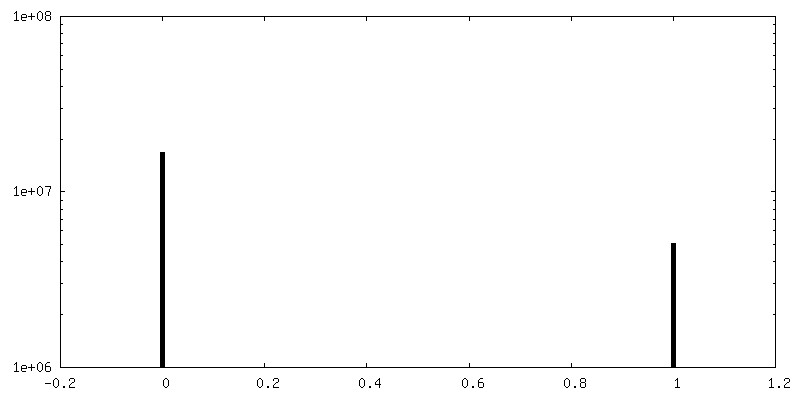
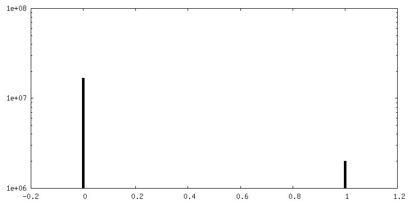
| 密度ヒストグラム |

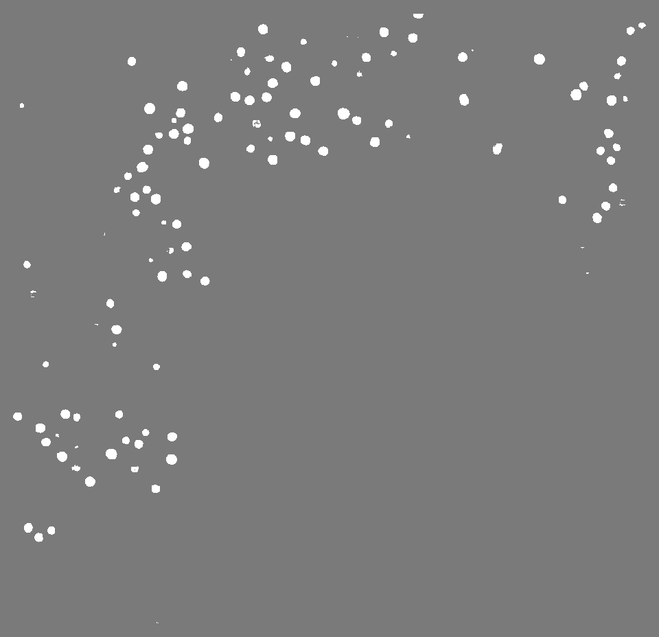
-追加マップ: Binary segmentation of ribosomes
| ファイル | emd_19781_additional_3.map | ||||||||||||
|---|---|---|---|---|---|---|---|---|---|---|---|---|---|
| 注釈 | Binary segmentation of ribosomes | ||||||||||||
| 投影像・断面図 |
| ||||||||||||
| 密度ヒストグラム |
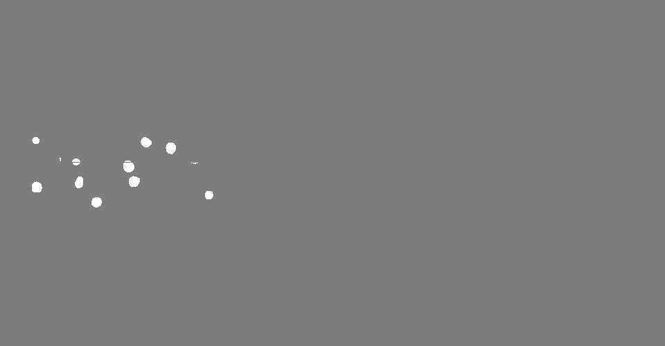
-追加マップ: Binary segmentation of ribosomes
| ファイル | emd_19781_additional_4.map | ||||||||||||
|---|---|---|---|---|---|---|---|---|---|---|---|---|---|
| 注釈 | Binary segmentation of ribosomes | ||||||||||||
| 投影像・断面図 |
| ||||||||||||
| 密度ヒストグラム |


- 試料の構成要素
-全体 : Dissociated C. elegans embryonic cells
| 全体 | 名称: Dissociated C. elegans embryonic cells |
|---|---|
| 要素 |
|
-超分子 #1: Dissociated C. elegans embryonic cells
| 超分子 | 名称: Dissociated C. elegans embryonic cells / タイプ: cell / ID: 1 / 親要素: 0 |
|---|---|
| 由来(天然) | 生物種: Caenorhabditis elegans (センチュウ) / 組織: Embryo |
-実験情報
-構造解析
| 手法 | クライオ電子顕微鏡法 |
|---|---|
 解析 解析 | 電子線トモグラフィー法 |
| 試料の集合状態 | cell |
-試料調製
| 緩衝液 | pH: 7.4 |
|---|---|
| グリッド | モデル: Quantifoil / 材質: GOLD / メッシュ: 200 / 支持フィルム - トポロジー: HOLEY / 前処理 - タイプ: GLOW DISCHARGE / 前処理 - 時間: 45 sec. / 前処理 - 雰囲気: AIR / 前処理 - 気圧: 0.037 kPa |
| 凍結 | 凍結剤: ETHANE / 装置: LEICA EM GP |
| 切片作成 | 集束イオンビーム - 装置: OTHER / 集束イオンビーム - イオン: OTHER / 集束イオンビーム - 電圧: 30 / 集束イオンビーム - 電流: 0.5 / 集束イオンビーム - 時間: 600 / 集束イオンビーム - 温度: 95 K / 集束イオンビーム - Initial thickness: 1000 / 集束イオンビーム - 最終 厚さ: 200 集束イオンビーム - 詳細: The value given for _em_focused_ion_beam.instrument is FEI Aquilos cryo-FIB. This is not in a list of allowed values {'OTHER', 'DB235'} so OTHER is written into the XML file. |
- 電子顕微鏡法
| 顕微鏡 | FEI TITAN KRIOS |
|---|---|
| 電子線 | 加速電圧: 300 kV / 電子線源: FIELD EMISSION GUN |
| 電子光学系 | 照射モード: FLOOD BEAM / 撮影モード: BRIGHT FIELDBright-field microscopy / Cs: 2.7 mm / 最大 デフォーカス(公称値): 4.5 µm / 最小 デフォーカス(公称値): 2.5 µm |
| 特殊光学系 | 位相板: VOLTA PHASE PLATE / エネルギーフィルター - スリット幅: 20 eV |
| 試料ステージ | 試料ホルダーモデル: FEI TITAN KRIOS AUTOGRID HOLDER ホルダー冷却材: NITROGEN |
| 撮影 | フィルム・検出器のモデル: GATAN K2 QUANTUM (4k x 4k) 検出モード: COUNTING / 平均電子線量: 120.0 e/Å2 |
| 実験機器 |  モデル: Titan Krios / 画像提供: FEI Company |
-画像解析
| 最終 再構成 | ソフトウェア - 名称: Warp (ver. 1.0.7) / 使用した粒子像数: 61 |
|---|
