E. coli / membrane protein / Ni-NTA resin / cytochrome bo3 quinol oxidase / ubiquinone-8 release / peptidisc / single particle analysis / cryo-EM
Function / homology
Function and homology information
cytochrome o ubiquinol oxidase complex / oxidoreduction-driven active transmembrane transporter activity / ubiquinol oxidase (H+-transporting) / cytochrome bo3 ubiquinol oxidase activity / aerobic electron transport chain / oxidoreductase activity, acting on diphenols and related substances as donors, oxygen as acceptor / ubiquinone binding / electron transport coupled proton transport / cytochrome-c oxidase activity / proton transmembrane transporter activity ...cytochrome o ubiquinol oxidase complex / oxidoreduction-driven active transmembrane transporter activity / ubiquinol oxidase (H+-transporting) / cytochrome bo3 ubiquinol oxidase activity / aerobic electron transport chain / oxidoreductase activity, acting on diphenols and related substances as donors, oxygen as acceptor / ubiquinone binding / electron transport coupled proton transport / cytochrome-c oxidase activity / proton transmembrane transporter activity / ATP synthesis coupled electron transport / respirasome / aerobic respiration / respiratory electron transport chain / electron transfer activity / copper ion binding / heme binding / plasma membrane Similarity search - Function
Cytochrome o ubiquinol oxidase, subunit III / Cytochrome o ubiquinol oxidase subunit IV / Cytochrome o ubiquinol oxidase, subunit I / Ubiquinol oxidase subunit III domain / Cytochrome C oxidase subunit IV, prokaryotes / COX aromatic rich motif / Prokaryotic Cytochrome C oxidase subunit IV / COX Aromatic Rich Motif / Cytochrome o ubiquinol oxidase subunit II / Ubiquinol oxidase subunit 2, cupredoxin domain ...Cytochrome o ubiquinol oxidase, subunit III / Cytochrome o ubiquinol oxidase subunit IV / Cytochrome o ubiquinol oxidase, subunit I / Ubiquinol oxidase subunit III domain / Cytochrome C oxidase subunit IV, prokaryotes / COX aromatic rich motif / Prokaryotic Cytochrome C oxidase subunit IV / COX Aromatic Rich Motif / Cytochrome o ubiquinol oxidase subunit II / Ubiquinol oxidase subunit 2, cupredoxin domain / Cytochrome c oxidase subunit III / Cytochrome c oxidase subunit III-like / Cytochrome c oxidase, subunit III, 4-helical bundle / Cytochrome c oxidase subunit III / Heme-copper oxidase subunit III family profile. / Cytochrome c oxidase subunit III-like superfamily / Cytochrome C oxidase subunit II, transmembrane domain / Cytochrome oxidase subunit II transmembrane region profile. / Cytochrome c/quinol oxidase subunit II / Cytochrome C oxidase subunit II, transmembrane domain superfamily / Cytochrome c oxidase, subunit I, copper-binding site / Heme-copper oxidase catalytic subunit, copper B binding region signature. / Cytochrome c oxidase-like, subunit I domain / Cytochrome oxidase subunit I profile. / Cytochrome c oxidase subunit I / Cytochrome c oxidase-like, subunit I superfamily / Cytochrome C and Quinol oxidase polypeptide I / Cytochrome C oxidase subunit II, periplasmic domain / Cytochrome c oxidase subunit II-like C-terminal / Cytochrome oxidase subunit II copper A binding domain profile. / Cupredoxin / Prokaryotic membrane lipoprotein lipid attachment site profile. Similarity search - Domain/homology
Netherlands Organisation for Scientific Research (NWO)
731.016.407
Netherlands
Netherlands Organisation for Scientific Research (NWO)
184.034.014
Netherlands
Health-Holland
LSHM21029
Netherlands
Citation
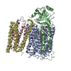
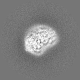
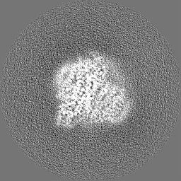
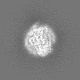
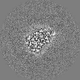
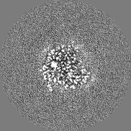
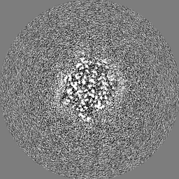
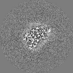
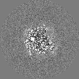
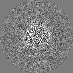
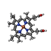
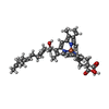
Journal: Biochim Biophys Acta Bioenerg / Year: 2024 Title: Cryo-EM structure of cytochrome bo quinol oxidase assembled in peptidiscs reveals an "open" conformation for potential ubiquinone-8 release. Authors: Ye Gao / Yue Zhang / Sneha Hakke / Ronny Mohren / Lyanne J P M Sijbers / Peter J Peters / Raimond B G Ravelli / Abstract: Cytochrome bo quinol oxidase belongs to the heme‑copper-oxidoreductase (HCO) superfamily, which is part of the respiratory chain and essential for cell survival. While the reaction mechanism of cyt ...Cytochrome bo quinol oxidase belongs to the heme‑copper-oxidoreductase (HCO) superfamily, which is part of the respiratory chain and essential for cell survival. While the reaction mechanism of cyt bo has been studied extensively over the last decades, specific details about its substrate binding and product release have remained unelucidated due to the lack of structural information. Here, we report a 2.8 Å cryo-electron microscopy structure of cyt bo from Escherichia coli assembled in peptidiscs. Our structural model shows a conformation for amino acids 1-41 of subunit I different from all previously published structures while the remaining parts of this enzyme are similar. Our new conformation shows a "U-shape" assembly in contrast to the transmembrane helix, named "TM0", in other reported structural models. However, TM0 blocks ubiquinone-8 (reaction product) release, suggesting that other cyt bo conformations should exist. Our structural model presents experimental evidence for an "open" conformation to facilitate substrate/product exchange. This work helps further understand the reaction cycle of this oxidase, which could be a benefit for potential drug/antibiotic design for health science.
Film or detector model: GATAN K3 BIOQUANTUM (6k x 4k) / Digitization - Dimensions - Width: 5760 pixel / Digitization - Dimensions - Height: 4092 pixel / Number grids imaged: 1 / Number real images: 8050 / Average exposure time: 2.1 sec. / Average electron dose: 50.0 e/Å2
Experimental equipment
Model: Titan Krios / Image courtesy: FEI Company
-
Image processing
Particle selection
Number selected: 1277595
Startup model
Type of model: INSILICO MODEL
Initial angle assignment
Type: MAXIMUM LIKELIHOOD / Software - Name: RELION
Final 3D classification
Number classes: 3 / Software - Name: RELION
Final angle assignment
Type: MAXIMUM LIKELIHOOD / Software - Name: RELION
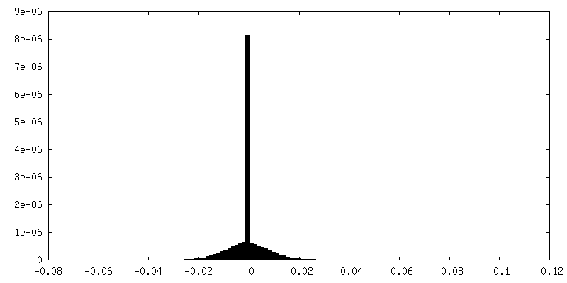
Final reconstruction
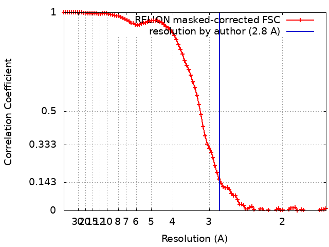
Number classes used: 1 / Applied symmetry - Point group: C1 (asymmetric) / Algorithm: FOURIER SPACE / Resolution.type: BY AUTHOR / Resolution: 2.8 Å / Resolution method: FSC 0.143 CUT-OFF / Software - Name: RELION / Number images used: 45014
In the structure databanks used in Yorodumi, some data are registered as the other names, "COVID-19 virus" and "2019-nCoV". Here are the details of the virus and the list of structure data.
Jan 31, 2019. EMDB accession codes are about to change! (news from PDBe EMDB page)
EMDB accession codes are about to change! (news from PDBe EMDB page)
The allocation of 4 digits for EMDB accession codes will soon come to an end. Whilst these codes will remain in use, new EMDB accession codes will include an additional digit and will expand incrementally as the available range of codes is exhausted. The current 4-digit format prefixed with “EMD-” (i.e. EMD-XXXX) will advance to a 5-digit format (i.e. EMD-XXXXX), and so on. It is currently estimated that the 4-digit codes will be depleted around Spring 2019, at which point the 5-digit format will come into force.
The EM Navigator/Yorodumi systems omit the EMD- prefix.
Related info.:Q: What is EMD? / ID/Accession-code notation in Yorodumi/EM Navigator
Yorodumi is a browser for structure data from EMDB, PDB, SASBDB, etc.
This page is also the successor to EM Navigator detail page, and also detail information page/front-end page for Omokage search.
The word "yorodu" (or yorozu) is an old Japanese word meaning "ten thousand". "mi" (miru) is to see.
Related info.:EMDB / PDB / SASBDB / Comparison of 3 databanks / Yorodumi Search / Aug 31, 2016. New EM Navigator & Yorodumi / Yorodumi Papers / Jmol/JSmol / Function and homology information / Changes in new EM Navigator and Yorodumi
 Movie
Movie Controller

 Yorodumi
 Open data
 Basic information
 Map data
 Sample
 Keywords
 E. coli /
  Function and homology information
 Authors
 Netherlands, 3 items
  Citation
 Structure visualization
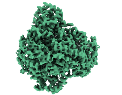
 Downloads & links
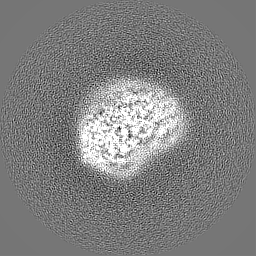
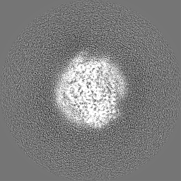
 emd_18594.png
 http://ftp.pdbj.org/pub/emdb/structures/EMD-18594
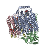


 Z (Sec.)
 Y (Row.)
 X (Col.)


 Sample components

 Processing
 Electron microscopy