 Movie
Movie Controller


+ Open data

- Basic information
| Entry | Database: PDB / ID: 1fft | ||||||
|---|---|---|---|---|---|---|---|
| Title | The structure of ubiquinol oxidase from Escherichia coli | ||||||
 Components Components | (UBIQUINOL OXIDASE ) x 4 ) x 4 | ||||||
 Keywords Keywords | OXIDOREDUCTASE / ELECTRON TRANSPORT / CYTOCHROME OXIDASE / MEMBRANE PROTEIN | ||||||
| Function / homology |  Function and homology information Function and homology informationcytochrome o ubiquinol oxidase complex / oxidoreduction-driven active transmembrane transporter activity / ubiquinol oxidase (H+-transporting) / cytochrome bo3 ubiquinol oxidase activity / aerobic electron transport chain / oxidoreductase activity, acting on diphenols and related substances as donors, oxygen as acceptor / ubiquinone binding / electron transport coupled proton transport / cytochrome-c oxidase activity / proton transmembrane transporter activity ...cytochrome o ubiquinol oxidase complex / oxidoreduction-driven active transmembrane transporter activity / ubiquinol oxidase (H+-transporting) / cytochrome bo3 ubiquinol oxidase activity / aerobic electron transport chain / oxidoreductase activity, acting on diphenols and related substances as donors, oxygen as acceptor / ubiquinone binding / electron transport coupled proton transport / cytochrome-c oxidase activity / proton transmembrane transporter activity / ATP synthesis coupled electron transport / respirasome / aerobic respiration / respiratory electron transport chain / electron transfer activity / copper ion binding / heme binding / plasma membraneSimilarity search - Function | ||||||
| Biological species |  Escherichia coli (E. coli) Escherichia coli (E. coli) | ||||||
| Method | X-RAY DIFFRACTION / SYNCHROTRON / Resolution: 3.5 Å | ||||||
 Authors Authors | Abramson, J. / Riistama, S. / Larsson, G. / Jasaitis, A. / Svensson-Ek, M. / Puustinen, A. / Iwata, S. / Wikstrom, M. | ||||||
 Citation Citation | Journal: Nat.Struct.Biol. / Year: 2000 Title: The structure of the ubiquinol oxidase from Escherichia coli and its ubiquinone binding site. Authors: Abramson, J. / Riistama, S. / Larsson, G. / Jasaitis, A. / Svensson-Ek, M. / Laakkonen, L. / Puustinen, A. / Iwata, S. / Wikstrom, M. #1: Journal: Acta Crystallogr.,Sect.D / Year: 2000Title: Purification, crystallization and preliminary crystallographic studies on an integral membrane protein, cytochrome bo3 ubiquinol oxidase from Escherichia coli. Authors: Abramson, J. / Larsson, G. / Byrne, B. / Puustinen, A. / Garcia-Horsman, A. / Iwata, S. | ||||||
| History |
|
- Structure visualization
| Structure viewer | Molecule: MolmilJmol/JSmol |
|---|
- Downloads & links
-Download
| PDBx/mmCIF format | 1fft.cif.gz | 371.5 KB | Display | PDBx/mmCIF format |
|---|---|---|---|---|
| PDB format | pdb1fft.ent.gz | 283.4 KB | Display | PDB format |
| PDBx/mmJSON format | 1fft.json.gz | Tree view | PDBx/mmJSON format | |
| Others |  Other downloads Other downloads |
-Validation report
| Arichive directory | https://data.pdbj.org/pub/pdb/validation_reports/ff/1fftftp://data.pdbj.org/pub/pdb/validation_reports/ff/1fft | HTTPS FTP |
|---|
-Related structure data
| Similar structure data |
|---|


-Links
 PDBj
PDBj


- Assembly
| Deposited unit | 
| ||||||||
|---|---|---|---|---|---|---|---|---|---|
| 1 | 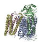
| ||||||||
| 2 | 
| ||||||||
| Unit cell |
| ||||||||
| Details | The biological assembly is a monomer composed of chains A, B, C, D, and E. |
-Components
-Protein , 4 types, 8 molecules AFBGCHDI
| #1: Protein | Mass: 74424.469 Da / Num. of mol.: 2 Source method: isolated from a genetically manipulated source Source: (gene. exp.) Escherichia coli (E. coli) / Plasmid: PBR322 / Production host: Escherichia coli (E. coli)References: UniProt: P0ABI8, Oxidoreductases; Acting on diphenols and related substances as donors; With oxygen as acceptor#2: Protein | Mass: 34947.203 Da / Num. of mol.: 2 Source method: isolated from a genetically manipulated source Source: (gene. exp.) Escherichia coli (E. coli) / Plasmid: PBR322 / Production host: Escherichia coli (E. coli)References: UniProt: P0ABJ1, Oxidoreductases; Acting on diphenols and related substances as donors; With oxygen as acceptor#3: Protein | Mass: 22642.566 Da / Num. of mol.: 2 Source method: isolated from a genetically manipulated source Source: (gene. exp.) Escherichia coli (E. coli) / Plasmid: PBR322 / Production host: Escherichia coli (E. coli)References: UniProt: P0ABJ3, Oxidoreductases; Acting on diphenols and related substances as donors; With oxygen as acceptor#4: Protein | Mass: 9294.448 Da / Num. of mol.: 2 Source method: isolated from a genetically manipulated source Source: (gene. exp.) Escherichia coli (E. coli) / Plasmid: PBR322 / Production host: Escherichia coli (E. coli)References: Oxidoreductases; Acting on diphenols and related substances as donors; With oxygen as acceptor |
|---|
-Non-polymers , 3 types, 6 molecules 
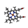
| #5: Chemical | Copper Mass: 63.546 Da / Num. of mol.: 2 / Source method: obtained synthetically / Formula: Cu Mass: 63.546 Da / Num. of mol.: 2 / Source method: obtained synthetically / Formula: Cu#6: Chemical | Heme B Mass: 616.487 Da / Num. of mol.: 2 / Source method: obtained synthetically / Formula: C34H32FeN4O4 Mass: 616.487 Da / Num. of mol.: 2 / Source method: obtained synthetically / Formula: C34H32FeN4O4#7: Chemical | Heme O Mass: 838.854 Da / Num. of mol.: 2 / Source method: obtained synthetically / Formula: C49H58FeN4O5 Mass: 838.854 Da / Num. of mol.: 2 / Source method: obtained synthetically / Formula: C49H58FeN4O5 |
|---|
-Experimental details
-Experiment
| Experiment | Method: X-RAY DIFFRACTION / Number of used crystals: 1 |
|---|
- Sample preparation
| Crystal | Density Matthews: 3.53 Å3/Da / Density % sol: 65.17 % | ||||||||||||||||||||||||||||||||||||||||||||||||
|---|---|---|---|---|---|---|---|---|---|---|---|---|---|---|---|---|---|---|---|---|---|---|---|---|---|---|---|---|---|---|---|---|---|---|---|---|---|---|---|---|---|---|---|---|---|---|---|---|---|
| Crystal grow | Temperature: 277 K / Method: vapor diffusion, hanging drop / pH: 7 Details: 9-10% PEG 1500, 100 mM NaCl, 100 mM MgCl2, 5% ethanol & 100 mM HEPES , pH 7.0, VAPOR DIFFUSION, HANGING DROP, temperature 4K | ||||||||||||||||||||||||||||||||||||||||||||||||
| Crystal grow | *PLUS pH: 7.5 | ||||||||||||||||||||||||||||||||||||||||||||||||
| Components of the solutions | *PLUS
|
-Data collection
| Diffraction | Mean temperature: 100 K |
|---|---|
| Diffraction source | Source: SYNCHROTRON / Site: ESRF  / Beamline: ID14-3 / Wavelength: 0.95 / Beamline: ID14-3 / Wavelength: 0.95 |
| Detector | Type: MAR CCD 165 mm / Detector: CCD / Date: Apr 11, 1998 |
| Radiation | Protocol: SINGLE WAVELENGTH / Monochromatic (M) / Laue (L): M / Scattering type: x-ray |
| Radiation wavelength | Wavelength: 0.95 Å / Relative weight: 1 |
| Reflection | Resolution: 3.5→40 Å / Num. obs: 44359 / % possible obs: 87 % / Observed criterion σ(I): -0.2 / Redundancy: 3.2 % / Biso Wilson estimate: 37.1 Å2 / Rmerge(I) obs: 0.123 / Net I/σ(I): 7.86 |
| Reflection shell | Resolution: 3.5→3.63 Å / Rmerge(I) obs: 0.399 / Num. unique all: 3956 / % possible all: 78.5 |
| Reflection shell | *PLUS % possible obs: 78.5 % / Num. unique obs: 3956 / Mean I/σ(I) obs: 1.47 |
- Processing
| Software |
| ||||||||||||
|---|---|---|---|---|---|---|---|---|---|---|---|---|---|
| Refinement | Resolution: 3.5→40 Å / Details: This is a non-refined structure. | ||||||||||||
| Refinement step | Cycle: LAST / Resolution: 3.5→40 Å
| ||||||||||||
| Refinement | *PLUS Highest resolution: 3.5 Å / Lowest resolution: 40 Å / Rfactor obs: 0.446 | ||||||||||||
| Solvent computation | *PLUS | ||||||||||||
| Displacement parameters | *PLUS | ||||||||||||
| LS refinement shell | *PLUS Rfactor obs: 0.482 |
