Movie
Movie Controller

[English] 日本語
 Yorodumi
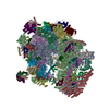
- EMDB-16898: 28S human mitochondrial small ribosomal subunit with mtRF1 and P-... -
+ Open data

- Basic information
| Entry |  | ||||||||||||
|---|---|---|---|---|---|---|---|---|---|---|---|---|---|
| Title | 28S human mitochondrial small ribosomal subunit with mtRF1 and P-site tRNA | ||||||||||||
 Map data Map data | Human 28S mitoribosomal small subunit | ||||||||||||
 Sample Sample |
| ||||||||||||
 Keywords Keywords |  human mitochondrial ribosome / release factor mtRF1 / non-canonical stop codon / RIBOSOME human mitochondrial ribosome / release factor mtRF1 / non-canonical stop codon / RIBOSOME | ||||||||||||
| Function / homology |  Function and homology informationmitochondrial ribosome binding / translation release factor activity / mitochondrial translational termination / mitochondrial ribosome assembly / translation release factor activity, codon specific / Mitochondrial translation elongation / positive regulation of mitochondrial translation / Mitochondrial translation termination / Mitochondrial translation initiation / negative regulation of mitotic nuclear division ...mitochondrial ribosome binding / translation release factor activity / mitochondrial translational termination / mitochondrial ribosome assembly / translation release factor activity, codon specific / Mitochondrial translation elongation / positive regulation of mitochondrial translation / Mitochondrial translation termination / Mitochondrial translation initiation / negative regulation of mitotic nuclear division / mitochondrial large ribosomal subunit / mitochondrial small ribosomal subunit / mitochondrial ribosome / mitochondrial translation / positive regulation of proteolysis / ribosomal small subunit binding / apoptotic signaling pathway / small ribosomal subunit rRNA binding / fibrillar center / ribosomal small subunit assembly / regulation of translation / cell junction / small ribosomal subunit / nuclear membrane / mitochondrial inner membrane / cell population proliferation / tRNA binding / rRNA binding / ribosome / mitochondrial matrix / structural constituent of ribosome / translation / protein domain specific binding / mRNA binding / intracellular membrane-bounded organelle / synapse / GTP binding / nucleolus / mitochondrion / RNA binding / nucleoplasm / nucleus / plasma membrane / cytosol / cytoplasm Function and homology informationmitochondrial ribosome binding / translation release factor activity / mitochondrial translational termination / mitochondrial ribosome assembly / translation release factor activity, codon specific / Mitochondrial translation elongation / positive regulation of mitochondrial translation / Mitochondrial translation termination / Mitochondrial translation initiation / negative regulation of mitotic nuclear division ...mitochondrial ribosome binding / translation release factor activity / mitochondrial translational termination / mitochondrial ribosome assembly / translation release factor activity, codon specific / Mitochondrial translation elongation / positive regulation of mitochondrial translation / Mitochondrial translation termination / Mitochondrial translation initiation / negative regulation of mitotic nuclear division / mitochondrial large ribosomal subunit / mitochondrial small ribosomal subunit / mitochondrial ribosome / mitochondrial translation / positive regulation of proteolysis / ribosomal small subunit binding / apoptotic signaling pathway / small ribosomal subunit rRNA binding / fibrillar center / ribosomal small subunit assembly / regulation of translation / cell junction / small ribosomal subunit / nuclear membrane / mitochondrial inner membrane / cell population proliferation / tRNA binding / rRNA binding / ribosome / mitochondrial matrix / structural constituent of ribosome / translation / protein domain specific binding / mRNA binding / intracellular membrane-bounded organelle / synapse / GTP binding / nucleolus / mitochondrion / RNA binding / nucleoplasm / nucleus / plasma membrane / cytosol / cytoplasmSimilarity search - Function | ||||||||||||
| Biological species |  Homo sapiens (human) Homo sapiens (human) | ||||||||||||
| Method | single particle reconstruction / cryo EM / Resolution: 3.0 Å | ||||||||||||
 Authors Authors | Saurer M / Leibundgut M / Scaiola A / Schoenhut T / Ban N | ||||||||||||
| Funding support |  Switzerland, European Union, 3 items Switzerland, European Union, 3 items
| ||||||||||||
 Citation Citation | Journal: Science / Year: 2023 Title: Molecular basis of translation termination at noncanonical stop codons in human mitochondria. Authors: Martin Saurer / Marc Leibundgut / Hima Priyanka Nadimpalli / Alain Scaiola / Tanja Schönhut / Richard G Lee / Stefan J Siira / Oliver Rackham / René Dreos / Tea Lenarčič / Eva Kummer / ...Authors: Martin Saurer / Marc Leibundgut / Hima Priyanka Nadimpalli / Alain Scaiola / Tanja Schönhut / Richard G Lee / Stefan J Siira / Oliver Rackham / René Dreos / Tea Lenarčič / Eva Kummer / David Gatfield / Aleksandra Filipovska / Nenad Ban /   Abstract: The genetic code that specifies the identity of amino acids incorporated into proteins during protein synthesis is almost universally conserved. Mitochondrial genomes feature deviations from the ...The genetic code that specifies the identity of amino acids incorporated into proteins during protein synthesis is almost universally conserved. Mitochondrial genomes feature deviations from the standard genetic code, including the reassignment of two arginine codons to stop codons. The protein required for translation termination at these noncanonical stop codons to release the newly synthesized polypeptides is not currently known. In this study, we used gene editing and ribosomal profiling in combination with cryo-electron microscopy to establish that mitochondrial release factor 1 (mtRF1) detects noncanonical stop codons in human mitochondria by a previously unknown mechanism of codon recognition. We discovered that binding of mtRF1 to the decoding center of the ribosome stabilizes a highly unusual conformation in the messenger RNA in which the ribosomal RNA participates in specific recognition of the noncanonical stop codons. | ||||||||||||
| History |
|
- Structure visualization
| Supplemental images |
|---|

- Downloads & links
-EMDB archive
| Map data | emd_16898.map.gz | 483.5 MB | EMDB map data format | |
|---|---|---|---|---|
| Header (meta data) | emd-16898-v30.xmlemd-16898.xml | 63.5 KB 63.5 KB | Display Display | EMDB header |
| FSC (resolution estimation) | emd_16898_fsc.xml | 17 KB | Display | FSC data file |
| Images | 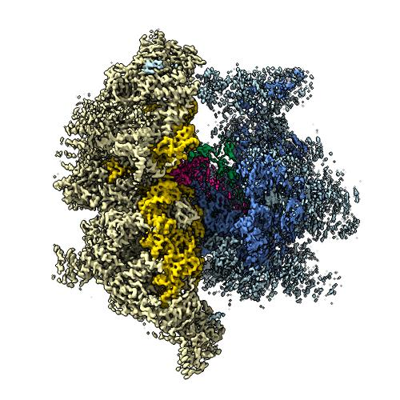 emd_16898.png emd_16898.png | 195.6 KB | ||
| Filedesc metadata | emd-16898.cif.gz | 15.2 KB | ||
| Others | emd_16898_half_map_1.map.gzemd_16898_half_map_2.map.gz | 475.6 MB 475.6 MB | ||
| Archive directory |  http://ftp.pdbj.org/pub/emdb/structures/EMD-16898ftp://ftp.pdbj.org/pub/emdb/structures/EMD-16898 http://ftp.pdbj.org/pub/emdb/structures/EMD-16898ftp://ftp.pdbj.org/pub/emdb/structures/EMD-16898 | HTTPS FTP |
-Related structure data
| Related structure data | 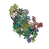 8oisMC  8oinC  8oipC  8oiqC  8oirC  8oitC C: citing same article ( M: atomic model generated by this map |
|---|---|
| Similar structure data |


-Links
| EMDB pages | EMDB (EBI/PDBe) / EMDataResource |
|---|---|
| Related items in Molecule of the Month |


-Map
| File | Download / File: emd_16898.map.gz / Format: CCP4 / Size: 512 MB / Type: IMAGE STORED AS FLOATING POINT NUMBER (4 BYTES) | ||||||||||||||||||||||||||||||||||||
|---|---|---|---|---|---|---|---|---|---|---|---|---|---|---|---|---|---|---|---|---|---|---|---|---|---|---|---|---|---|---|---|---|---|---|---|---|---|
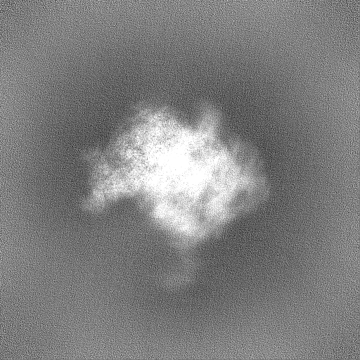
| Annotation | Human 28S mitoribosomal small subunit | ||||||||||||||||||||||||||||||||||||
| Projections & slices | Image control
Images are generated by Spider. | ||||||||||||||||||||||||||||||||||||
| Voxel size | X=Y=Z: 1.06 Å | ||||||||||||||||||||||||||||||||||||
| Density |
| ||||||||||||||||||||||||||||||||||||
| Symmetry | Space group: 1 | ||||||||||||||||||||||||||||||||||||
| Details | EMDB XML:
|
 Z (Sec.)
 Y (Row.)
 X (Col.)


-Supplemental data
-Half map: half map B
| File | emd_16898_half_map_1.map | ||||||||||||
|---|---|---|---|---|---|---|---|---|---|---|---|---|---|
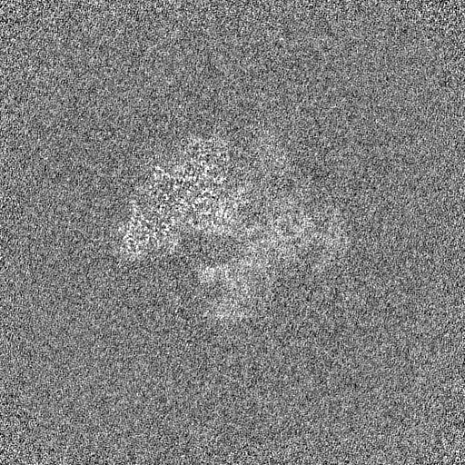
| Annotation | half map B | ||||||||||||
| Projections & Slices |
| ||||||||||||
| Density Histograms |


-Half map: half map A
| File | emd_16898_half_map_2.map | ||||||||||||
|---|---|---|---|---|---|---|---|---|---|---|---|---|---|
| Annotation | half map A | ||||||||||||
| Projections & Slices |
| ||||||||||||
| Density Histograms |


- Sample components
+Entire : 28S human mitochondrial small ribosomal subunit with mtRF1, mRNA,...
+Supramolecule #1: 28S human mitochondrial small ribosomal subunit with mtRF1, mRNA,...
+Macromolecule #1: 39S ribosomal protein L19, mitochondrial
+Macromolecule #2: 39S ribosomal protein L55, mitochondrial
+Macromolecule #4: 28S ribosomal protein S35, mitochondrial
+Macromolecule #5: 28S ribosomal protein S24, mitochondrial
+Macromolecule #6: Aurora kinase A-interacting protein
+Macromolecule #7: 28S ribosomal protein S6, mitochondrial
+Macromolecule #8: 28S ribosomal protein S7, mitochondrial
+Macromolecule #10: 28S ribosomal protein S10, mitochondrial
+Macromolecule #12: 28S ribosomal protein S12, mitochondrial
+Macromolecule #13: 28S ribosomal protein S14, mitochondrial
+Macromolecule #14: 28S ribosomal protein S15, mitochondrial
+Macromolecule #15: 28S ribosomal protein S16, mitochondrial
+Macromolecule #16: 28S ribosomal protein S17, mitochondrial
+Macromolecule #17: 28S ribosomal protein S18b, mitochondrial
+Macromolecule #18: 28S ribosomal protein S18c, mitochondrial
+Macromolecule #19: 28S ribosomal protein S21, mitochondrial
+Macromolecule #20: 28S ribosomal protein S22, mitochondrial
+Macromolecule #21: 28S ribosomal protein S23, mitochondrial
+Macromolecule #22: 28S ribosomal protein S25, mitochondrial
+Macromolecule #23: 28S ribosomal protein S26, mitochondrial
+Macromolecule #24: 28S ribosomal protein S27, mitochondrial
+Macromolecule #25: 28S ribosomal protein S28, mitochondrial
+Macromolecule #26: 28S ribosomal protein S29, mitochondrial
+Macromolecule #27: 28S ribosomal protein S31, mitochondrial
+Macromolecule #28: 28S ribosomal protein S33, mitochondrial
+Macromolecule #29: Peptide chain release factor 1, mitochondrial,mtRF1(AAQ)
+Macromolecule #30: 28S ribosomal protein S2, mitochondrial
+Macromolecule #31: Coiled-coil-helix-coiled-coil-helix domain-containing protein 1
+Macromolecule #32: 28S ribosomal protein S5, mitochondrial
+Macromolecule #33: Pentatricopeptide repeat domain-containing protein 3, mitochondrial
+Macromolecule #34: 28S ribosomal protein S9, mitochondrial
+Macromolecule #35: 28S ribosomal protein S11, mitochondrial
+Macromolecule #36: 28S ribosomal protein S34, mitochondrial
+Macromolecule #3: 12S rRNA
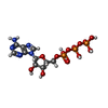
+Macromolecule #9: P-site Met-tRNA(Met)
+Macromolecule #11: mRNA
+Macromolecule #37: POTASSIUM ION
+Macromolecule #38: MAGNESIUM ION
+Macromolecule #39: METHIONINE

+Macromolecule #40: ZINC ION
+Macromolecule #41: FE-S-O HYBRID CLUSTER

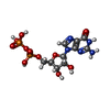
+Macromolecule #42: ADENOSINE-5'-TRIPHOSPHATE
+Macromolecule #43: GUANOSINE-5'-DIPHOSPHATE
+Macromolecule #44: water

-Experimental details
-Structure determination
| Method | cryo EM |
|---|---|
 Processing Processing | single particle reconstruction |
| Aggregation state | particle |
-Sample preparation
| Buffer | pH: 7.5 Component:
| |||||||||||||||
|---|---|---|---|---|---|---|---|---|---|---|---|---|---|---|---|---|
| Grid | Model: Quantifoil R2/2 / Material: COPPER / Support film - #0 - Film type ID: 1 / Support film - #0 - Material: CARBON / Support film - #0 - topology: HOLEY / Support film - #1 - Film type ID: 2 / Support film - #1 - Material: CARBON / Support film - #1 - topology: CONTINUOUS / Pretreatment - Type: GLOW DISCHARGE | |||||||||||||||
| Vitrification | Cryogen name: ETHANE-PROPANE / Chamber humidity: 100 % / Chamber temperature: 277.15 K / Instrument: FEI VITROBOT MARK IV | |||||||||||||||
| Details | Affinity-purified 55S mitoribosome. Refinement focused on 28S small subunit. |
- Electron microscopy
| Microscope | FEI TITAN KRIOS |
|---|---|
| Electron beam | Acceleration voltage: 300 kV / Electron source: FIELD EMISSION GUN |
| Electron optics | C2 aperture diameter: 100.0 µm / Illumination mode: FLOOD BEAM / Imaging mode: BRIGHT FIELDBright-field microscopy / Cs: 2.7 mm / Nominal defocus max: 3.0 µm / Nominal defocus min: 0.6 µm / Nominal magnification: 81000 |
| Specialist optics | Energy filter - Slit width: 20 eV |
| Sample stage | Specimen holder model: FEI TITAN KRIOS AUTOGRID HOLDER / Cooling holder cryogen: NITROGEN |
| Image recording | Film or detector model: GATAN K3 (6k x 4k) / Number grids imaged: 2 / Number real images: 43882 / Average electron dose: 60.0 e/Å2 |
| Experimental equipment |  Model: Titan Krios / Image courtesy: FEI Company |
-Image processing
| Particle selection | Number selected: 5423665 Details: Particles were picked with blob picker in Cryosparc. |
|---|---|
| Startup model | Type of model: EMDB MAP EMDB ID: Details: low-pass filtered to 40A |
| Initial angle assignment | Type: MAXIMUM LIKELIHOOD / Software - Name: cryoSPARC (ver. 4) Details: Homogeneous refinement (Cryosparc) of all particles with EMD-2914 (low-pass filtered to 40A) as initial reference |
| Final angle assignment | Type: MAXIMUM LIKELIHOOD / Software - Name: cryoSPARC (ver. 4) |
| Final reconstruction | Resolution.type: BY AUTHOR / Resolution: 3.0 Å / Resolution method: FSC 0.143 CUT-OFF / Software - Name: cryoSPARC (ver. 4) / Number images used: 41288 |
| Details | Micrographs were motion corrected and CTF parameters were estimated using Cryosparc. |
| FSC plot (resolution estimation) |  |

-Atomic model buiding 1
| Initial model |
| |||||||||
|---|---|---|---|---|---|---|---|---|---|---|
| Refinement | Space: REAL / Protocol: FLEXIBLE FIT | |||||||||
| Output model | PDB-8ois: |