[English] 日本語
Yorodumi
- EMDB-13160: V. nigripulchritudo ExoY-G-actin-complex -

+
Open data


ID or keywords:

Loading...

-
Basic information

Entry
Database: EMDB / ID: EMD-13160
TitleV. nigripulchritudo ExoY-G-actin-complex
Map dataDeepEMhanced map
Sample
  • Complex: V. nigripulchritudo ExoY-G-actin-complex
    • Protein or peptide: ExoY
    • Protein or peptide: Actin, alpha skeletal muscle
Function / homology
Function and homology information


calcium- and calmodulin-responsive adenylate cyclase activity / positive regulation of norepinephrine uptake / synapse maturation / modification of postsynaptic actin cytoskeleton / negative regulation of actin filament bundle assembly / cellular response to cytochalasin B / adenyl-nucleotide exchange factor activity / bBAF complex / npBAF complex / postsynaptic actin cytoskeleton organization ...calcium- and calmodulin-responsive adenylate cyclase activity / positive regulation of norepinephrine uptake / synapse maturation / modification of postsynaptic actin cytoskeleton / negative regulation of actin filament bundle assembly / cellular response to cytochalasin B / adenyl-nucleotide exchange factor activity / bBAF complex / npBAF complex / postsynaptic actin cytoskeleton organization / regulation of transepithelial transport / brahma complex / nBAF complex / structural constituent of postsynaptic actin cytoskeleton / morphogenesis of a polarized epithelium / GBAF complex / positive regulation of actin filament bundle assembly / postsynaptic actin cytoskeleton / protein localization to adherens junction / Formation of annular gap junctions / negative regulation of actin filament polymerization / regulation of G0 to G1 transition / dense body / Gap junction degradation / Tat protein binding / Signaling by ROBO receptors / Cell-extracellular matrix interactions / Folding of actin by CCT/TriC / regulation of actin filament polymerization / regulation of double-strand break repair / regulation of nucleotide-excision repair / RSC-type complex / apical protein localization / Prefoldin mediated transfer of substrate to CCT/TriC / adherens junction assembly / RHOF GTPase cycle / Adherens junctions interactions / positive regulation of ATP-dependent activity / PCP/CE pathway / proline-rich region binding / tight junction / Sensory processing of sound by outer hair cells of the cochlea / positive regulation of ruffle assembly / SWI/SNF complex / Interaction between L1 and Ankyrins / Sensory processing of sound by inner hair cells of the cochlea / regulation of mitotic metaphase/anaphase transition / regulation of norepinephrine uptake / positive regulation of double-strand break repair / positive regulation of T cell differentiation / negative regulation of stress fiber assembly / host cell cytosol / NuA4 histone acetyltransferase complex / regulation of synaptic vesicle endocytosis / apical junction complex / establishment or maintenance of cell polarity / maintenance of blood-brain barrier / detection of maltose stimulus / positive regulation of actin filament polymerization / positive regulation of stem cell population maintenance / maltose binding / maltose transport complex / positive regulation of double-strand break repair via homologous recombination / positive regulation of epithelial cell migration / cortical cytoskeleton / maltose transport / maltodextrin transmembrane transport / nitric-oxide synthase binding / Recycling pathway of L1 / regulation of cyclin-dependent protein serine/threonine kinase activity / regulation of G1/S transition of mitotic cell cycle / negative regulation of cell differentiation / brush border / calyx of Held / kinesin binding / carbohydrate transmembrane transporter activity / ATP-binding cassette (ABC) transporter complex, substrate-binding subunit-containing / actin monomer binding / carbohydrate transport / : / EPH-ephrin mediated repulsion of cells / RHO GTPases Activate WASPs and WAVEs / RHO GTPases activate IQGAPs / positive regulation of myoblast differentiation / regulation of protein localization to plasma membrane / EPHB-mediated forward signaling / phosphatidylinositol-4,5-bisphosphate binding / substantia nigra development / phosphotyrosine residue binding / ATP-binding cassette (ABC) transporter complex / cell chemotaxis / axonogenesis / negative regulation of protein binding / neural tube closure / Translocation of SLC2A4 (GLUT4) to the plasma membrane / cell motility / RHO GTPases Activate Formins / actin filament / regulation of transmembrane transporter activity / positive regulation of cell differentiation
Similarity search - Function
Peptidase C58, YopT-type domain / Yersinia/Haemophilus virulence surface antigen / Profilin1/2/3, vertebrate / Anthrax toxin, edema factor, central / Anthrax toxin, edema factor, C-terminal / Anthrax toxin, edema factor, central domain superfamily / Anthrax toxin LF subunit / : / Profilin conserved site / Profilin signature. ...Peptidase C58, YopT-type domain / Yersinia/Haemophilus virulence surface antigen / Profilin1/2/3, vertebrate / Anthrax toxin, edema factor, central / Anthrax toxin, edema factor, C-terminal / Anthrax toxin, edema factor, central domain superfamily / Anthrax toxin LF subunit / : / Profilin conserved site / Profilin signature. / Profilin / Profilin / Dermonecrotic/RTX toxin, membrane localization domain / Profilin / Membrane Localization Domain / Profilin superfamily / Serine aminopeptidase, S33 / CGT/MARTX, cysteine protease (CPD) domain / CGT/MARTX, cysteine protease (CPD) domain superfamily / Peptidase C80 family / CGT/MARTX cysteine protease (CPD) domain profile. / Serine aminopeptidase, S33 / Maltose/Cyclodextrin ABC transporter, substrate-binding protein / Solute-binding family 1, conserved site / Bacterial extracellular solute-binding proteins, family 1 signature. / Bacterial extracellular solute-binding protein / Actins signature 1. / Actin, conserved site / Actins signature 2. / Actin/actin-like conserved site / Actins and actin-related proteins signature. / Bacterial extracellular solute-binding protein / Actin / Actin family / Actin / ATPase, nucleotide binding domain / Alpha/Beta hydrolase fold
Similarity search - Domain/homology
RTX-toxin / Profilin-1 / Maltose/maltodextrin-binding periplasmic protein / Actin, cytoplasmic 1
Similarity search - Component
Biological speciesVibrio nigripulchritudo (bacteria) / Oryctolagus cuniculus (rabbit)
Methodsingle particle reconstruction / cryo EM / Resolution: 4.2 Å
AuthorsBelyy A / Merino F / Raunser S
Funding support Germany, 1 items
OrganizationGrant numberCountry
Max Planck Society Germany
Citation
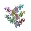
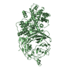
Journal: Nat Commun / Year: 2021
Title: Mechanism of actin-dependent activation of nucleotidyl cyclase toxins from bacterial human pathogens.
Authors: Alexander Belyy / Felipe Merino / Undine Mechold / Stefan Raunser /
Abstract: Bacterial human pathogens secrete initially inactive nucleotidyl cyclases that become potent enzymes by binding to actin inside eukaryotic host cells. The underlying molecular mechanism of this ...Bacterial human pathogens secrete initially inactive nucleotidyl cyclases that become potent enzymes by binding to actin inside eukaryotic host cells. The underlying molecular mechanism of this activation is, however, unclear. Here, we report structures of ExoY from Pseudomonas aeruginosa and Vibrio vulnificus bound to their corresponding activators F-actin and profilin-G-actin. The structures reveal that in contrast to the apo-state, two flexible regions become ordered and interact strongly with actin. The specific stabilization of these regions results in an allosteric stabilization of the nucleotide binding pocket and thereby to an activation of the enzyme. Differences in the sequence and conformation of the actin-binding regions are responsible for the selective binding to either F- or G-actin. Other nucleotidyl cyclase toxins that bind to calmodulin rather than actin undergo a similar disordered-to-ordered transition during activation, suggesting that the allosteric activation-by-stabilization mechanism of ExoY is conserved in these enzymes, albeit the different activator.
History
DepositionJul 1, 2021-
Header (metadata) releaseNov 17, 2021-
Map releaseNov 17, 2021-
UpdateDec 1, 2021-
Current statusDec 1, 2021Processing site: PDBe / Status: Released

-
Structure visualization

Movie
  • Surface view with section colored by density value
  • Surface level: 0.15
  • Imaged by UCSF Chimera
  • Download
  • Surface view colored by height
  • Surface level: 0.15
  • Imaged by UCSF Chimera
  • Download
Movie viewer
Structure viewerEM map:
SurfViewMolmilJmol/JSmol
Supplemental images

Downloads & links

-
Map

FileDownload / File: emd_13160.map.gz / Format: CCP4 / Size: 83.7 MB / Type: IMAGE STORED AS FLOATING POINT NUMBER (4 BYTES)
AnnotationDeepEMhanced map
Voxel sizeX=Y=Z: 0.85 Å
Density
Contour LevelBy AUTHOR: 0.15 / Movie #1: 0.15
Minimum - Maximum-0.0017831661 - 2.4429297
Average (Standard dev.)0.0010925975 (±0.024843154)
SymmetrySpace group: 1
Details

EMDB XML:

Map geometry
Axis orderXYZ
Origin000
Dimensions280280280
Spacing280280280
CellA=B=C: 238.0 Å
α=β=γ: 90.0 °

CCP4 map header:

modeImage stored as Reals
Å/pix. X/Y/Z0.850.850.85
M x/y/z280280280
origin x/y/z0.0000.0000.000
length x/y/z238.000238.000238.000
α/β/γ90.00090.00090.000
start NX/NY/NZ000
NX/NY/NZ300300300
MAP C/R/S123
start NC/NR/NS000
NC/NR/NS280280280
D min/max/mean-0.0022.4430.001

-
Supplemental data

-
Sample components

-
Entire : V. nigripulchritudo ExoY-G-actin-complex

EntireName: V. nigripulchritudo ExoY-G-actin-complex
Components
  • Complex: V. nigripulchritudo ExoY-G-actin-complex
    • Protein or peptide: ExoY
    • Protein or peptide: Actin, alpha skeletal muscle

-
Supramolecule #1: V. nigripulchritudo ExoY-G-actin-complex

SupramoleculeName: V. nigripulchritudo ExoY-G-actin-complex / type: complex / ID: 1 / Parent: 0 / Macromolecule list: all

-
Macromolecule #1: ExoY

MacromoleculeName: ExoY / type: protein_or_peptide / ID: 1 / Enantiomer: LEVO
Source (natural)Organism: Vibrio nigripulchritudo (bacteria)
Recombinant expressionOrganism: Escherichia coli (E. coli)
SequenceString: MGYNYGQALQ EAQLDIATMK PRQRVTANEL QLGDDNAITN AVTSEQEATP NQDGSHKTYQ SRDLVLEPIQ HPKSIELGMP EVDQSVLAEV AERENVIIGV RPVDEKSKSL IASKMYSSKG LFVKAKSSDW GPMSGFIPVD QSFAKASARR DLEKFNEYAE QSILSGNAVS ...String:
MGYNYGQALQ EAQLDIATMK PRQRVTANEL QLGDDNAITN AVTSEQEATP NQDGSHKTYQ SRDLVLEPIQ HPKSIELGMP EVDQSVLAEV AERENVIIGV RPVDEKSKSL IASKMYSSKG LFVKAKSSDW GPMSGFIPVD QSFAKASARR DLEKFNEYAE QSILSGNAVS ANLYLNQVRI EELVSKYESL TPLELDVDSG MYKTTATNGD QTIPFFLNKV TVDDKELWQV HYLREGELAP FKVIGDPVSK QPMTADYDLL TVMYTYGDLG PQDKVKQPLT WEQWKESVTY EDLSPKYKAR YDNQALYEKQ DGASLGMVSD RLKELKDVIN TSLGRTDGLE MVHHGADDAN PYAVMADNFP ATFFVPKHFF DDDGLGEGKG SIQTYFNVNE QGAVVIQNPQ EFSNFQQVAI NASYRASLND KWNSGLDSPL FTTKRKLSHD YLDARDEVAK KLGLTESSKL NGLG

-
Macromolecule #2: Actin, alpha skeletal muscle

MacromoleculeName: Actin, alpha skeletal muscle / type: protein_or_peptide / ID: 2 / Enantiomer: LEVO
Source (natural)Organism: Oryctolagus cuniculus (rabbit)
SequenceString: MCDEDETTAL VCDNGSGLVK AGFAGDDAPR AVFPSIVGRP RHQGVMVGMG QKDSYVGDEA QSKRGILTLK YPIEHGIITN WDDMEKIWH HTFYNELRVA PEEHPTLLTE APLNPKANRE KMTQIMFETF NVPAMYVAIQ AVLSLYASGR TTGIVLDSGD G VTHNVPIY ...String:
MCDEDETTAL VCDNGSGLVK AGFAGDDAPR AVFPSIVGRP RHQGVMVGMG QKDSYVGDEA QSKRGILTLK YPIEHGIITN WDDMEKIWH HTFYNELRVA PEEHPTLLTE APLNPKANRE KMTQIMFETF NVPAMYVAIQ AVLSLYASGR TTGIVLDSGD G VTHNVPIY EGYALPHAIM RLDLAGRDLT DYLMKILTER GYSFVTTAER EIVRDIKEKL CYVALDFENE MATAASSSSL EK SYELPDG QVITIGNERF RCPETLFQPS FIGMESAGIH ETTYNSIMKC DIDIRKDLYA NNVMSGGTTM YPGIADRMQK EIT ALAPST MKIKIIAPPE RKYSVWIGGS ILASLSTFQQ MWITKQEYDE AGPSIVHRKC F

-
Experimental details

-
Structure determination

Methodcryo EM
Processingsingle particle reconstruction
Aggregation stateparticle

-
Sample preparation

BufferpH: 8
GridModel: Quantifoil R2/1 / Material: COPPER / Mesh: 300
VitrificationCryogen name: ETHANE / Chamber humidity: 100 % / Instrument: FEI VITROBOT MARK III

-
Electron microscopy

MicroscopeFEI TITAN KRIOS
Electron beamAcceleration voltage: 300 kV / Electron source: FIELD EMISSION GUN
Electron opticsIllumination mode: SPOT SCAN / Imaging mode: BRIGHT FIELDBright-field microscopy / Nominal defocus max: 2.5 µm / Nominal defocus min: 1.0 µm
Image recordingFilm or detector model: GATAN K2 SUMMIT (4k x 4k) / Detector mode: COUNTING / Number grids imaged: 1 / Number real images: 10659 / Average exposure time: 8.0 sec. / Average electron dose: 80.0 e/Å2
Experimental equipment
Model: Titan Krios / Image courtesy: FEI Company

-
Image processing

Particle selectionNumber selected: 1865213
CTF correctionSoftware - Name: CTFFIND
Initial angle assignmentType: MAXIMUM LIKELIHOOD / Software - Name: SPHIRE
Final angle assignmentType: MAXIMUM LIKELIHOOD / Software - Name: SPHIRE
Final reconstructionApplied symmetry - Point group: C1 (asymmetric) / Algorithm: FOURIER SPACE / Resolution.type: BY AUTHOR / Resolution: 4.2 Å / Resolution method: FSC 0.143 CUT-OFF / Software - Name: SPHIRE / Number images used: 236009
FSC plot (resolution estimation)

+
About Yorodumi

-
News

-
Feb 9, 2022. New format data for meta-information of EMDB entries

New format data for meta-information of EMDB entries

  • Version 3 of the EMDB header file is now the official format.
  • The previous official version 1.9 will be removed from the archive.

Related info.:EMDB header

External links:wwPDB to switch to version 3 of the EMDB data model

-
Aug 12, 2020. Covid-19 info

Covid-19 info

URL: https://pdbj.org/emnavi/covid19.php

New page: Covid-19 featured information page in EM Navigator.

Related info.:Covid-19 info / Mar 5, 2020. Novel coronavirus structure data

+
Mar 5, 2020. Novel coronavirus structure data

Novel coronavirus structure data

Related info.:Yorodumi Speices / Aug 12, 2020. Covid-19 info

External links:COVID-19 featured content - PDBj / Molecule of the Month (242):Coronavirus Proteases

+
Jan 31, 2019. EMDB accession codes are about to change! (news from PDBe EMDB page)

EMDB accession codes are about to change! (news from PDBe EMDB page)

  • The allocation of 4 digits for EMDB accession codes will soon come to an end. Whilst these codes will remain in use, new EMDB accession codes will include an additional digit and will expand incrementally as the available range of codes is exhausted. The current 4-digit format prefixed with “EMD-” (i.e. EMD-XXXX) will advance to a 5-digit format (i.e. EMD-XXXXX), and so on. It is currently estimated that the 4-digit codes will be depleted around Spring 2019, at which point the 5-digit format will come into force.
  • The EM Navigator/Yorodumi systems omit the EMD- prefix.

Related info.:Q: What is EMD? / ID/Accession-code notation in Yorodumi/EM Navigator

External links:EMDB Accession Codes are Changing Soon! / Contact to PDBj

+
Jul 12, 2017. Major update of PDB

Major update of PDB

  • wwPDB released updated PDB data conforming to the new PDBx/mmCIF dictionary.
  • This is a major update changing the version number from 4 to 5, and with Remediation, in which all the entries are updated.
  • In this update, many items about electron microscopy experimental information are reorganized (e.g. em_software).
  • Now, EM Navigator and Yorodumi are based on the updated data.

External links:wwPDB Remediation / Enriched Model Files Conforming to OneDep Data Standards Now Available in the PDB FTP Archive

-
Yorodumi

Thousand views of thousand structures

  • Yorodumi is a browser for structure data from EMDB, PDB, SASBDB, etc.
  • This page is also the successor to EM Navigator detail page, and also detail information page/front-end page for Omokage search.
  • The word "yorodu" (or yorozu) is an old Japanese word meaning "ten thousand". "mi" (miru) is to see.

Related info.:EMDB / PDB / SASBDB / Comparison of 3 databanks / Yorodumi Search / Aug 31, 2016. New EM Navigator & Yorodumi / Yorodumi Papers / Jmol/JSmol / Function and homology information / Changes in new EM Navigator and Yorodumi

Read more