Movie
Movie Controller


+ Open data

- Basic information
| Entry | Database: EMDB / ID: EMD-13158 | |||||||||
|---|---|---|---|---|---|---|---|---|---|---|
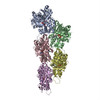
| Title | Structure of the P. aeruginosa ExoY-F-actin complex | |||||||||
 Map data Map data | Locally filtered map | |||||||||
 Sample Sample |
| |||||||||
| Function / homology |  Function and homology information Function and homology informationcalcium- and calmodulin-responsive adenylate cyclase activity / cytoskeletal motor activator activity / detection of maltose stimulus /  maltose binding / tropomyosin binding / maltose transport complex / myosin heavy chain binding / maltose transport / mesenchyme migration / maltodextrin transmembrane transport ...calcium- and calmodulin-responsive adenylate cyclase activity / cytoskeletal motor activator activity / detection of maltose stimulus / maltose binding / tropomyosin binding / maltose transport complex / myosin heavy chain binding / maltose transport / mesenchyme migration / maltodextrin transmembrane transport / troponin I binding / actin filament bundle / filamentous actin / actin filament bundle assembly / skeletal muscle thin filament assembly / striated muscle thin filament / carbohydrate transmembrane transporter activity / ATP-binding cassette (ABC) transporter complex, substrate-binding subunit-containing / skeletal muscle myofibril / actin monomer binding / carbohydrate transport / skeletal muscle fiber development / stress fiber / titin binding / actin filament polymerization / ATP-binding cassette (ABC) transporter complex / cell chemotaxis / filopodium / actin filament / Hydrolases; Acting on acid anhydrides; Acting on acid anhydrides to facilitate cellular and subcellular movement / calcium-dependent protein binding / lamellipodium / cell body / outer membrane-bounded periplasmic space / periplasmic space / hydrolase activity / protein domain specific binding / DNA damage response / calcium ion binding / positive regulation of gene expression / magnesium ion binding / extracellular region / ATP binding / membrane / identical protein binding / cytoplasm maltose binding / tropomyosin binding / maltose transport complex / myosin heavy chain binding / maltose transport / mesenchyme migration / maltodextrin transmembrane transport ...calcium- and calmodulin-responsive adenylate cyclase activity / cytoskeletal motor activator activity / detection of maltose stimulus / maltose binding / tropomyosin binding / maltose transport complex / myosin heavy chain binding / maltose transport / mesenchyme migration / maltodextrin transmembrane transport / troponin I binding / actin filament bundle / filamentous actin / actin filament bundle assembly / skeletal muscle thin filament assembly / striated muscle thin filament / carbohydrate transmembrane transporter activity / ATP-binding cassette (ABC) transporter complex, substrate-binding subunit-containing / skeletal muscle myofibril / actin monomer binding / carbohydrate transport / skeletal muscle fiber development / stress fiber / titin binding / actin filament polymerization / ATP-binding cassette (ABC) transporter complex / cell chemotaxis / filopodium / actin filament / Hydrolases; Acting on acid anhydrides; Acting on acid anhydrides to facilitate cellular and subcellular movement / calcium-dependent protein binding / lamellipodium / cell body / outer membrane-bounded periplasmic space / periplasmic space / hydrolase activity / protein domain specific binding / DNA damage response / calcium ion binding / positive regulation of gene expression / magnesium ion binding / extracellular region / ATP binding / membrane / identical protein binding / cytoplasmSimilarity search - Function | |||||||||
| Biological species |  Pseudomonas aeruginosa PAO1 (bacteria) / Pseudomonas aeruginosa PAO1 (bacteria) /  Rabbit (rabbit) / Rabbit (rabbit) /  Amanita phalloides (death cap) Amanita phalloides (death cap) | |||||||||
| Method | single particle reconstruction / cryo EM / Resolution: 3.2 Å | |||||||||
 Authors Authors | Belyy A / Merino F / Raunser S | |||||||||
| Funding support |  Germany, 1 items Germany, 1 items
| |||||||||
 Citation Citation | Journal: Nat Commun / Year: 2021 Title: Mechanism of actin-dependent activation of nucleotidyl cyclase toxins from bacterial human pathogens. Authors: Alexander Belyy / Felipe Merino / Undine Mechold / Stefan Raunser /  Abstract: Bacterial human pathogens secrete initially inactive nucleotidyl cyclases that become potent enzymes by binding to actin inside eukaryotic host cells. The underlying molecular mechanism of this ...Bacterial human pathogens secrete initially inactive nucleotidyl cyclases that become potent enzymes by binding to actin inside eukaryotic host cells. The underlying molecular mechanism of this activation is, however, unclear. Here, we report structures of ExoY from Pseudomonas aeruginosa and Vibrio vulnificus bound to their corresponding activators F-actin and profilin-G-actin. The structures reveal that in contrast to the apo-state, two flexible regions become ordered and interact strongly with actin. The specific stabilization of these regions results in an allosteric stabilization of the nucleotide binding pocket and thereby to an activation of the enzyme. Differences in the sequence and conformation of the actin-binding regions are responsible for the selective binding to either F- or G-actin. Other nucleotidyl cyclase toxins that bind to calmodulin rather than actin undergo a similar disordered-to-ordered transition during activation, suggesting that the allosteric activation-by-stabilization mechanism of ExoY is conserved in these enzymes, albeit the different activator. | |||||||||
| History |
|
- Structure visualization
| Movie |
Movie viewer |
|---|---|
| Structure viewer | EM map: SurfViewMolmilJmol/JSmol |
| Supplemental images |

- Downloads & links
-EMDB archive
| Map data | emd_13158.map.gz | 6.8 MB | EMDB map data format | |
|---|---|---|---|---|
| Header (meta data) | emd-13158-v30.xmlemd-13158.xml | 17.7 KB 17.7 KB | Display Display | EMDB header |
| FSC (resolution estimation) | emd_13158_fsc.xml | 11.7 KB | Display | FSC data file |
| Images |  emd_13158.png emd_13158.png | 153.2 KB | ||
| Others | emd_13158_additional_1.map.gz | 91.9 MB | ||
| Archive directory |  http://ftp.pdbj.org/pub/emdb/structures/EMD-13158ftp://ftp.pdbj.org/pub/emdb/structures/EMD-13158 http://ftp.pdbj.org/pub/emdb/structures/EMD-13158ftp://ftp.pdbj.org/pub/emdb/structures/EMD-13158 | HTTPS FTP |
-Related structure data
| Related structure data | 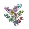 7p1gMC  7p1hC M: atomic model generated by this map C: citing same article ( |
|---|---|
| Similar structure data |


-Links
| EMDB pages | EMDB (EBI/PDBe) / EMDataResource |
|---|---|
| Related items in Molecule of the Month |


-Map
| File | Download / File: emd_13158.map.gz / Format: CCP4 / Size: 103 MB / Type: IMAGE STORED AS FLOATING POINT NUMBER (4 BYTES) | ||||||||||||||||||||||||||||||||||||||||||||||||||||||||||||
|---|---|---|---|---|---|---|---|---|---|---|---|---|---|---|---|---|---|---|---|---|---|---|---|---|---|---|---|---|---|---|---|---|---|---|---|---|---|---|---|---|---|---|---|---|---|---|---|---|---|---|---|---|---|---|---|---|---|---|---|---|---|
| Annotation | Locally filtered map | ||||||||||||||||||||||||||||||||||||||||||||||||||||||||||||
| Voxel size | X=Y=Z: 1.1 Å | ||||||||||||||||||||||||||||||||||||||||||||||||||||||||||||
| Density |
| ||||||||||||||||||||||||||||||||||||||||||||||||||||||||||||
| Symmetry | Space group: 1 | ||||||||||||||||||||||||||||||||||||||||||||||||||||||||||||
| Details | EMDB XML:
CCP4 map header:
| ||||||||||||||||||||||||||||||||||||||||||||||||||||||||||||
-Supplemental data
-Additional map: DeepEMhanced map
| File | emd_13158_additional_1.map | ||||||||||||
|---|---|---|---|---|---|---|---|---|---|---|---|---|---|
| Annotation | DeepEMhanced map | ||||||||||||
| Projections & Slices |
| ||||||||||||
| Density Histograms |
 Z
Z Y
Y X
X


- Sample components
-Entire : The complex of P. aeruginosa ExoY with F-actin
| Entire | Name: The complex of P. aeruginosa ExoY with F-actin |
|---|---|
| Components |
|
-Supramolecule #1: The complex of P. aeruginosa ExoY with F-actin
| Supramolecule | Name: The complex of P. aeruginosa ExoY with F-actin / type: complex / ID: 1 / Parent: 0 / Macromolecule list: #1-#3 |
|---|
-Macromolecule #1: Maltose/maltodextrin-binding periplasmic protein,Adenylate cyclas...
| Macromolecule | Name: Maltose/maltodextrin-binding periplasmic protein,Adenylate cyclase ExoY type: protein_or_peptide / ID: 1 / Number of copies: 5 / Enantiomer: LEVO |
|---|---|
| Source (natural) | Organism: Pseudomonas aeruginosa PAO1 (bacteria) |
| Molecular weight | Theoretical: 84.941797 KDa |
| Recombinant expression | Organism: Escherichia coli BL21(DE3) (bacteria) |
| Sequence | String: MGSSHHHHHH SSGLVPRGSH MKIEEGKLVI WINGDKGYNG LAEVGKKFEK DTGIKVTVEH PDKLEEKFPQ VAATGDGPDI IFWAHDRFG GYAQSGLLAE ITPDKAFQDK LYPFTWDAVR YNGKLIAYPI AVEALSLIYN KDLLPNPPKT WEEIPALDKE L KAKGKSAL ...String: MGSSHHHHHH SSGLVPRGSH MKIEEGKLVI WINGDKGYNG LAEVGKKFEK DTGIKVTVEH PDKLEEKFPQ VAATGDGPDI IFWAHDRFG GYAQSGLLAE ITPDKAFQDK LYPFTWDAVR YNGKLIAYPI AVEALSLIYN KDLLPNPPKT WEEIPALDKE L KAKGKSAL MFNLQEPYFT WPLIAADGGY AFKYENGKYD IKDVGVDNAG AKAGLTFLVD LIKNKHMNAD TDYSIAEAAF NK GETAMTI NGPWAWSNID TSKVNYGVTV LPTFKGQPSK PFVGVLSAGI NAASPNKELA KEFLENYLLT DEGLEAVNKD KPL GAVALK SYEEELVKDP RIAATMENAQ KGEIMPNIPQ MSAFWYAVRT AVINAASGRQ TVDEALKDAQ TNSGSSGSSG RIDG HRQVV SNATAQPGPL LRPADMQARA LQDLFDAQGV GVPVEHALRM QAVARQTNTV FGIRPVERIV TTLIEEGFPT KGFSV KGKS SNWGPQAGFI CVDQHLSKRE DRDTAEIRKL NLAVAKGMDG GAYTQTDLRI SRQRLAELVR NFGLVADGVG PVRLLT AQG PSGKRYEFEA RQEPDGLYRI SRLGRSEAVQ VLASPACGLA MTADYDLFLV APSIEAHGSG GLDARRNTAV RYTPLGA KD PLSEDGFYGR EDMARGNITP RTRQLVDALN DCLGRGEHRE MFHHSDDAGN PGSHMGDNFP ATFYLPRAME HRVGEESV R FDEVCVVADR KSFSLLVECI KGNGYHFTAH PDWNVPLRPS FQEALDFFQR KV |
-Macromolecule #2: Actin, alpha skeletal muscle
| Macromolecule | Name: Actin, alpha skeletal muscle / type: protein_or_peptide / ID: 2 / Number of copies: 5 / Enantiomer: LEVO |
|---|---|
| Source (natural) | Organism: Rabbit (rabbit) |
| Molecular weight | Theoretical: 42.096953 KDa |
| Sequence | String: MCDEDETTAL VCDNGSGLVK AGFAGDDAPR AVFPSIVGRP RHQGVMVGMG QKDSYVGDEA QSKRGILTLK YPIEHGIITN WDDMEKIWH HTFYNELRVA PEEHPTLLTE APLNPKANRE KMTQIMFETF NVPAMYVAIQ AVLSLYASGR TTGIVLDSGD G VTHNVPIY ...String: MCDEDETTAL VCDNGSGLVK AGFAGDDAPR AVFPSIVGRP RHQGVMVGMG QKDSYVGDEA QSKRGILTLK YPIEHGIITN WDDMEKIWH HTFYNELRVA PEEHPTLLTE APLNPKANRE KMTQIMFETF NVPAMYVAIQ AVLSLYASGR TTGIVLDSGD G VTHNVPIY EGYALPHAIM RLDLAGRDLT DYLMKILTER GYSFVTTAER EIVRDIKEKL CYVALDFENE MATAASSSSL EK SYELPDG QVITIGNERF RCPETLFQPS FIGMESAGIH ETTYNSIMKC DIDIRKDLYA NNVMSGGTTM YPGIADRMQK EIT ALAPST MKIKIIAPPE RKYSVWIGGS ILASLSTFQQ MWITKQEYDE AGPSIVHRKC F |
-Macromolecule #3: Phalloidin
| Macromolecule | Name: Phalloidin / type: protein_or_peptide / ID: 3 / Number of copies: 5 / Enantiomer: LEVO |
|---|---|
| Source (natural) | Organism: Amanita phalloides (death cap) |
| Molecular weight | Theoretical: 808.899 Da |
| Sequence | String: W(EEP)A(DTH)C(HYP)A |
-Macromolecule #4: MAGNESIUM ION
| Macromolecule | Name: MAGNESIUM ION / type: ligand / ID: 4 / Number of copies: 10 / Formula: MG |
|---|---|
| Molecular weight | Theoretical: 24.305 Da |
-Macromolecule #5: 3'-DEOXY-GUANOSINE-5'-TRIPHOSPHATE
| Macromolecule | Name: 3'-DEOXY-GUANOSINE-5'-TRIPHOSPHATE / type: ligand / ID: 5 / Number of copies: 5 / Formula: GH3 |
|---|---|
| Molecular weight | Theoretical: 507.181 Da |
| Chemical component information |  ChemComp-GH3: |
-Macromolecule #6: ADENOSINE-5'-DIPHOSPHATE
| Macromolecule | Name: ADENOSINE-5'-DIPHOSPHATE / type: ligand / ID: 6 / Number of copies: 5 / Formula: ADP |
|---|---|
| Molecular weight | Theoretical: 427.201 Da |
| Chemical component information |  ChemComp-ADP: |
-Macromolecule #7: PHOSPHATE ION
| Macromolecule | Name: PHOSPHATE ION / type: ligand / ID: 7 / Number of copies: 5 / Formula: PO4 |
|---|---|
| Molecular weight | Theoretical: 94.971 Da |
| Chemical component information |  ChemComp-PO4: |
-Experimental details
-Structure determination
| Method | cryo EM |
|---|---|
 Processing Processing | single particle reconstruction |
| Aggregation state | filament |
-Sample preparation
| Buffer | pH: 8 |
|---|---|
| Grid | Model: Quantifoil R2/1 / Material: COPPER / Mesh: 300 |
| Vitrification | Cryogen name: ETHANE / Chamber humidity: 100 % / Instrument: FEI VITROBOT MARK III |
- Electron microscopy
| Microscope | FEI TITAN KRIOS |
|---|---|
| Electron beam | Acceleration voltage: 300 kV / Electron source: FIELD EMISSION GUN |
| Electron optics | Illumination mode: SPOT SCAN / Imaging mode: BRIGHT FIELDBright-field microscopy / Nominal defocus max: 3.5 µm / Nominal defocus min: 0.4 µm |
| Image recording | Film or detector model: FEI FALCON III (4k x 4k) / Detector mode: INTEGRATING / Number grids imaged: 1 / Number real images: 12437 / Average exposure time: 1.5 sec. / Average electron dose: 93.0 e/Å2 |
| Experimental equipment |  Model: Titan Krios / Image courtesy: FEI Company |
-Image processing
| Particle selection | Number selected: 2249589 |
|---|---|
| CTF correction | Software - Name: CTFFIND |
| Startup model | Type of model: PDB ENTRY PDB model - PDB ID: |
| Initial angle assignment | Type: MAXIMUM LIKELIHOOD / Software - Name: SPHIRE |
| Final 3D classification | Number classes: 3 / Software - Name: RELION (ver. 3) |
| Final angle assignment | Type: MAXIMUM LIKELIHOOD / Software - Name: SPHIRE |
| Final reconstruction | Applied symmetry - Point group: C1 (asymmetric) / Algorithm: FOURIER SPACE / Resolution.type: BY AUTHOR / Resolution: 3.2 Å / Resolution method: FSC 0.143 CUT-OFF / Software - Name: SPHIRE / Number images used: 1535755 |
| FSC plot (resolution estimation) |  |