Movie
Movie Controller


+ Open data

- Basic information
| Entry | Database: EMDB / ID: EMD-11950 | |||||||||
|---|---|---|---|---|---|---|---|---|---|---|
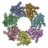
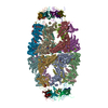
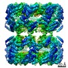
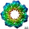
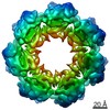
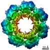
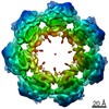
| Title | Structure of the human mitochondrial HSPD1 single ring | |||||||||
 Map data Map data | final map filtered by local resolution | |||||||||
 Sample Sample |
| |||||||||
 Keywords Keywords | HSPD1 /  Hsp60 / chaperonin / CHAPERONE Hsp60 / chaperonin / CHAPERONE | |||||||||
| Function / homology |  Function and homology information Function and homology informationcoated vesicle / isotype switching to IgG isotypes / mitochondrial unfolded protein response / lipopolysaccharide receptor complex / apolipoprotein A-I binding / TFAP2A acts as a transcriptional repressor during retinoic acid induced cell differentiation / protein import into mitochondrial intermembrane space / migrasome / high-density lipoprotein particle binding / positive regulation of T cell mediated immune response to tumor cell ...coated vesicle / isotype switching to IgG isotypes / mitochondrial unfolded protein response / lipopolysaccharide receptor complex / apolipoprotein A-I binding / TFAP2A acts as a transcriptional repressor during retinoic acid induced cell differentiation / protein import into mitochondrial intermembrane space / migrasome / high-density lipoprotein particle binding / positive regulation of T cell mediated immune response to tumor cell / chaperonin ATPase / Mitochondrial protein import / positive regulation of macrophage activation / cellular response to interleukin-7 / biological process involved in interaction with symbiont / MyD88-dependent toll-like receptor signaling pathway / 'de novo' protein folding / sperm plasma membrane / B cell activation / DNA replication origin binding / B cell proliferation / apoptotic mitochondrial changes / positive regulation of interferon-alpha production / apolipoprotein binding / positive regulation of interleukin-10 production / protein maturation / response to unfolded protein / chaperone-mediated protein complex assembly / clathrin-coated pit / isomerase activity / sperm midpiece / response to cold / positive regulation of interleukin-12 production / T cell activation / secretory granule / lipopolysaccharide binding / ATP-dependent protein folding chaperone / positive regulation of interleukin-6 production / activation of cysteine-type endopeptidase activity involved in apoptotic process / positive regulation of T cell activation / unfolded protein binding / positive regulation of type II interferon production / p53 binding / protein folding / single-stranded DNA binding / double-stranded RNA binding / protein-folding chaperone binding / protein refolding / mitochondrial inner membrane / protein stabilization / early endosome / mitochondrial matrix / positive regulation of apoptotic process / ubiquitin protein ligase binding / negative regulation of apoptotic process / enzyme binding / cell surface / ATP hydrolysis activity / protein-containing complex / mitochondrion / extracellular space / RNA binding / extracellular exosome / ATP binding / membrane / plasma membrane / cytosol / cytoplasmSimilarity search - Function | |||||||||
| Biological species |  Homo sapiens (human) Homo sapiens (human) | |||||||||
| Method | single particle reconstruction / cryo EM / Resolution: 3.5 Å | |||||||||
 Authors Authors | Klebl DP / Feasey MC / Muench SP | |||||||||
| Funding support |  United Kingdom, 2 items United Kingdom, 2 items
| |||||||||
 Citation Citation | Journal: iScience / Year: 2021 Title: Cryo-EM structure of human mitochondrial HSPD1. Authors: David P Klebl / Matthew C Feasey / Emma L Hesketh / Neil A Ranson / Heiko Wurdak / Frank Sobott / Robin S Bon / Stephen P Muench /  Abstract: Chaperonins play an important role in folding newly synthesized or translocated proteins in all organisms. The bacterial chaperonin GroEL has served as a model system for the understanding of these ...Chaperonins play an important role in folding newly synthesized or translocated proteins in all organisms. The bacterial chaperonin GroEL has served as a model system for the understanding of these proteins. In comparison, its human homolog, known as mitochondrial heat shock protein family member D1 (HSPD1) is poorly understood. Here, we present the structure of HSPD1 in the apo state determined by cryo-electron microscopy (cryo-EM). Unlike GroEL, HSPD1 forms mostly single ring assemblies in the absence of co-chaperonin (HSPE1). Comparison with GroEL shows a rotation and increased flexibility of the apical domain. Together with published structures of the HSPD1/HSPE1 co-chaperonin complex, this work gives insight into the structural changes that occur during the catalytic cycle. This new understanding of HSPD1 structure and its rearrangements upon complex formation may provide new insights for the development of HSPD1-targeting treatments against a diverse range of diseases including glioblastoma. | |||||||||
| History |
|
- Structure visualization
| Movie |
Movie viewer |
|---|---|
| Structure viewer | EM map: SurfViewMolmilJmol/JSmol |
| Supplemental images |

- Downloads & links
-EMDB archive
| Map data | emd_11950.map.gz | 39.2 MB | EMDB map data format | |
|---|---|---|---|---|
| Header (meta data) | emd-11950-v30.xmlemd-11950.xml | 18 KB 18 KB | Display Display | EMDB header |
| FSC (resolution estimation) | emd_11950_fsc.xml | 9.1 KB | Display | FSC data file |
| Images |  emd_11950.png emd_11950.png | 161.8 KB | ||
| Filedesc metadata | emd-11950.cif.gz | 5.7 KB | ||
| Others | emd_11950_additional_1.map.gzemd_11950_half_map_1.map.gzemd_11950_half_map_2.map.gz | 48.8 MB 49.4 MB 49.4 MB | ||
| Archive directory |  http://ftp.pdbj.org/pub/emdb/structures/EMD-11950ftp://ftp.pdbj.org/pub/emdb/structures/EMD-11950 http://ftp.pdbj.org/pub/emdb/structures/EMD-11950ftp://ftp.pdbj.org/pub/emdb/structures/EMD-11950 | HTTPS FTP |
-Related structure data
| Related structure data | 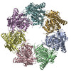 7azpMC C: citing same article ( M: atomic model generated by this map |
|---|---|
| Similar structure data |


-Links
| EMDB pages | EMDB (EBI/PDBe) / EMDataResource |
|---|---|
| Related items in Molecule of the Month |

-Map
| File | Download / File: emd_11950.map.gz / Format: CCP4 / Size: 64 MB / Type: IMAGE STORED AS FLOATING POINT NUMBER (4 BYTES) | ||||||||||||||||||||||||||||||||||||||||||||||||||||||||||||
|---|---|---|---|---|---|---|---|---|---|---|---|---|---|---|---|---|---|---|---|---|---|---|---|---|---|---|---|---|---|---|---|---|---|---|---|---|---|---|---|---|---|---|---|---|---|---|---|---|---|---|---|---|---|---|---|---|---|---|---|---|---|
| Annotation | final map filtered by local resolution | ||||||||||||||||||||||||||||||||||||||||||||||||||||||||||||
| Voxel size | X=Y=Z: 1.065 Å | ||||||||||||||||||||||||||||||||||||||||||||||||||||||||||||
| Density |
| ||||||||||||||||||||||||||||||||||||||||||||||||||||||||||||
| Symmetry | Space group: 1 | ||||||||||||||||||||||||||||||||||||||||||||||||||||||||||||
| Details | EMDB XML:
CCP4 map header:
| ||||||||||||||||||||||||||||||||||||||||||||||||||||||||||||
-Supplemental data
-Additional map: unsharpened map
| File | emd_11950_additional_1.map | ||||||||||||
|---|---|---|---|---|---|---|---|---|---|---|---|---|---|
| Annotation | unsharpened map | ||||||||||||
| Projections & Slices |
| ||||||||||||
| Density Histograms |
 Z
Z Y
Y X
X


-Half map: #2
| File | emd_11950_half_map_1.map | ||||||||||||
|---|---|---|---|---|---|---|---|---|---|---|---|---|---|
| Projections & Slices |
| ||||||||||||
| Density Histograms |


-Half map: #1
| File | emd_11950_half_map_2.map | ||||||||||||
|---|---|---|---|---|---|---|---|---|---|---|---|---|---|
| Projections & Slices |
| ||||||||||||
| Density Histograms |


- Sample components
-Entire : human mitochondrial heat shock protein family member D1 (HSPD1)
| Entire | Name: human mitochondrial heat shock protein family member D1 (HSPD1) |
|---|---|
| Components |
|
-Supramolecule #1: human mitochondrial heat shock protein family member D1 (HSPD1)
| Supramolecule | Name: human mitochondrial heat shock protein family member D1 (HSPD1) type: complex / ID: 1 / Parent: 0 / Macromolecule list: all Details: produced by heterologous expression, mature HSPD1, apo state |
|---|---|
| Source (natural) | Organism: Homo sapiens (human) |
-Macromolecule #1: 60 kDa heat shock protein, mitochondrial
| Macromolecule | Name: 60 kDa heat shock protein, mitochondrial / type: protein_or_peptide / ID: 1 / Number of copies: 7 / Enantiomer: LEVO / EC number: chaperonin ATPase |
|---|---|
| Source (natural) | Organism: Homo sapiens (human) |
| Molecular weight | Theoretical: 58.306977 KDa |
| Recombinant expression | Organism:  Escherichia coli BL21 (bacteria) Escherichia coli BL21 (bacteria) |
| Sequence | String: SNAAKDVKFG ADARALMLQG VDLLADAVAV TMGPKGRTVI IEQSWGSPKV TKDGVTVAKS IDLKDKYKNI GAKLVQDVAN NTNEEAGDG TTTATVLARS IAKEGFEKIS KGANPVEIRR GVMLAVDAVI AELKKQSKPV TTPEEIAQVA TISANGDKEI G NIISDAMK ...String: SNAAKDVKFG ADARALMLQG VDLLADAVAV TMGPKGRTVI IEQSWGSPKV TKDGVTVAKS IDLKDKYKNI GAKLVQDVAN NTNEEAGDG TTTATVLARS IAKEGFEKIS KGANPVEIRR GVMLAVDAVI AELKKQSKPV TTPEEIAQVA TISANGDKEI G NIISDAMK KVGRKGVITV KDGKTLNDEL EIIEGMKFDR GYISPYFINT SKGQKCEFQD AYVLLSEKKI SSIQSIVPAL EI ANAHRKP LVIIAEDVDG EALSTLVLNR LKVGLQVVAV KAPGFGDNRK NQLKDMAIAT GGAVFGEEGL TLNLEDVQPH DLG KVGEVI VTKDDAMLLK GKGDKAQIEK RIQEIIEQLD VTTSEYEKEK LNERLAKLSD GVAVLKVGGT SDVEVNEKKD RVTD ALNAT RAAVEEGIVL GGGCALLRCI PALDSLTPAN EDQKIGIEII KRTLKIPAMT IAKNAGVEGS LIVEKIMQSS SEVGY DAMA GDFVNMVEKG IIDPTKVVRT ALLDAAGVAS LLTTAEVVVT EIPKEEKDPG MGAMGGMGGG MGGGMF UniProtKB: 60 kDa heat shock protein, mitochondrial |
-Experimental details
-Structure determination
| Method | cryo EM |
|---|---|
 Processing Processing | single particle reconstruction |
| Aggregation state | particle |
-Sample preparation
| Concentration | 2.3 mg/mL |
|---|---|
| Buffer | pH: 7.7 |
| Vitrification | Cryogen name: ETHANE / Chamber humidity: 90 % / Chamber temperature: 293 K / Instrument: FEI VITROBOT MARK IV / Details: blot time 6 s blot force 6. |
- Electron microscopy
| Microscope | FEI TITAN KRIOS |
|---|---|
| Electron beam | Acceleration voltage: 300 kV / Electron source: FIELD EMISSION GUN |
| Electron optics | Illumination mode: FLOOD BEAM / Imaging mode: BRIGHT FIELDBright-field microscopy |
| Image recording | Film or detector model: FEI FALCON III (4k x 4k) / Detector mode: COUNTING / Number grids imaged: 1 / Average exposure time: 70.0 sec. / Average electron dose: 38.5 e/Å2 |
| Experimental equipment |  Model: Titan Krios / Image courtesy: FEI Company |
-Image processing
| Startup model | Type of model: NONE |
|---|---|
| Initial angle assignment | Type: MAXIMUM LIKELIHOOD / Software - Name: RELION |
| Final angle assignment | Type: MAXIMUM LIKELIHOOD / Software - Name: RELION |
| Final reconstruction | Resolution.type: BY AUTHOR / Resolution: 3.5 Å / Resolution method: FSC 0.143 CUT-OFF / Software - Name: RELION / Number images used: 124784 |
| FSC plot (resolution estimation) |  |