 Movie
Movie Controller

[English] 日本語
 Yorodumi
- EMDB-40979: Cryo-EM structure of the RBD-ACE2 interface of the SARS-CoV-2 tri... -
+ Open data

- Basic information
| Entry |  | |||||||||
|---|---|---|---|---|---|---|---|---|---|---|
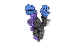
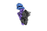
| Title | Cryo-EM structure of the RBD-ACE2 interface of the SARS-CoV-2 trimeric spike protein bound to ACE2 receptor after local refinement at upRBD conformation | |||||||||
 Map data Map data | Cryo-EM Map of the RBD-ACE2 interface of the SARS-CoV-2 trimeric spike protein bound to ACE2 receptor after local refinement | |||||||||
 Sample Sample |
| |||||||||
 Keywords Keywords |  Coronavirus / Spike / ACE2 / Mink / PROTEIN BINDING / VIRAL PROTEIN Coronavirus / Spike / ACE2 / Mink / PROTEIN BINDING / VIRAL PROTEIN | |||||||||
| Function / homology |  Function and homology informationHydrolases; Acting on peptide bonds (peptidases) / peptidyl-dipeptidase activity / carboxypeptidase activity / cilium / metallopeptidase activity / Maturation of spike protein / viral translation / Translation of Structural Proteins / Virion Assembly and Release / host cell surface ...Hydrolases; Acting on peptide bonds (peptidases) / peptidyl-dipeptidase activity / carboxypeptidase activity / cilium / metallopeptidase activity / Maturation of spike protein / viral translation / Translation of Structural Proteins / Virion Assembly and Release / host cell surface / host extracellular space / suppression by virus of host tetherin activity / Induction of Cell-Cell Fusion / structural constituent of virion / entry receptor-mediated virion attachment to host cell / host cell endoplasmic reticulum-Golgi intermediate compartment membrane / receptor-mediated endocytosis of virus by host cell / Attachment and Entry / membrane fusion / positive regulation of viral entry into host cell / receptor-mediated virion attachment to host cell / receptor ligand activity / host cell surface receptor binding / apical plasma membrane / fusion of virus membrane with host plasma membrane / fusion of virus membrane with host endosome membrane / viral envelope / symbiont-mediated suppression of host type I interferon-mediated signaling pathway / virion attachment to host cell / SARS-CoV-2 activates/modulates innate and adaptive immune responses / host cell plasma membrane / virion membrane / proteolysis / extracellular region / membrane / identical protein binding / metal ion binding / plasma membrane / cytoplasm Function and homology informationHydrolases; Acting on peptide bonds (peptidases) / peptidyl-dipeptidase activity / carboxypeptidase activity / cilium / metallopeptidase activity / Maturation of spike protein / viral translation / Translation of Structural Proteins / Virion Assembly and Release / host cell surface ...Hydrolases; Acting on peptide bonds (peptidases) / peptidyl-dipeptidase activity / carboxypeptidase activity / cilium / metallopeptidase activity / Maturation of spike protein / viral translation / Translation of Structural Proteins / Virion Assembly and Release / host cell surface / host extracellular space / suppression by virus of host tetherin activity / Induction of Cell-Cell Fusion / structural constituent of virion / entry receptor-mediated virion attachment to host cell / host cell endoplasmic reticulum-Golgi intermediate compartment membrane / receptor-mediated endocytosis of virus by host cell / Attachment and Entry / membrane fusion / positive regulation of viral entry into host cell / receptor-mediated virion attachment to host cell / receptor ligand activity / host cell surface receptor binding / apical plasma membrane / fusion of virus membrane with host plasma membrane / fusion of virus membrane with host endosome membrane / viral envelope / symbiont-mediated suppression of host type I interferon-mediated signaling pathway / virion attachment to host cell / SARS-CoV-2 activates/modulates innate and adaptive immune responses / host cell plasma membrane / virion membrane / proteolysis / extracellular region / membrane / identical protein binding / metal ion binding / plasma membrane / cytoplasmSimilarity search - Function | |||||||||
| Biological species |   Severe acute respiratory syndrome coronavirus 2 / Severe acute respiratory syndrome coronavirus 2 /  Neovison vison (American mink) / Neovison vison (American mink) /  Homo sapiens (human) Homo sapiens (human) | |||||||||
| Method | single particle reconstruction / cryo EM / Resolution: 3.87 Å | |||||||||
 Authors Authors | Ahn HM / Calderon B / Fan X / Gao Y / Horgan N / Zhou B / Liang B | |||||||||
| Funding support |  United States, 1 items United States, 1 items
| |||||||||
 Citation Citation | Journal: J Med Virol / Year: 2023 Title: Structural basis of the American mink ACE2 binding by Y453F trimeric spike glycoproteins of SARS-CoV-2. Authors: Hyunjun Ahn / Brenda M Calderon / Xiaoyu Fan / Yunrong Gao / Natalie L Horgan / Nannan Jiang / Dylan S Blohm / Jaber Hossain / Nicole Wedad K Rayyan / Sarah H Osman / Xudong Lin / Michael ...Authors: Hyunjun Ahn / Brenda M Calderon / Xiaoyu Fan / Yunrong Gao / Natalie L Horgan / Nannan Jiang / Dylan S Blohm / Jaber Hossain / Nicole Wedad K Rayyan / Sarah H Osman / Xudong Lin / Michael Currier / John Steel / David E Wentworth / Bin Zhou / Bo Liang / Abstract: Severe Acute Respiratory Syndrome Coronavirus 2 (SARS-CoV-2) enters the host cell by binding to angiotensin-converting enzyme 2 (ACE2). While evolutionarily conserved, ACE2 receptors differ across ...Severe Acute Respiratory Syndrome Coronavirus 2 (SARS-CoV-2) enters the host cell by binding to angiotensin-converting enzyme 2 (ACE2). While evolutionarily conserved, ACE2 receptors differ across various species and differential interactions with Spike (S) glycoproteins of SARS-CoV-2 viruses impact species specificity. Reverse zoonoses led to SARS-CoV-2 outbreaks on multiple American mink (Mustela vison) farms during the pandemic and gave rise to mink-associated S substitutions known for transmissibility between mink and zoonotic transmission to humans. In this study, we used bio-layer interferometry (BLI) to discern the differences in binding affinity between multiple human and mink-derived S glycoproteins of SARS-CoV-2 and their respective ACE2 receptors. Further, we conducted a structural analysis of a mink variant S glycoprotein and American mink ACE2 (mvACE2) using cryo-electron microscopy (cryo-EM), revealing four distinct conformations. We discovered a novel intermediary conformation where the mvACE2 receptor is bound to the receptor-binding domain (RBD) of the S glycoprotein in a "down" position, approximately 34° lower than previously reported "up" RBD. Finally, we compared residue interactions in the S-ACE2 complex interface of S glycoprotein conformations with varying RBD orientations. These findings provide valuable insights into the molecular mechanisms of SARS-CoV-2 entry. | |||||||||
| History |
|
- Structure visualization
| Supplemental images |
|---|

- Downloads & links
-EMDB archive
| Map data | emd_40979.map.gz | 482.6 MB | EMDB map data format | |
|---|---|---|---|---|
| Header (meta data) | emd-40979-v30.xmlemd-40979.xml | 18.2 KB 18.2 KB | Display Display | EMDB header |
| FSC (resolution estimation) | emd_40979_fsc.xml | 16.9 KB | Display | FSC data file |
| Images | 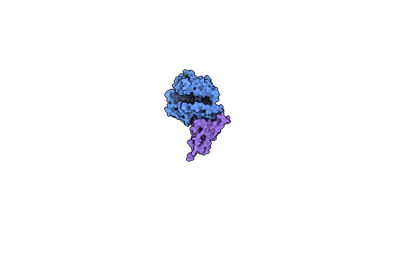 emd_40979.png emd_40979.png | 12 KB | ||
| Filedesc metadata | emd-40979.cif.gz | 6.6 KB | ||
| Others | emd_40979_half_map_1.map.gzemd_40979_half_map_2.map.gz | 474.6 MB 474.7 MB | ||
| Archive directory |  http://ftp.pdbj.org/pub/emdb/structures/EMD-40979ftp://ftp.pdbj.org/pub/emdb/structures/EMD-40979 http://ftp.pdbj.org/pub/emdb/structures/EMD-40979ftp://ftp.pdbj.org/pub/emdb/structures/EMD-40979 | HTTPS FTP |
-Related structure data
| Related structure data |  8t23MC  8t20C  8t21C  8t22C  8t25C  8tazC M: atomic model generated by this map C: citing same article ( |
|---|---|
| Similar structure data |


-Links
| EMDB pages | EMDB (EBI/PDBe) / EMDataResource |
|---|---|
| Related items in Molecule of the Month |


-Map
| File | Download / File: emd_40979.map.gz / Format: CCP4 / Size: 512 MB / Type: IMAGE STORED AS FLOATING POINT NUMBER (4 BYTES) | ||||||||||||||||||||
|---|---|---|---|---|---|---|---|---|---|---|---|---|---|---|---|---|---|---|---|---|---|
| Annotation | Cryo-EM Map of the RBD-ACE2 interface of the SARS-CoV-2 trimeric spike protein bound to ACE2 receptor after local refinement | ||||||||||||||||||||
| Voxel size | X=Y=Z: 1.11 Å | ||||||||||||||||||||
| Density |
| ||||||||||||||||||||
| Symmetry | Space group: 1 | ||||||||||||||||||||
| Details | EMDB XML:
|
-Supplemental data
-Half map: Half map of the density of the RBD-ACE2...
| File | emd_40979_half_map_1.map | ||||||||||||
|---|---|---|---|---|---|---|---|---|---|---|---|---|---|
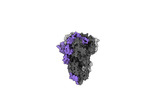
| Annotation | Half map of the density of the RBD-ACE2 interface of the SARS-CoV-2 trimeric spike protein bound to ACE2 receptor after local refinement. | ||||||||||||
| Projections & Slices |
| ||||||||||||
| Density Histograms |
 Z
Z Y
Y X
X


-Half map: Half map of the density of the RBD-ACE2...
| File | emd_40979_half_map_2.map | ||||||||||||
|---|---|---|---|---|---|---|---|---|---|---|---|---|---|
| Annotation | Half map of the density of the RBD-ACE2 interface of the SARS-CoV-2 trimeric spike protein bound to ACE2 receptor after local refinement. | ||||||||||||
| Projections & Slices |
| ||||||||||||
| Density Histograms |


- Sample components
-Entire : mink variant spike RBD-ACE2 interface after local refinement
| Entire | Name: mink variant spike RBD-ACE2 interface after local refinement |
|---|---|
| Components |
|
-Supramolecule #1: mink variant spike RBD-ACE2 interface after local refinement
| Supramolecule | Name: mink variant spike RBD-ACE2 interface after local refinement type: complex / ID: 1 / Parent: 0 / Macromolecule list: #1-#2 |
|---|---|
| Source (natural) | Organism: Severe acute respiratory syndrome coronavirus 2 |
-Macromolecule #1: Angiotensin-converting enzyme
| Macromolecule | Name: Angiotensin-converting enzyme / type: protein_or_peptide / ID: 1 / Number of copies: 1 / Enantiomer: LEVO |
|---|---|
| Source (natural) | Organism: Neovison vison (American mink) |
| Molecular weight | Theoretical: 89.3755 KDa |
| Recombinant expression | Organism: Homo sapiens (human) |
| Sequence | String: MLGSSWLLLS LAALTAAQST TEDLAKTFLE KFNYEAEELS YQNSLASWNY NTNITDENIQ KMNIAGAKWS AFYEEESQHA KTYPLEEIQ DPIIKRQLRA LQQSGSSVLS ADKRERLNTI LNAMSTIYST GKACNPNNPQ ECLLLEPGLD DIMENSKDYN E RLWAWEGW ...String: MLGSSWLLLS LAALTAAQST TEDLAKTFLE KFNYEAEELS YQNSLASWNY NTNITDENIQ KMNIAGAKWS AFYEEESQHA KTYPLEEIQ DPIIKRQLRA LQQSGSSVLS ADKRERLNTI LNAMSTIYST GKACNPNNPQ ECLLLEPGLD DIMENSKDYN E RLWAWEGW RSEVGKQLRP LYEEYVALKN EMARANNYED YGDYWRGDYE EEWADGYNYS RNQLIEDVEH TFTQIKPLYE HL HAYVRAK LMDAYPSRIS PTGCLPAHLL GDMWGRFWTN LYPLMVPFGQ KPNIDVTDAM VNQSWDARRI FKEAEKFFVS VGL PNMTEG FWQNSMLTEP GDNRKVVCHP TAWDLGKHDF RIKMCTKVTM DDFLTAHHEM GHIQYDMAYA AQPFLLRNGA NEGF HEAVG EIMSLSAATP NHLKNIGLLP PDFSEDSETD INFLLKQALT IVGTLPFTYM LEKWRWMVFK GEIPKEQWMQ KWWEM KRDI VGVVEPLPHD ETYCDPAALF HVANDYSFIR YYTRTIYQFQ FQEALCQIAK HEGPLYKCDI SNSREAGQKL HEMLSL GRS KPWTFALERV VGAKTMDVRP LLNYFEPLFT WLKEQNRNSF VGWNTDWSPY ADQSIKVRIS LKSALGEKAY EWNDNEM YF FQSSIAYAMR EYFSKVKKQT IPFVDKDVRV SDLKPRISFN FIVTSPENMS DIIPRADVEE AIRKSRGRIN DAFRLDDN S LEFLGIQPTL EPPYQPPVGS GSGSGHHHHH HGSGSGLNDI FEAQKIEWHE UniProtKB: Angiotensin-converting enzyme |
-Macromolecule #2: Spike glycoprotein
| Macromolecule | Name: Spike glycoprotein / type: protein_or_peptide / ID: 2 / Number of copies: 1 / Enantiomer: LEVO |
|---|---|
| Source (natural) | Organism: Homo sapiens (human) |
| Molecular weight | Theoretical: 23.370246 KDa |
| Recombinant expression | Organism: Homo sapiens (human) |
| Sequence | String: QPTESIVRFP NITNLCPFGE VFNATRFASV YAWNRKRISN CVADYSVLYN SASFSTFKCY GVSPTKLNDL CFTNVYADSF VIRGDEVRQ IAPGQTGKIA DYNYKLPDDF TGCVIAWNSN NLDSKVGGNY NYLFRLFRKS NLKPFERDIS TEIYQAGSTP C NGVEGFNC ...String: QPTESIVRFP NITNLCPFGE VFNATRFASV YAWNRKRISN CVADYSVLYN SASFSTFKCY GVSPTKLNDL CFTNVYADSF VIRGDEVRQ IAPGQTGKIA DYNYKLPDDF TGCVIAWNSN NLDSKVGGNY NYLFRLFRKS NLKPFERDIS TEIYQAGSTP C NGVEGFNC YFPLQSYGFQ PTNGVGYQPY RVVVLSFELL HAPATVCGPK UniProtKB: Spike glycoprotein |
-Macromolecule #5: 2-acetamido-2-deoxy-beta-D-glucopyranose
| Macromolecule | Name: 2-acetamido-2-deoxy-beta-D-glucopyranose / type: ligand / ID: 5 / Number of copies: 2 / Formula: NAG |
|---|---|
| Molecular weight | Theoretical: 221.208 Da |
| Chemical component information |  ChemComp-NAG: |
-Experimental details
-Structure determination
| Method | cryo EM |
|---|---|
 Processing Processing | single particle reconstruction |
| Aggregation state | particle |
-Sample preparation
| Concentration | 1.5 mg/mL | |||||||||
|---|---|---|---|---|---|---|---|---|---|---|
| Buffer | pH: 8 Component:
Details: 20 mM Tris pH 8.0, 200 mM NaCl | |||||||||
| Grid | Material: COPPER / Support film - Material: CARBON / Support film - topology: HOLEY / Pretreatment - Type: GLOW DISCHARGE | |||||||||
| Vitrification | Cryogen name: ETHANE / Chamber humidity: 100 % / Instrument: FEI VITROBOT MARK IV |
- Electron microscopy
| Microscope | FEI TITAN KRIOS |
|---|---|
| Electron beam | Acceleration voltage: 300 kV / Electron source: FIELD EMISSION GUN |
| Electron optics | C2 aperture diameter: 100.0 µm / Illumination mode: OTHER / Imaging mode: OTHER / Nominal defocus max: 1.75 µm / Nominal defocus min: 0.75 µm / Nominal magnification: 81000 |
| Sample stage | Specimen holder model: FEI TITAN KRIOS AUTOGRID HOLDER / Cooling holder cryogen: NITROGEN |
| Image recording | Film or detector model: GATAN K3 (6k x 4k) / Number real images: 11392 / Average exposure time: 4.0 sec. / Average electron dose: 49.98 e/Å2 |
| Experimental equipment |  Model: Titan Krios / Image courtesy: FEI Company |
-Image processing
| Startup model | Type of model: PDB ENTRY PDB model - PDB ID: |
|---|---|
| Initial angle assignment | Type: MAXIMUM LIKELIHOOD |
| Final angle assignment | Type: MAXIMUM LIKELIHOOD |
| Final reconstruction | Resolution.type: BY AUTHOR / Resolution: 3.87 Å / Resolution method: FSC 0.143 CUT-OFF / Number images used: 512722 |
| FSC plot (resolution estimation) |  |

