Movie
Movie Controller
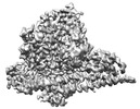
 Structure viewers
 About Yorodumi Papers


+Search query
-Structure paper
| Title | Cryo-EM structures of cancer-specific helical and kinase domain mutations of PI3Kα. |
|---|---|
| Journal, issue, pages | Proc Natl Acad Sci U S A, Vol. 119, Issue 46, Page e2215621119, Year 2022 |
| Publish date | Nov 16, 2022 |
 Authors Authors | Xiao Liu / Qingtong Zhou / Jonathan R Hart / Yingna Xu / Su Yang / Dehua Yang / Peter K Vogt / Ming-Wei Wang /    |
| PubMed Abstract | Phosphoinositide 3-kinases (PI3Ks) are a family of lipid kinases that perform multiple and important cellular functions. The protein investigated here belongs to class IA of the PI3Ks; it is a dimer ...Phosphoinositide 3-kinases (PI3Ks) are a family of lipid kinases that perform multiple and important cellular functions. The protein investigated here belongs to class IA of the PI3Ks; it is a dimer consisting of a catalytic subunit, p110α, and a regulatory subunit, p85α, and is referred to as PI3Kα. The catalytic subunit p110α is frequently mutated in cancer. The mutations induce a gain of function and constitute a driving force in cancer development. About 80% of these mutations lead to single-amino-acid substitutions in one of three sites of p110α: two in the helical domain of the protein (E542K and E545K) and one at the C-terminus of the kinase domain (H1047R). Here, we report the cryo-electron microscopy structures of these mutants in complex with the p110α-specific inhibitor BYL-719. The H1047R mutant rotates its sidechain to a new position and weakens the kα11 activation loop interaction, thereby reducing the inhibitory effect of p85α on p110α. E542K and E545K completely abolish the tight interaction between the helical domain of p110α and the N-terminal SH2 domain of p85α and lead to the disruption of all p85α binding and a dramatic increase in flexibility of the adaptor-binding domain (ABD) in p110α. Yet, the dimerization of PI3Kα is preserved through the ABD-p85α interaction. The local and global structural features induced by these mutations provide molecular insights into the activation of PI3Kα, deepen our understanding of the oncogenic mechanism of this important signaling molecule, and may facilitate the development of mutant-specific inhibitors. |
 External links External links | Proc Natl Acad Sci U S A / PubMed:36343266 / PubMed Central |
| Methods | EM (single particle) |
| Resolution | 2.62 - 2.77 Å |
| Structure data | EMDB-34271, PDB-8gua: EMDB-34272, PDB-8gub: EMDB-34273, PDB-8gud: |
| Chemicals |  ChemComp-1LT: |
| Source |
|
 Keywords Keywords |  STRUCTURAL PROTEIN / Phosphoinositide 3-kinase (PI3K) / helical domain / mutation / cancers / kinase domain STRUCTURAL PROTEIN / Phosphoinositide 3-kinase (PI3K) / helical domain / mutation / cancers / kinase domain |