 Movie
Movie Controller

[English] 日本語
 Yorodumi
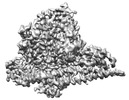
- EMDB-34273: Cryo-EM structure of cancer-specific PI3Kalpha mutant E545K in co... -
+ Open data

- Basic information
| Entry |  | ||||||||||||||||||||||||||||||||||||||||||
|---|---|---|---|---|---|---|---|---|---|---|---|---|---|---|---|---|---|---|---|---|---|---|---|---|---|---|---|---|---|---|---|---|---|---|---|---|---|---|---|---|---|---|---|
| Title | Cryo-EM structure of cancer-specific PI3Kalpha mutant E545K in complex with BYL-719 | ||||||||||||||||||||||||||||||||||||||||||
 Map data Map data | |||||||||||||||||||||||||||||||||||||||||||
 Sample Sample |
| ||||||||||||||||||||||||||||||||||||||||||
| Function / homology |  Function and homology information Function and homology informationresponse to muscle inactivity / negative regulation of actin filament depolymerization / response to L-leucine / regulation of actin filament organization / response to butyrate / autosome genomic imprinting / IRS-mediated signalling / cellular response to hydrostatic pressure / PI3K events in ERBB4 signaling / Activated NTRK2 signals through PI3K ...response to muscle inactivity / negative regulation of actin filament depolymerization / response to L-leucine / regulation of actin filament organization / response to butyrate / autosome genomic imprinting / IRS-mediated signalling / cellular response to hydrostatic pressure / PI3K events in ERBB4 signaling / Activated NTRK2 signals through PI3K / positive regulation of protein localization to membrane / Activated NTRK3 signals through PI3K / negative regulation of fibroblast apoptotic process / cardiac muscle cell contraction / phosphatidylinositol 3-kinase complex, class IB / vasculature development / Signaling by cytosolic FGFR1 fusion mutants /  regulation of cellular respiration / anoikis / phosphatidylinositol 3-kinase complex / Nephrin family interactions / Costimulation by the CD28 family / relaxation of cardiac muscle / 1-phosphatidylinositol-4-phosphate 3-kinase activity / 1-phosphatidylinositol-4,5-bisphosphate 3-kinase activity / MET activates PI3K/AKT signaling / PI3K/AKT activation / phosphatidylinositol-4,5-bisphosphate 3-kinase / vascular endothelial growth factor signaling pathway / phosphatidylinositol 3-kinase complex, class IA / phosphatidylinositol 3-kinase / phosphatidylinositol-3-phosphate biosynthetic process / negative regulation of macroautophagy / 1-phosphatidylinositol-3-kinase activity / Signaling by ALK / PI-3K cascade:FGFR3 / Erythropoietin activates Phosphoinositide-3-kinase (PI3K) / protein kinase activator activity / response to dexamethasone / PI-3K cascade:FGFR2 / PI-3K cascade:FGFR4 / PI-3K cascade:FGFR1 / phosphatidylinositol phosphate biosynthetic process / CD28 dependent PI3K/Akt signaling / Synthesis of PIPs at the plasma membrane / PI3K events in ERBB2 signaling / negative regulation of anoikis / intercalated disc / RET signaling / regulation of multicellular organism growth / insulin receptor substrate binding / Interleukin-3, Interleukin-5 and GM-CSF signaling / PI3K Cascade / positive regulation of TOR signaling / endothelial cell migration / RAC2 GTPase cycle / GAB1 signalosome / Role of phospholipids in phagocytosis / Role of LAT2/NTAL/LAB on calcium mobilization / adipose tissue development / Interleukin receptor SHC signaling / positive regulation of lamellipodium assembly / phagocytosis / Signaling by PDGFRA transmembrane, juxtamembrane and kinase domain mutants / Signaling by PDGFRA extracellular domain mutants / energy homeostasis / Signaling by FGFR4 in disease / cardiac muscle contraction / Signaling by FLT3 ITD and TKD mutants / Signaling by FGFR3 in disease / Tie2 Signaling / GPVI-mediated activation cascade / Signaling by FGFR2 in disease / RAC1 GTPase cycle / T cell costimulation / response to muscle stretch / Signaling by FLT3 fusion proteins / FLT3 Signaling / Signaling by FGFR1 in disease / Downstream signal transduction / insulin-like growth factor receptor signaling pathway / Signaling by phosphorylated juxtamembrane, extracellular and kinase domain KIT mutants / liver development / response to activity / phosphatidylinositol 3-kinase/protein kinase B signal transduction / Regulation of signaling by CBL / cellular response to glucose stimulus / Signaling by ALK fusions and activated point mutants / positive regulation of smooth muscle cell proliferation / regulation of protein phosphorylation / Constitutive Signaling by EGFRvIII / Signaling by ERBB2 ECD mutants / epidermal growth factor receptor signaling pathway / Signaling by ERBB2 KD Mutants / Signaling by SCF-KIT / platelet activation / VEGFA-VEGFR2 Pathway / cellular response to insulin stimulus / Constitutive Signaling by Aberrant PI3K in Cancer / glucose metabolic process regulation of cellular respiration / anoikis / phosphatidylinositol 3-kinase complex / Nephrin family interactions / Costimulation by the CD28 family / relaxation of cardiac muscle / 1-phosphatidylinositol-4-phosphate 3-kinase activity / 1-phosphatidylinositol-4,5-bisphosphate 3-kinase activity / MET activates PI3K/AKT signaling / PI3K/AKT activation / phosphatidylinositol-4,5-bisphosphate 3-kinase / vascular endothelial growth factor signaling pathway / phosphatidylinositol 3-kinase complex, class IA / phosphatidylinositol 3-kinase / phosphatidylinositol-3-phosphate biosynthetic process / negative regulation of macroautophagy / 1-phosphatidylinositol-3-kinase activity / Signaling by ALK / PI-3K cascade:FGFR3 / Erythropoietin activates Phosphoinositide-3-kinase (PI3K) / protein kinase activator activity / response to dexamethasone / PI-3K cascade:FGFR2 / PI-3K cascade:FGFR4 / PI-3K cascade:FGFR1 / phosphatidylinositol phosphate biosynthetic process / CD28 dependent PI3K/Akt signaling / Synthesis of PIPs at the plasma membrane / PI3K events in ERBB2 signaling / negative regulation of anoikis / intercalated disc / RET signaling / regulation of multicellular organism growth / insulin receptor substrate binding / Interleukin-3, Interleukin-5 and GM-CSF signaling / PI3K Cascade / positive regulation of TOR signaling / endothelial cell migration / RAC2 GTPase cycle / GAB1 signalosome / Role of phospholipids in phagocytosis / Role of LAT2/NTAL/LAB on calcium mobilization / adipose tissue development / Interleukin receptor SHC signaling / positive regulation of lamellipodium assembly / phagocytosis / Signaling by PDGFRA transmembrane, juxtamembrane and kinase domain mutants / Signaling by PDGFRA extracellular domain mutants / energy homeostasis / Signaling by FGFR4 in disease / cardiac muscle contraction / Signaling by FLT3 ITD and TKD mutants / Signaling by FGFR3 in disease / Tie2 Signaling / GPVI-mediated activation cascade / Signaling by FGFR2 in disease / RAC1 GTPase cycle / T cell costimulation / response to muscle stretch / Signaling by FLT3 fusion proteins / FLT3 Signaling / Signaling by FGFR1 in disease / Downstream signal transduction / insulin-like growth factor receptor signaling pathway / Signaling by phosphorylated juxtamembrane, extracellular and kinase domain KIT mutants / liver development / response to activity / phosphatidylinositol 3-kinase/protein kinase B signal transduction / Regulation of signaling by CBL / cellular response to glucose stimulus / Signaling by ALK fusions and activated point mutants / positive regulation of smooth muscle cell proliferation / regulation of protein phosphorylation / Constitutive Signaling by EGFRvIII / Signaling by ERBB2 ECD mutants / epidermal growth factor receptor signaling pathway / Signaling by ERBB2 KD Mutants / Signaling by SCF-KIT / platelet activation / VEGFA-VEGFR2 Pathway / cellular response to insulin stimulus / Constitutive Signaling by Aberrant PI3K in Cancer / glucose metabolic processSimilarity search - Function | ||||||||||||||||||||||||||||||||||||||||||
| Biological species |  Homo sapiens (human) Homo sapiens (human) | ||||||||||||||||||||||||||||||||||||||||||
| Method | single particle reconstruction / cryo EM / Resolution: 2.62 Å | ||||||||||||||||||||||||||||||||||||||||||
 Authors Authors | Liu X / Zhou Q / Hart JR / Xu Y / Yang S / Yang D / Vogt PK / Wang M-W | ||||||||||||||||||||||||||||||||||||||||||
| Funding support |  China, China,  United States, 13 items United States, 13 items
| ||||||||||||||||||||||||||||||||||||||||||
 Citation Citation | Journal: Proc Natl Acad Sci U S A / Year: 2022 Title: Cryo-EM structures of cancer-specific helical and kinase domain mutations of PI3Kα. Authors: Xiao Liu / Qingtong Zhou / Jonathan R Hart / Yingna Xu / Su Yang / Dehua Yang / Peter K Vogt / Ming-Wei Wang /  Abstract: Phosphoinositide 3-kinases (PI3Ks) are a family of lipid kinases that perform multiple and important cellular functions. The protein investigated here belongs to class IA of the PI3Ks; it is a dimer ...Phosphoinositide 3-kinases (PI3Ks) are a family of lipid kinases that perform multiple and important cellular functions. The protein investigated here belongs to class IA of the PI3Ks; it is a dimer consisting of a catalytic subunit, p110α, and a regulatory subunit, p85α, and is referred to as PI3Kα. The catalytic subunit p110α is frequently mutated in cancer. The mutations induce a gain of function and constitute a driving force in cancer development. About 80% of these mutations lead to single-amino-acid substitutions in one of three sites of p110α: two in the helical domain of the protein (E542K and E545K) and one at the C-terminus of the kinase domain (H1047R). Here, we report the cryo-electron microscopy structures of these mutants in complex with the p110α-specific inhibitor BYL-719. The H1047R mutant rotates its sidechain to a new position and weakens the kα11 activation loop interaction, thereby reducing the inhibitory effect of p85α on p110α. E542K and E545K completely abolish the tight interaction between the helical domain of p110α and the N-terminal SH2 domain of p85α and lead to the disruption of all p85α binding and a dramatic increase in flexibility of the adaptor-binding domain (ABD) in p110α. Yet, the dimerization of PI3Kα is preserved through the ABD-p85α interaction. The local and global structural features induced by these mutations provide molecular insights into the activation of PI3Kα, deepen our understanding of the oncogenic mechanism of this important signaling molecule, and may facilitate the development of mutant-specific inhibitors. | ||||||||||||||||||||||||||||||||||||||||||
| History |
|
- Structure visualization
| Supplemental images |
|---|

- Downloads & links
-EMDB archive
| Map data | emd_34273.map.gz | 59.7 MB | EMDB map data format | |
|---|---|---|---|---|
| Header (meta data) | emd-34273-v30.xmlemd-34273.xml | 18.5 KB 18.5 KB | Display Display | EMDB header |
| FSC (resolution estimation) | emd_34273_fsc.xml | 9.1 KB | Display | FSC data file |
| Images |  emd_34273.png emd_34273.png | 61.5 KB | ||
| Others | emd_34273_half_map_1.map.gzemd_34273_half_map_2.map.gz | 59.4 MB 59.4 MB | ||
| Archive directory |  http://ftp.pdbj.org/pub/emdb/structures/EMD-34273ftp://ftp.pdbj.org/pub/emdb/structures/EMD-34273 http://ftp.pdbj.org/pub/emdb/structures/EMD-34273ftp://ftp.pdbj.org/pub/emdb/structures/EMD-34273 | HTTPS FTP |
-Related structure data
| Related structure data |  8gudMC  8guaC  8gubC M: atomic model generated by this map C: citing same article ( |
|---|---|
| Similar structure data |

-Links
| EMDB pages | EMDB (EBI/PDBe) / EMDataResource |
|---|---|
| Related items in Molecule of the Month |


-Map
| File | Download / File: emd_34273.map.gz / Format: CCP4 / Size: 64 MB / Type: IMAGE STORED AS FLOATING POINT NUMBER (4 BYTES) | ||||||||||||||||||||
|---|---|---|---|---|---|---|---|---|---|---|---|---|---|---|---|---|---|---|---|---|---|
| Voxel size | X=Y=Z: 1.071 Å | ||||||||||||||||||||
| Density |
| ||||||||||||||||||||
| Symmetry | Space group: 1 | ||||||||||||||||||||
| Details | EMDB XML:
|
-Supplemental data
-Half map: #2
| File | emd_34273_half_map_1.map | ||||||||||||
|---|---|---|---|---|---|---|---|---|---|---|---|---|---|
| Projections & Slices |
| ||||||||||||
| Density Histograms |
 Z
Z Y
Y X
X


-Half map: #1
| File | emd_34273_half_map_2.map | ||||||||||||
|---|---|---|---|---|---|---|---|---|---|---|---|---|---|
| Projections & Slices |
| ||||||||||||
| Density Histograms |


- Sample components
-Entire : Cryo-EM structure of PI3Kalpha mutant E545K in complex with BYL-719
| Entire | Name: Cryo-EM structure of PI3Kalpha mutant E545K in complex with BYL-719 |
|---|---|
| Components |
|
-Supramolecule #1: Cryo-EM structure of PI3Kalpha mutant E545K in complex with BYL-719
| Supramolecule | Name: Cryo-EM structure of PI3Kalpha mutant E545K in complex with BYL-719 type: complex / Chimera: Yes / ID: 1 / Parent: 0 / Macromolecule list: #1 |
|---|---|
| Source (natural) | Organism: Homo sapiens (human) |
| Recombinant expression | Organism:  Trichoplusia ni (cabbage looper) / Recombinant strain: Sf-9 Trichoplusia ni (cabbage looper) / Recombinant strain: Sf-9 |
-Macromolecule #1: Phosphatidylinositol 4,5-bisphosphate 3-kinase catalytic subunit ...
| Macromolecule | Name: Phosphatidylinositol 4,5-bisphosphate 3-kinase catalytic subunit alpha isoform type: protein_or_peptide / ID: 1 / Number of copies: 1 / Enantiomer: LEVO / EC number: phosphatidylinositol 3-kinase |
|---|---|
| Source (natural) | Organism: Homo sapiens (human) |
| Molecular weight | Theoretical: 127.822641 KDa |
| Recombinant expression | Organism: Trichoplusia ni (cabbage looper) |
| Sequence | String: MSYYHHHHHH DYDIPTTENL YFQGAMGSMP PRPSSGELWG IHLMPPRILV ECLLPNGMIV TLECLREATL ITIKHELFKE ARKYPLHQL LQDESSYIFV SVTQEAEREE FFDETRRLCD LRLFQPFLKV IEPVGNREEK ILNREIGFAI GMPVCEFDMV K DPEVQDFR ...String: MSYYHHHHHH DYDIPTTENL YFQGAMGSMP PRPSSGELWG IHLMPPRILV ECLLPNGMIV TLECLREATL ITIKHELFKE ARKYPLHQL LQDESSYIFV SVTQEAEREE FFDETRRLCD LRLFQPFLKV IEPVGNREEK ILNREIGFAI GMPVCEFDMV K DPEVQDFR RNILNVCKEA VDLRDLNSPH SRAMYVYPPN VESSPELPKH IYNKLDKGQI IVVIWVIVSP NNDKQKYTLK IN HDCVPEQ VIAEAIRKKT RSMLLSSEQL KLCVLEYQGK YILKVCGCDE YFLEKYPLSQ YKYIRSCIML GRMPNLMLMA KES LYSQLP MDCFTMPSYS RRISTATPYM NGETSTKSLW VINSALRIKI LCATYVNVNI RDIDKIYVRT GIYHGGEPLC DNVN TQRVP CSNPRWNEWL NYDIYIPDLP RAARLCLSIC SVKGRKGAKE EHCPLAWGNI NLFDYTDTLV SGKMALNLWP VPHGL EDLL NPIGVTGSNP NKETPCLELE FDWFSSVVKF PDMSVIEEHA NWSVSREAGF SYSHAGLSNR LARDNELREN DKEQLK AIS TRDPLSEITK QEKDFLWSHR HYCVTIPEIL PKLLLSVKWN SRDEVAQMYC LVKDWPPIKP EQAMELLDCN YPDPMVR GF AVRCLEKYLT DDKLSQYLIQ LVQVLKYEQY LDNLLVRFLL KKALTNQRIG HFFFWHLKSE MHNKTVSQRF GLLLESYC R ACGMYLKHLN RQVEAMEKLI NLTDILKQEK KDETQKVQMK FLVEQMRRPD FMDALQGFLS PLNPAHQLGN LRLEECRIM SSAKRPLWLN WENPDIMSEL LFQNNEIIFK NGDDLRQDML TLQIIRIMEN IWQNQGLDLR MLPYGCLSIG DCVGLIEVVR NSHTIMQIQ CKGGLKGALQ FNSHTLHQWL KDKNKGEIYD AAIDLFTRSC AGYCVATFIL GIGDRHNSNI MVKDDGQLFH I DFGHFLDH KKKKFGYKRE RVPFVLTQDF LIVISKGAQE CTKTREFERF QEMCYKAYLA IRQHANLFIN LFSMMLGSGM PE LQSFDDI AYIRKTLALD KTEQEALEYF MKQMNDAHHG GWTTKMDWIF HTIKQHALN |
-Macromolecule #2: (2S)-N~1~-{4-methyl-5-[2-(1,1,1-trifluoro-2-methylpropan-2-yl)pyr...
| Macromolecule | Name: (2S)-N~1~-{4-methyl-5-[2-(1,1,1-trifluoro-2-methylpropan-2-yl)pyridin-4-yl]-1,3-thiazol-2-yl}pyrrolidine-1,2-dicarboxamide type: ligand / ID: 2 / Number of copies: 1 / Formula: 1LT |
|---|---|
| Molecular weight | Theoretical: 441.47 Da |
| Chemical component information |  ChemComp-1LT: |
-Experimental details
-Structure determination
| Method | cryo EM |
|---|---|
 Processing Processing | single particle reconstruction |
| Aggregation state | particle |
-Sample preparation
| Concentration | 1.0 mg/mL |
|---|---|
| Buffer | pH: 7.6 |
| Vitrification | Cryogen name: ETHANE |
- Electron microscopy
| Microscope | FEI TITAN KRIOS |
|---|---|
| Electron beam | Acceleration voltage: 300 kV / Electron source: OTHER |
| Electron optics | Illumination mode: OTHER / Imaging mode: BRIGHT FIELDBright-field microscopy / Nominal defocus max: 2.5 µm / Nominal defocus min: 1.5 µm |
| Image recording | Film or detector model: GATAN K3 (6k x 4k) / Number real images: 5144 / Average electron dose: 70.0 e/Å2 |
| Experimental equipment |  Model: Titan Krios / Image courtesy: FEI Company |
-Image processing
| Initial angle assignment | Type: OTHER |
|---|---|
| Final angle assignment | Type: OTHER |
| Final reconstruction | Resolution.type: BY AUTHOR / Resolution: 2.62 Å / Resolution method: FSC 0.143 CUT-OFF / Number images used: 753253 |
| FSC plot (resolution estimation) |  |

