Movie
Movie Controller
 Structure viewers
 About Yorodumi Papers


+Search query
-Structure paper
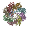
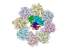
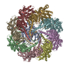
| Title | Structural visualization of the tubulin folding pathway directed by human chaperonin TRiC/CCT. |
|---|---|
| Journal, issue, pages | Cell, Vol. 185, Issue 25, Page 4770-4787.e20, Year 2022 |
| Publish date | Dec 8, 2022 |
 Authors Authors | Daniel Gestaut / Yanyan Zhao / Junsun Park / Boxue Ma / Alexander Leitner / Miranda Collier / Grigore Pintilie / Soung-Hun Roh / Wah Chiu / Judith Frydman /    |
| PubMed Abstract | The ATP-dependent ring-shaped chaperonin TRiC/CCT is essential for cellular proteostasis. To uncover why some eukaryotic proteins can only fold with TRiC assistance, we reconstituted the folding of ...The ATP-dependent ring-shaped chaperonin TRiC/CCT is essential for cellular proteostasis. To uncover why some eukaryotic proteins can only fold with TRiC assistance, we reconstituted the folding of β-tubulin using human prefoldin and TRiC. We find unstructured β-tubulin is delivered by prefoldin to the open TRiC chamber followed by ATP-dependent chamber closure. Cryo-EM resolves four near-atomic-resolution structures containing progressively folded β-tubulin intermediates within the closed TRiC chamber, culminating in native tubulin. This substrate folding pathway appears closely guided by site-specific interactions with conserved regions in the TRiC chamber. Initial electrostatic interactions between the TRiC interior wall and both the folded tubulin N domain and its C-terminal E-hook tail establish the native substrate topology, thus enabling C-domain folding. Intrinsically disordered CCT C termini within the chamber promote subsequent folding of tubulin's core and middle domains and GTP-binding. Thus, TRiC's chamber provides chemical and topological directives that shape the folding landscape of its obligate substrates. |
 External links External links | Cell / PubMed:36493755 / PubMed Central |
| Methods | EM (single particle) |
| Resolution | 2.9 - 3.85 Å |
| Structure data | EMDB-26089, PDB-7trg: EMDB-26120, PDB-7ttn: EMDB-26123, PDB-7ttt: EMDB-26131, PDB-7tub:  EMDB-32822: An apo TRiC map EMDB-32823, PDB-7wu7: |
| Chemicals |  ChemComp-MG:  ChemComp-ADP:  ChemComp-AF3:  ChemComp-HOH: |
| Source |
|
 Keywords Keywords |  CHAPERONE / Human chaperonin TRiC with beta-tubulin folding intermediate I / Human chaperonin TRiC with beta-tubulin folding intermediate II / Human chaperonin TRiC with beta-tubulin folding intermediate III / Human chaperonin TRiC with beta-tubulin folding intermediate IV / chapronin complex CHAPERONE / Human chaperonin TRiC with beta-tubulin folding intermediate I / Human chaperonin TRiC with beta-tubulin folding intermediate II / Human chaperonin TRiC with beta-tubulin folding intermediate III / Human chaperonin TRiC with beta-tubulin folding intermediate IV / chapronin complex |