 Movie
Movie Controller


+ Open data

- Basic information
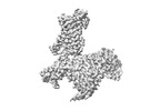
| Entry | Database: PDB / ID: 8x16 | ||||||
|---|---|---|---|---|---|---|---|
| Title | Cryo-EM structure of adenosine receptor A3AR bound to CF101 | ||||||
 Components Components |
| ||||||
 Keywords Keywords |  MEMBRANE PROTEIN/IMMUNE SYSTEM / GPCR / adenosine A3 receptor / ligand selectivity / CF101 / MEMBRANE PROTEIN / MEMBRANE PROTEIN-IMMUNE SYSTEM complex MEMBRANE PROTEIN/IMMUNE SYSTEM / GPCR / adenosine A3 receptor / ligand selectivity / CF101 / MEMBRANE PROTEIN / MEMBRANE PROTEIN-IMMUNE SYSTEM complex | ||||||
| Function / homology |  Function and homology information Function and homology informationAdenylate cyclase inhibitory pathway / regulation of norepinephrine secretion / Adenosine P1 receptors / G protein-coupled adenosine receptor activity / G protein-coupled adenosine receptor signaling pathway / G-protein activation / Activation of the phototransduction cascade / : / Glucagon-type ligand receptors / Thromboxane signalling through TP receptor ...Adenylate cyclase inhibitory pathway / regulation of norepinephrine secretion / Adenosine P1 receptors / G protein-coupled adenosine receptor activity / G protein-coupled adenosine receptor signaling pathway / G-protein activation / Activation of the phototransduction cascade / : / Glucagon-type ligand receptors / Thromboxane signalling through TP receptor / Sensory perception of sweet, bitter, and umami (glutamate) taste / G beta:gamma signalling through PI3Kgamma / G beta:gamma signalling through CDC42 / Cooperation of PDCL (PhLP1) and TRiC/CCT in G-protein beta folding / Ca2+ pathway / Activation of G protein gated Potassium channels / Synthesis, secretion, and inactivation of Glucagon-like Peptide-1 (GLP-1) / Inhibition of voltage gated Ca2+ channels via Gbeta/gamma subunits / G alpha (z) signalling events / Vasopressin regulates renal water homeostasis via Aquaporins / Glucagon-like Peptide-1 (GLP1) regulates insulin secretion / Adrenaline,noradrenaline inhibits insulin secretion / ADP signalling through P2Y purinoceptor 12 / G alpha (q) signalling events / G alpha (i) signalling events / Thrombin signalling through proteinase activated receptors (PARs) / Activation of G protein gated Potassium channels / G-protein activation / G beta:gamma signalling through PI3Kgamma / Prostacyclin signalling through prostacyclin receptor / G beta:gamma signalling through PLC beta / ADP signalling through P2Y purinoceptor 1 / Thromboxane signalling through TP receptor / Presynaptic function of Kainate receptors / G beta:gamma signalling through CDC42 / Inhibition of voltage gated Ca2+ channels via Gbeta/gamma subunits / Glucagon-type ligand receptors / alkylglycerophosphoethanolamine phosphodiesterase activity / Adrenaline,noradrenaline inhibits insulin secretion / G alpha (12/13) signalling events / G beta:gamma signalling through BTK / ADP signalling through P2Y purinoceptor 12 / Cooperation of PDCL (PhLP1) and TRiC/CCT in G-protein beta folding / Thrombin signalling through proteinase activated receptors (PARs) / Ca2+ pathway / regulation of heart contraction / G alpha (z) signalling events / Extra-nuclear estrogen signaling / G alpha (s) signalling events / G alpha (q) signalling events / photoreceptor outer segment membrane / G alpha (i) signalling events / Glucagon-like Peptide-1 (GLP1) regulates insulin secretion / spectrin binding / Vasopressin regulates renal water homeostasis via Aquaporins / negative regulation of NF-kappaB transcription factor activity / photoreceptor outer segment / positive regulation of protein localization to cell cortex / regulation of cAMP-mediated signaling / D2 dopamine receptor binding / G protein-coupled serotonin receptor binding / regulation of mitotic spindle organization / cellular response to forskolin / cardiac muscle cell apoptotic process / activation of adenylate cyclase activity / presynaptic modulation of chemical synaptic transmission / photoreceptor inner segment / negative regulation of cell migration / G protein-coupled receptor binding / G-protein beta/gamma-subunit complex binding / Schaffer collateral - CA1 synapse / adenylate cyclase-modulating G protein-coupled receptor signaling pathway / response to wounding / cellular response to catecholamine stimulus / adenylate cyclase-activating dopamine receptor signaling pathway / cellular response to prostaglandin E stimulus / sensory perception of taste / GDP binding / G-protein beta-subunit binding / heterotrimeric G-protein complex / signaling receptor complex adaptor activity / retina development in camera-type eye / GTPase binding / presynaptic membrane / cell body / phospholipase C-activating G protein-coupled receptor signaling pathway / cell cortex / midbody / positive regulation of cytosolic calcium ion concentration / G alpha (i) signalling events / cellular response to hypoxia / cell population proliferation / inflammatory response / cell cycle / G protein-coupled receptor signaling pathway / cell division / negative regulation of cell population proliferation / GTPase activity / centrosome / synapseSimilarity search - Function | ||||||
| Biological species |  Homo sapiens (human) Homo sapiens (human) Bos taurus (cattle)Rattus norvegicus (Norway rat)Mus musculus (house mouse) Bos taurus (cattle)Rattus norvegicus (Norway rat)Mus musculus (house mouse) | ||||||
| Method | ELECTRON MICROSCOPY / single particle reconstruction / cryo EM / Resolution: 3.29 Å | ||||||
 Authors Authors | Cai, H. / Xu, Y. / Xu, H.E. | ||||||
| Funding support |  China, 1items China, 1items
| ||||||
 Citation Citation | Journal: Nat Commun / Year: 2024 Title: Cryo-EM structures of adenosine receptor AAR bound to selective agonists. Authors: Hongmin Cai / Shimeng Guo / Youwei Xu / Jun Sun / Junrui Li / Zhikan Xia / Yi Jiang / Xin Xie / H Eric Xu / Abstract: The adenosine A receptor (AAR), a key member of the G protein-coupled receptor family, is a promising therapeutic target for inflammatory and cancerous conditions. The selective AAR agonists, CF101 ...The adenosine A receptor (AAR), a key member of the G protein-coupled receptor family, is a promising therapeutic target for inflammatory and cancerous conditions. The selective AAR agonists, CF101 and CF102, are clinically significant, yet their recognition mechanisms remained elusive. Here we report the cryogenic electron microscopy structures of the full-length human AAR bound to CF101 and CF102 with heterotrimeric G protein in complex at 3.3-3.2 Å resolution. These agonists reside in the orthosteric pocket, forming conserved interactions via their adenine moieties, while their 3-iodobenzyl groups exhibit distinct orientations. Functional assays reveal the critical role of extracellular loop 3 in AAR's ligand selectivity and receptor activation. Key mutations, including His, Ser, and Ser, in a unique sub-pocket of AAR, significantly impact receptor activation. Comparative analysis with the inactive AAR structure highlights a conserved receptor activation mechanism. Our findings provide comprehensive insights into the molecular recognition and signaling of AAR, paving the way for designing subtype-selective adenosine receptor ligands. | ||||||
| History |
|
- Structure visualization
| Structure viewer | Molecule: MolmilJmol/JSmol |
|---|
- Downloads & links
-Download
| PDBx/mmCIF format | 8x16.cif.gz | 209.8 KB | Display | PDBx/mmCIF format |
|---|---|---|---|---|
| PDB format | pdb8x16.ent.gz | 161.7 KB | Display | PDB format |
| PDBx/mmJSON format | 8x16.json.gz | Tree view | PDBx/mmJSON format | |
| Others |  Other downloads Other downloads |
-Validation report
| Arichive directory | https://data.pdbj.org/pub/pdb/validation_reports/x1/8x16ftp://data.pdbj.org/pub/pdb/validation_reports/x1/8x16 | HTTPS FTP |
|---|
-Related structure data
| Related structure data |  37985MC  8x17C M: map data used to model this data C: citing same article ( |
|---|---|
| Similar structure data |
-Links
 PDBj
PDBj


- Assembly
| Deposited unit | 
|
|---|---|
| 1 |
|
-Components
-Guanine nucleotide-binding protein ... , 3 types, 3 molecules ABC
| #2: Protein | Mass: 40445.059 Da / Num. of mol.: 1 Source method: isolated from a genetically manipulated source Source: (gene. exp.) Bos taurus (cattle) / Gene: GNAI1 / Production host:  Trichoplusia ni (cabbage looper) / References: UniProt: P63097 Trichoplusia ni (cabbage looper) / References: UniProt: P63097 |
|---|---|
| #3: Protein | Mass: 37915.496 Da / Num. of mol.: 1 Source method: isolated from a genetically manipulated source Source: (gene. exp.) Rattus norvegicus (Norway rat) / Gene: Gnb1 / Production host: Trichoplusia ni (cabbage looper) / References: UniProt: P54311 |
| #4: Protein | Mass: 7861.143 Da / Num. of mol.: 1 Source method: isolated from a genetically manipulated source Source: (gene. exp.) Bos taurus (cattle) / Gene: GNG2 / Production host: Trichoplusia ni (cabbage looper) / References: UniProt: P63212 |
-Protein / Antibody / Non-polymers , 3 types, 3 molecules RH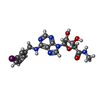
| #1: Protein | Mass: 36214.465 Da / Num. of mol.: 1 Source method: isolated from a genetically manipulated source Source: (gene. exp.) Homo sapiens (human) / Gene: ADORA3 / Production host: Trichoplusia ni (cabbage looper) / References: UniProt: P0DMS8 |
|---|---|
| #5: Antibody | Mass: 26277.299 Da / Num. of mol.: 1 Source method: isolated from a genetically manipulated source Source: (gene. exp.) Mus musculus (house mouse) / Production host: Trichoplusia ni (cabbage looper) |
| #6: Chemical | ChemComp-Q8L /  Mass: 510.286 Da / Num. of mol.: 1 / Source method: obtained synthetically / Formula: C18H19IN6O4 / Feature type: SUBJECT OF INVESTIGATION Mass: 510.286 Da / Num. of mol.: 1 / Source method: obtained synthetically / Formula: C18H19IN6O4 / Feature type: SUBJECT OF INVESTIGATION |
-Details
| Has ligand of interest | Y |
|---|
-Experimental details
-Experiment
| Experiment | Method: ELECTRON MICROSCOPY |
|---|---|
| EM experiment | Aggregation state: PARTICLE / 3D reconstruction method: single particle reconstruction |
- Sample preparation
| Component |
| ||||||||||||||||||||||||||||||||||||
|---|---|---|---|---|---|---|---|---|---|---|---|---|---|---|---|---|---|---|---|---|---|---|---|---|---|---|---|---|---|---|---|---|---|---|---|---|---|
| Molecular weight | Value: 0.16 MDa / Experimental value: NO | ||||||||||||||||||||||||||||||||||||
| Source (natural) |
| ||||||||||||||||||||||||||||||||||||
| Source (recombinant) | Organism: Trichoplusia ni (cabbage looper) | ||||||||||||||||||||||||||||||||||||
| Buffer solution | pH: 7.5 | ||||||||||||||||||||||||||||||||||||
| Specimen | Embedding applied: NO / Shadowing applied: NO / Staining applied: NO / Vitrification applied: YES | ||||||||||||||||||||||||||||||||||||
| Vitrification | Cryogen name: ETHANE |
- Electron microscopy imaging
| Experimental equipment |  Model: Titan Krios / Image courtesy: FEI Company |
|---|---|
| Microscopy | Model: FEI TITAN KRIOS |
| Electron gun | Electron source: FIELD EMISSION GUN / Accelerating voltage: 300 kV / Illumination mode: FLOOD BEAM |
| Electron lens | Mode: BRIGHT FIELDBright-field microscopy / Nominal defocus max: 5000 nm / Nominal defocus min: 1200 nm |
| Image recording | Electron dose: 50 e/Å2 / Film or detector model: GATAN K3 (6k x 4k) |
- Processing
| CTF correction | Type: NONE |
|---|---|
| 3D reconstruction | Resolution: 3.29 Å / Resolution method: FSC 0.143 CUT-OFF / Num. of particles: 271323 / Symmetry type: POINT |
