 Movie
Movie Controller


+ Open data

- Basic information
| Entry | Database: PDB / ID: 8uej | ||||||
|---|---|---|---|---|---|---|---|
| Title | ssRNA phage PhiCb5 virion | ||||||
 Components Components |
| ||||||
 Keywords Keywords |  VIRUS / ssRNA phage VIRUS / ssRNA phage | ||||||
| Function / homology | Assembly protein / Phage maturation protein / virion attachment to host cell pilus / virion component / viral capsid / Maturation protein / Coat protein Function and homology information Function and homology information | ||||||
| Biological species |  Caulobacter phage phiCb5 (virus) Caulobacter phage phiCb5 (virus) | ||||||
| Method | ELECTRON MICROSCOPY / single particle reconstruction / cryo EM / Resolution: 2.7 Å | ||||||
 Authors Authors | Wang, Y. / Zhang, J. | ||||||
| Funding support |  United States, 1items United States, 1items
| ||||||
 Citation Citation | Journal: Sci Adv / Year: 2024 Title: Structural mechanisms of Tad pilus assembly and its interaction with an RNA virus. Authors: Yuhang Wang / Matthew Theodore / Zhongliang Xing / Utkarsh Narsaria / Zihao Yu / Lanying Zeng / Junjie Zhang / Abstract: Tad (tight adherence) pili, part of the type IV pili family, are crucial for mechanosensing, surface adherence, bacteriophage (phage) adsorption, and cell-cycle regulation. Unlike other type IV ... Tad (tight adherence) pili, part of the type IV pili family, are crucial for mechanosensing, surface adherence, bacteriophage (phage) adsorption, and cell-cycle regulation. Unlike other type IV pilins, Tad pilins lack the typical globular β sheet domain responsible for pilus assembly and phage binding. The mechanisms of Tad pilus assembly and its interaction with phage ΦCb5 have been elusive. Using cryo-electron microscopy, we unveiled the Tad pilus assembly mechanism, featuring a unique network of hydrogen bonds at its core. We then identified the Tad pilus binding to the ΦCb5 maturation protein (Mat) through its β region. Notably, the amino terminus of ΦCb5 Mat is exposed outside the capsid and phage/pilus interface, enabling the attachment of fluorescent and affinity tags. These engineered ΦCb5 virions can be efficiently assembled and purified in , maintaining infectivity against , which presents promising applications, including RNA delivery and phage display. | ||||||
| History |
|
- Structure visualization
| Structure viewer | Molecule: MolmilJmol/JSmol |
|---|
- Downloads & links
-Download
| PDBx/mmCIF format | 8uej.cif.gz | 3.5 MB | Display | PDBx/mmCIF format |
|---|---|---|---|---|
| PDB format | pdb8uej.ent.gz | Display | PDB format | |
| PDBx/mmJSON format | 8uej.json.gz | Tree view | PDBx/mmJSON format | |
| Others |  Other downloads Other downloads |
-Validation report
| Arichive directory | https://data.pdbj.org/pub/pdb/validation_reports/ue/8uejftp://data.pdbj.org/pub/pdb/validation_reports/ue/8uej | HTTPS FTP |
|---|
-Related structure data
| Related structure data |  42163MC  8u2bC 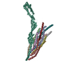 8ucrC C: citing same article ( M: map data used to model this data |
|---|---|
| Similar structure data |


-Links
 PDBj
PDBj- Assembly
| Deposited unit | 
|
|---|---|
| 1 |
|
-Components
| #1: Protein | Mass: 13498.981 Da / Num. of mol.: 178 / Source method: isolated from a natural source / Source: (natural) Caulobacter phage phiCb5 (virus) / References: UniProt: D7RIC2#2: Protein | | Mass: 40725.336 Da / Num. of mol.: 1 / Source method: isolated from a natural source / Source: (natural) Caulobacter phage phiCb5 (virus) / References: UniProt: D7RIC1#3: Chemical | ChemComp-CA /   Mass: 40.078 Da / Num. of mol.: 236 / Source method: obtained synthetically / Formula: Ca / Feature type: SUBJECT OF INVESTIGATION Mass: 40.078 Da / Num. of mol.: 236 / Source method: obtained synthetically / Formula: Ca / Feature type: SUBJECT OF INVESTIGATIONHas ligand of interest | Y | |
|---|
-Experimental details
-Experiment
| Experiment | Method: ELECTRON MICROSCOPY |
|---|---|
| EM experiment | Aggregation state: PARTICLE / 3D reconstruction method: single particle reconstruction |
- Sample preparation
| Component | Name: A complex of phiCb5 maturation protein and phiCb5 shell Type: COMPLEX Details: A complex of phiCb5 maturation protein and phiCb5 shell Entity ID: #1-#2 / Source: NATURAL | ||||||||||||||||||||
|---|---|---|---|---|---|---|---|---|---|---|---|---|---|---|---|---|---|---|---|---|---|
| Molecular weight | Value: 2.1 MDa / Experimental value: NO | ||||||||||||||||||||
| Source (natural) | Organism:  Caulobacter vibrioides (bacteria) Caulobacter vibrioides (bacteria) | ||||||||||||||||||||
| Buffer solution | pH: 7.5 / Details: 20mM tris, 2mM MgCl2, 3mM CaCl2 | ||||||||||||||||||||
| Buffer component |
| ||||||||||||||||||||
| Specimen | Embedding applied: NO / Shadowing applied: NO / Staining applied: NO / Vitrification applied: YES | ||||||||||||||||||||
| Specimen support | Grid type: C-flat-2/1 | ||||||||||||||||||||
| Vitrification | Cryogen name: ETHANE |
- Electron microscopy imaging
| Experimental equipment |  Model: Titan Krios / Image courtesy: FEI Company |
|---|---|
| Microscopy | Model: FEI TITAN KRIOS |
| Electron gun | Electron source: FIELD EMISSION GUN / Accelerating voltage: 300 kV / Illumination mode: FLOOD BEAM |
| Electron lens | Mode: BRIGHT FIELDBright-field microscopy / Nominal defocus max: 25000 nm / Nominal defocus min: 5000 nm |
| Image recording | Electron dose: 50 e/Å2 / Film or detector model: GATAN K3 (6k x 4k) |
- Processing
| EM software | Name: PHENIX / Version: 1.17.1_3660: / Category: model refinement | ||||||||||||||||||||||||
|---|---|---|---|---|---|---|---|---|---|---|---|---|---|---|---|---|---|---|---|---|---|---|---|---|---|
| CTF correction | Type: PHASE FLIPPING AND AMPLITUDE CORRECTION | ||||||||||||||||||||||||
| 3D reconstruction | Resolution: 2.7 Å / Resolution method: FSC 0.143 CUT-OFF / Num. of particles: 272204 / Symmetry type: POINT | ||||||||||||||||||||||||
| Refine LS restraints |
|
