 Movie
Movie Controller

[English] 日本語
 Yorodumi
- PDB-8gdr: SARS-Cov2 S protein structure in complex with neutralizing monocl... -
+ Open data

- Basic information
| Entry | Database: PDB / ID: 8gdr | ||||||
|---|---|---|---|---|---|---|---|
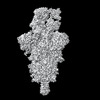
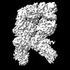
| Title | SARS-Cov2 S protein structure in complex with neutralizing monoclonal antibody 002-S21B10 | ||||||
 Components Components |
| ||||||
 Keywords Keywords |  VIRAL PROTEIN / antibody / spike / SARS-CoV-2 / viral protein-immune system complex VIRAL PROTEIN / antibody / spike / SARS-CoV-2 / viral protein-immune system complex | ||||||
| Function / homology |  Function and homology information Function and homology informationMaturation of spike protein / viral translation / Translation of Structural Proteins / Virion Assembly and Release / host cell surface / host extracellular space / suppression by virus of host tetherin activity / Induction of Cell-Cell Fusion / structural constituent of virion / entry receptor-mediated virion attachment to host cell ...Maturation of spike protein / viral translation / Translation of Structural Proteins / Virion Assembly and Release / host cell surface / host extracellular space / suppression by virus of host tetherin activity / Induction of Cell-Cell Fusion / structural constituent of virion / entry receptor-mediated virion attachment to host cell / host cell endoplasmic reticulum-Golgi intermediate compartment membrane / receptor-mediated endocytosis of virus by host cell / Attachment and Entry / membrane fusion / positive regulation of viral entry into host cell / receptor-mediated virion attachment to host cell / receptor ligand activity / host cell surface receptor binding / fusion of virus membrane with host plasma membrane / fusion of virus membrane with host endosome membrane / viral envelope / symbiont-mediated suppression of host type I interferon-mediated signaling pathway / virion attachment to host cell / SARS-CoV-2 activates/modulates innate and adaptive immune responses / host cell plasma membrane / virion membrane / membrane / identical protein binding / plasma membraneSimilarity search - Function | ||||||
| Biological species |  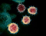 Severe acute respiratory syndrome coronavirus 2 Severe acute respiratory syndrome coronavirus 2 Homo sapiens (human) Homo sapiens (human) | ||||||
| Method | ELECTRON MICROSCOPY / single particle reconstruction / cryo EM / Resolution: 3.6 Å | ||||||
 Authors Authors | Patel, A. / Ortlund, E.A. | ||||||
| Funding support |  United States, 1items United States, 1items
| ||||||
 Citation Citation | Journal: To Be Published Title: Elucidating the mechanism of SARS-CoV-2 Omicron variant escape from a RBD class-3 human antibody Authors: Patel, A. / Kumar, S. / Lai, L. / Chakravarthy, C. / Valanparambil, R. / Keen, M. / Laughlin, Z.T. / Frank, F. / Cheedarla, N. / Verkerke, H.P. / Neish, A.S. / Roback, J.D. / Davis, C.W. / ...Authors: Patel, A. / Kumar, S. / Lai, L. / Chakravarthy, C. / Valanparambil, R. / Keen, M. / Laughlin, Z.T. / Frank, F. / Cheedarla, N. / Verkerke, H.P. / Neish, A.S. / Roback, J.D. / Davis, C.W. / Wrammert, J. / Ahmed, R. / Suthar, M.S. / Murali-Krishna, K. / Chandele, A. / Ortlund, E.A. | ||||||
| History |
|
- Structure visualization
| Structure viewer | Molecule: MolmilJmol/JSmol |
|---|
- Downloads & links
-Download
| PDBx/mmCIF format | 8gdr.cif.gz | 722.5 KB | Display | PDBx/mmCIF format |
|---|---|---|---|---|
| PDB format | pdb8gdr.ent.gz | 588 KB | Display | PDB format |
| PDBx/mmJSON format | 8gdr.json.gz | Tree view | PDBx/mmJSON format | |
| Others |  Other downloads Other downloads |
-Validation report
| Arichive directory | https://data.pdbj.org/pub/pdb/validation_reports/gd/8gdrftp://data.pdbj.org/pub/pdb/validation_reports/gd/8gdr | HTTPS FTP |
|---|
-Related structure data
| Related structure data | 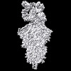 29950MC M: map data used to model this data C: citing same article ( |
|---|---|
| Similar structure data |
-Links
 PDBj
PDBj


- Assembly
| Deposited unit | 
|
|---|---|
| 1 |
|
-Components
| #1: Protein | Spike protein / S glycoprotein / E2 / Peplomer protein Mass: 128766.031 Da / Num. of mol.: 3 Source method: isolated from a genetically manipulated source Source: (gene. exp.) Severe acute respiratory syndrome coronavirus 2Gene: S, 2 / Production host: Homo sapiens (human) / References: UniProt: P0DTC2#2: Antibody | Mass: 24113.283 Da / Num. of mol.: 2 Source method: isolated from a genetically manipulated source Source: (gene. exp.) Homo sapiens (human) / Production host: Homo sapiens (human)#3: Antibody | Mass: 22861.320 Da / Num. of mol.: 2 Source method: isolated from a genetically manipulated source Source: (gene. exp.) Homo sapiens (human) / Production host: Homo sapiens (human)#4: Polysaccharide | 2-acetamido-2-deoxy-beta-D-glucopyranose-(1-4)-2-acetamido-2-deoxy-beta-D-glucopyranose / Mass: 424.401 Da / Num. of mol.: 4Source method: isolated from a genetically manipulated source #5: Sugar | ChemComp-NAG / N-Acetylglucosamine  Type: D-saccharide, beta linking / Mass: 221.208 Da / Num. of mol.: 28 Type: D-saccharide, beta linking / Mass: 221.208 Da / Num. of mol.: 28Source method: isolated from a genetically manipulated source Formula: C8H15NO6 / Feature type: SUBJECT OF INVESTIGATION Has ligand of interest | Y | |
|---|
-Experimental details
-Experiment
| Experiment | Method: ELECTRON MICROSCOPY |
|---|---|
| EM experiment | Aggregation state: PARTICLE / 3D reconstruction method: single particle reconstruction |
- Sample preparation
| Component |
| |||||||||||||||||||||||||
|---|---|---|---|---|---|---|---|---|---|---|---|---|---|---|---|---|---|---|---|---|---|---|---|---|---|---|
| Source (natural) |
| |||||||||||||||||||||||||
| Source (recombinant) |
| |||||||||||||||||||||||||
| Buffer solution | pH: 7.4 | |||||||||||||||||||||||||
| Buffer component |
| |||||||||||||||||||||||||
| Specimen | Conc.: 0.7 mg/ml / Embedding applied: NO / Shadowing applied: NO / Staining applied: NO / Vitrification applied: YES | |||||||||||||||||||||||||
| Specimen support | Grid material: COPPER / Grid mesh size: 400 divisions/in. / Grid type: C-flat-1.2/1.3 | |||||||||||||||||||||||||
| Vitrification | Instrument: FEI VITROBOT MARK IV / Cryogen name: ETHANE / Humidity: 100 % / Chamber temperature: 295 K |
- Electron microscopy imaging
| Experimental equipment |  Model: Titan Krios / Image courtesy: FEI Company |
|---|---|
| Microscopy | Model: TFS KRIOS |
| Electron gun | Electron source: FIELD EMISSION GUN / Accelerating voltage: 300 kV / Illumination mode: FLOOD BEAM |
| Electron lens | Mode: BRIGHT FIELDBright-field microscopy / Nominal magnification: 81000 X / Nominal defocus max: 2400 nm / Nominal defocus min: 1000 nm / Cs: 2.7 mm |
| Specimen holder | Cryogen: NITROGEN |
| Image recording | Electron dose: 54.37 e/Å2 / Film or detector model: GATAN K3 (6k x 4k) / Num. of grids imaged: 1 / Num. of real images: 9054 |
| EM imaging optics | Energyfilter slit width: 20 eV |
- Processing
| Software | Name: PHENIX / Version: 1.20.1_4487: / Classification: refinement | ||||||||||||||||||||||||||||||||||||||||
|---|---|---|---|---|---|---|---|---|---|---|---|---|---|---|---|---|---|---|---|---|---|---|---|---|---|---|---|---|---|---|---|---|---|---|---|---|---|---|---|---|---|
| EM software |
| ||||||||||||||||||||||||||||||||||||||||
| CTF correction | Type: PHASE FLIPPING AND AMPLITUDE CORRECTION | ||||||||||||||||||||||||||||||||||||||||
| Particle selection | Num. of particles selected: 1021598 | ||||||||||||||||||||||||||||||||||||||||
| 3D reconstruction | Resolution: 3.6 Å / Resolution method: FSC 0.5 CUT-OFF / Num. of particles: 326424 / Symmetry type: POINT | ||||||||||||||||||||||||||||||||||||||||
| Atomic model building | B value: 100.25 / Protocol: RIGID BODY FIT / Space: REAL / Target criteria: Correlation coefficient | ||||||||||||||||||||||||||||||||||||||||
| Refine LS restraints |
|
