 Movie
Movie Controller

[English] 日本語
 Yorodumi
- PDB-8e59: Human L-type voltage-gated calcium channel Cav1.3 in the presence... -
+ Open data

- Basic information
| Entry | Database: PDB / ID: 8.0E+59 | ||||||
|---|---|---|---|---|---|---|---|
| Title | Human L-type voltage-gated calcium channel Cav1.3 in the presence of Amiodarone at 3.1 Angstrom resolution | ||||||
 Components Components |
| ||||||
 Keywords Keywords |  TRANSPORT PROTEIN / Cav1.3 / Channels / Calcium Ion-Selective TRANSPORT PROTEIN / Cav1.3 / Channels / Calcium Ion-Selective | ||||||
| Function / homology |  Function and homology information Function and homology informationvoltage-gated calcium channel activity involved SA node cell action potential / voltage-gated calcium channel activity involved in cardiac muscle cell action potential / membrane depolarization during SA node cell action potential / regulation of membrane repolarization during action potential / Presynaptic depolarization and calcium channel opening / regulation of potassium ion transmembrane transporter activity / positive regulation of high voltage-gated calcium channel activity / regulation of atrial cardiac muscle cell membrane repolarization / calcium ion transmembrane transport via high voltage-gated calcium channel / membrane depolarization during bundle of His cell action potential ...voltage-gated calcium channel activity involved SA node cell action potential / voltage-gated calcium channel activity involved in cardiac muscle cell action potential / membrane depolarization during SA node cell action potential / regulation of membrane repolarization during action potential / Presynaptic depolarization and calcium channel opening / regulation of potassium ion transmembrane transporter activity / positive regulation of high voltage-gated calcium channel activity / regulation of atrial cardiac muscle cell membrane repolarization / calcium ion transmembrane transport via high voltage-gated calcium channel / membrane depolarization during bundle of His cell action potential / positive regulation of adenylate cyclase activity / L-type voltage-gated calcium channel complex / membrane depolarization during cardiac muscle cell action potential / cardiac muscle cell action potential involved in contraction / positive regulation of calcium ion transport / high voltage-gated calcium channel activity / regulation of potassium ion transmembrane transport / NCAM1 interactions / regulation of ventricular cardiac muscle cell membrane repolarization / calcium ion import / Sensory processing of sound by inner hair cells of the cochlea / calcium ion transport into cytosol / regulation of calcium ion transmembrane transport via high voltage-gated calcium channel / ankyrin binding / neuromuscular junction development / voltage-gated calcium channel complex / neuronal dense core vesicle / regulation of heart rate by cardiac conduction / alpha-actinin binding / calcium channel regulator activity / regulation of calcium ion transport / calcium ion import across plasma membrane / voltage-gated calcium channel activity / sarcoplasmic reticulum / Regulation of insulin secretion / protein localization to plasma membrane / sensory perception of sound / calcium ion transmembrane transport / adenylate cyclase-modulating G protein-coupled receptor signaling pathway / calcium channel activity / Adrenaline,noradrenaline inhibits insulin secretion / Z disc / cellular response to amyloid-beta / calcium ion transport / T cell receptor signaling pathway / chemical synaptic transmission / synapse / extracellular exosome / membrane / metal ion binding / plasma membrane / cytosolSimilarity search - Function | ||||||
| Biological species |  Homo sapiens (human) Homo sapiens (human) | ||||||
| Method | ELECTRON MICROSCOPY / single particle reconstruction / cryo EM / Resolution: 3.1 Å | ||||||
 Authors Authors | Gao, S. / Yao, X. / Yan, N. | ||||||
| Funding support |  United States, 1items United States, 1items
| ||||||
 Citation Citation | Journal: Cell / Year: 2022 Title: Structural basis for the severe adverse interaction of sofosbuvir and amiodarone on L-type Ca channels. Authors: Xia Yao / Shuai Gao / Jixin Wang / Zhangqiang Li / Jian Huang / Yan Wang / Zhifei Wang / Jiaofeng Chen / Xiao Fan / Weipeng Wang / Xueqin Jin / Xiaojing Pan / Yong Yu / Armando Lagrutta / Nieng Yan /  Abstract: Drug-drug interaction of the antiviral sofosbuvir and the antiarrhythmics amiodarone has been reported to cause fatal heartbeat slowing. Sofosbuvir and its analog, MNI-1, were reported to potentiate ...Drug-drug interaction of the antiviral sofosbuvir and the antiarrhythmics amiodarone has been reported to cause fatal heartbeat slowing. Sofosbuvir and its analog, MNI-1, were reported to potentiate the inhibition of cardiomyocyte calcium handling by amiodarone, which functions as a multi-channel antagonist, and implicate its inhibitory effect on L-type Ca channels, but the molecular mechanism has remained unclear. Here we present systematic cryo-EM structural analysis of Ca1.1 and Ca1.3 treated with amiodarone or sofosbuvir alone, or sofosbuvir/MNI-1 combined with amiodarone. Whereas amiodarone alone occupies the dihydropyridine binding site, sofosbuvir is not found in the channel when applied on its own. In the presence of amiodarone, sofosbuvir/MNI-1 is anchored in the central cavity of the pore domain through specific interaction with amiodarone and directly obstructs the ion permeation path. Our study reveals the molecular basis for the physical, pharmacodynamic interaction of two drugs on the scaffold of Ca channels. | ||||||
| History |
|
- Structure visualization
| Structure viewer | Molecule: MolmilJmol/JSmol |
|---|
- Downloads & links
-Download
| PDBx/mmCIF format | 8e59.cif.gz | 529.7 KB | Display | PDBx/mmCIF format |
|---|---|---|---|---|
| PDB format | pdb8e59.ent.gz | 416 KB | Display | PDB format |
| PDBx/mmJSON format | 8e59.json.gz | Tree view | PDBx/mmJSON format | |
| Others |  Other downloads Other downloads |
-Validation report
| Arichive directory | https://data.pdbj.org/pub/pdb/validation_reports/e5/8e59ftp://data.pdbj.org/pub/pdb/validation_reports/e5/8e59 | HTTPS FTP |
|---|
-Related structure data
| Related structure data | 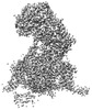 27907MC  8e56C 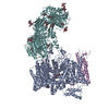 8e57C  8e58C  8e5aC  8e5bC M: map data used to model this data C: citing same article ( |
|---|---|
| Similar structure data |


-Links
 PDBj
PDBj


- Assembly
| Deposited unit | 
|
|---|---|
| 1 |
|
-Components
-Voltage-dependent L-type calcium channel subunit ... , 2 types, 2 molecules AC
| #1: Protein | Mass: 245417.734 Da / Num. of mol.: 1 Source method: isolated from a genetically manipulated source Source: (gene. exp.) Homo sapiens (human) / Gene: CACNA1D, CACH3, CACN4, CACNL1A2, CCHL1A2 / Production host: Homo sapiens (human) / References: UniProt: Q01668 |
|---|---|
| #3: Protein | Mass: 54607.852 Da / Num. of mol.: 1 Source method: isolated from a genetically manipulated source Source: (gene. exp.) Homo sapiens (human) / Gene: CACNB3, CACNLB3 / Production host: Homo sapiens (human) / References: UniProt: P54284 |
-Protein , 1 types, 1 molecules D
| #2: Protein | Voltage-gated calcium channel / Voltage-gated calcium channel subunit alpha-2/delta-1 Mass: 124692.469 Da / Num. of mol.: 1 Source method: isolated from a genetically manipulated source Source: (gene. exp.) Homo sapiens (human) / Gene: CACNA2D1, CACNL2A, CCHL2A, MHS3 / Production host: Homo sapiens (human) / References: UniProt: P54289 |
|---|
-Sugars , 4 types, 7 molecules 
| #4: Polysaccharide | 2-acetamido-2-deoxy-beta-D-glucopyranose-(1-4)-2-acetamido-2-deoxy-beta-D-glucopyranose-(1-4)-2- ...2-acetamido-2-deoxy-beta-D-glucopyranose-(1-4)-2-acetamido-2-deoxy-beta-D-glucopyranose-(1-4)-2-acetamido-2-deoxy-beta-D-glucopyranose  , Oligosaccharide / Class: Inhibitor / Mass: 627.594 Da / Num. of mol.: 1 , Oligosaccharide / Class: Inhibitor / Mass: 627.594 Da / Num. of mol.: 1Source method: isolated from a genetically manipulated source Details: oligosaccharide / References: triacetyl-beta-chitotriose | ||||
|---|---|---|---|---|---|
| #5: Polysaccharide | / Mass: 424.401 Da / Num. of mol.: 3 Source method: isolated from a genetically manipulated source #6: Polysaccharide | 2-acetamido-2-deoxy-beta-D-glucopyranose-(1-4)-2-acetamido-2-deoxy-beta-D-glucopyranose-(1-4)-2- ...2-acetamido-2-deoxy-beta-D-glucopyranose-(1-4)-2-acetamido-2-deoxy-beta-D-glucopyranose-(1-4)-2-acetamido-2-deoxy-beta-D-glucopyranose-(1-4)-2-acetamido-2-deoxy-beta-D-glucopyranose | / Mass: 830.786 Da / Num. of mol.: 1Source method: isolated from a genetically manipulated source #10: Sugar | N-Acetylglucosamine Type: D-saccharide, beta linking / Mass: 221.208 Da / Num. of mol.: 2 / Source method: obtained synthetically / Formula: C8H15NO6 Type: D-saccharide, beta linking / Mass: 221.208 Da / Num. of mol.: 2 / Source method: obtained synthetically / Formula: C8H15NO6 |
-Non-polymers , 3 types, 4 molecules 

| #7: Chemical | ChemComp-BBI / (Amiodarone Mass: 645.312 Da / Num. of mol.: 1 / Source method: obtained synthetically / Formula: C25H29I2NO3 / Feature type: SUBJECT OF INVESTIGATION / Comment: medication, antiarrhythmic*YM Mass: 645.312 Da / Num. of mol.: 1 / Source method: obtained synthetically / Formula: C25H29I2NO3 / Feature type: SUBJECT OF INVESTIGATION / Comment: medication, antiarrhythmic*YM | ||
|---|---|---|---|
| #8: Chemical |  Mass: 40.078 Da / Num. of mol.: 2 / Source method: obtained synthetically / Formula: Ca Mass: 40.078 Da / Num. of mol.: 2 / Source method: obtained synthetically / Formula: Ca#9: Chemical | ChemComp-3PE / | Phosphatidylethanolamine Mass: 748.065 Da / Num. of mol.: 1 / Source method: obtained synthetically / Formula: C41H82NO8P / Comment: phospholipid*YM Mass: 748.065 Da / Num. of mol.: 1 / Source method: obtained synthetically / Formula: C41H82NO8P / Comment: phospholipid*YM |
-Details
| Has ligand of interest | Y |
|---|
-Experimental details
-Experiment
| Experiment | Method: ELECTRON MICROSCOPY |
|---|---|
| EM experiment | Aggregation state: PARTICLE / 3D reconstruction method: single particle reconstruction |
- Sample preparation
| Component | Name: Cav1.3 / Type: COMPLEX / Entity ID: #1-#3 / Source: RECOMBINANT |
|---|---|
| Molecular weight | Experimental value: NO |
| Source (natural) | Organism: Homo sapiens (human) |
| Source (recombinant) | Organism: Homo sapiens (human) |
| Buffer solution | pH: 7.4 |
| Specimen | Embedding applied: NO / Shadowing applied: NO / Staining applied: NO / Vitrification applied: YES |
| Vitrification | Cryogen name: ETHANE / Humidity: 100 % / Chamber temperature: 281 K |
- Electron microscopy imaging
| Experimental equipment |  Model: Titan Krios / Image courtesy: FEI Company |
|---|---|
| Microscopy | Model: FEI TITAN KRIOS |
| Electron gun | Electron source: FIELD EMISSION GUN / Accelerating voltage: 300 kV / Illumination mode: FLOOD BEAM |
| Electron lens | Mode: BRIGHT FIELDBright-field microscopy / Nominal defocus max: 2100 nm / Nominal defocus min: 1900 nm |
| Image recording | Electron dose: 50 e/Å2 / Film or detector model: GATAN K2 SUMMIT (4k x 4k) |
- Processing
| Software | Name: PHENIX / Version: 1.20.1_4487: / Classification: refinement | ||||||||||||||||||||||||
|---|---|---|---|---|---|---|---|---|---|---|---|---|---|---|---|---|---|---|---|---|---|---|---|---|---|
| CTF correction | Type: PHASE FLIPPING AND AMPLITUDE CORRECTION | ||||||||||||||||||||||||
| 3D reconstruction | Resolution: 3.1 Å / Resolution method: FSC 0.143 CUT-OFF / Num. of particles: 86836 / Symmetry type: POINT | ||||||||||||||||||||||||
| Refine LS restraints |
|
