 Movie
Movie Controller

[English] 日本語
 Yorodumi
- PDB-7yed: In situ structure of polymerase complex of mammalian reovirus in ... -
+ Open data

- Basic information
| Entry | Database: PDB / ID: 7yed | |||||||||
|---|---|---|---|---|---|---|---|---|---|---|
| Title | In situ structure of polymerase complex of mammalian reovirus in the elongation state | |||||||||
 Components Components |
| |||||||||
 Keywords Keywords |  VIRAL PROTEIN/RNA / mammalian reovirus / cryo-em / RNA dependent RNA polymerase / transcription / VIRAL PROTEIN / VIRAL PROTEIN-RNA complex VIRAL PROTEIN/RNA / mammalian reovirus / cryo-em / RNA dependent RNA polymerase / transcription / VIRAL PROTEIN / VIRAL PROTEIN-RNA complex | |||||||||
| Function / homology |  Function and homology information Function and homology informationviral inner capsid / host cytoskeleton / viral outer capsid / 7-methylguanosine mRNA capping / viral genome replication / viral capsid / mRNA guanylyltransferase activity / viral nucleocapsid / mRNA 5'-cap (guanine-N7-)-methyltransferase activity / host cell cytoplasm ...viral inner capsid / host cytoskeleton / viral outer capsid / 7-methylguanosine mRNA capping / viral genome replication / viral capsid / mRNA guanylyltransferase activity / viral nucleocapsid / mRNA 5'-cap (guanine-N7-)-methyltransferase activity / host cell cytoplasm / RNA helicase activity / hydrolase activity / RNA-directed RNA polymerase / RNA-dependent RNA polymerase activity / GTP binding / structural molecule activity / RNA binding / ATP bindingSimilarity search - Function | |||||||||
| Biological species |  Mammalian orthoreovirus 3 Mammalian orthoreovirus 3 | |||||||||
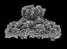
| Method | ELECTRON MICROSCOPY / single particle reconstruction / cryo EM / Resolution: 3 Å | |||||||||
 Authors Authors | Bao, K.Y. / Zhang, X.L. / Li, D.Y. / Zhu, P. | |||||||||
| Funding support |  China, 2items China, 2items
| |||||||||
 Citation Citation | Journal: Proc Natl Acad Sci U S A / Year: 2022 Title: In situ structures of polymerase complex of mammalian reovirus illuminate RdRp activation and transcription regulation. Authors: Keyan Bao / Xueli Zhang / Dongyu Li / Wei Sun / Zhenzhao Sun / Jingfei Wang / Ping Zhu / Abstract: Mammalian reovirus (reovirus) is a multilayered, turreted member of characterized by transcription of dsRNA genome within the innermost capsid shell. Here, we present high-resolution in situ ...Mammalian reovirus (reovirus) is a multilayered, turreted member of characterized by transcription of dsRNA genome within the innermost capsid shell. Here, we present high-resolution in situ structures of reovirus transcriptase complex in an intact double-layered virion, and in the uncoated single-layered core particles in the unloaded, reloaded, pre-elongation, and elongation states, respectively, obtained by cryo-electron microscopy and sub-particle reconstructions. At the template entry of RNA-dependent RNA polymerase (RdRp), the RNA-loading region gets flexible after uncoating resulting in the unloading of terminal genomic RNA and inactivity of transcription. However, upon adding transcriptional substrates, the RNA-loading region is recovered leading the RNAs loaded again. The priming loop in RdRp was found to play a critical role in regulating transcription, which hinders the elongation of transcript in virion and triggers the rearrangement of RdRp C-terminal domain (CTD) during elongation, resulting in splitting of template-transcript hybrid and opening of transcript exit. With the integration of these structures, a transcriptional model of reovirus with five states is proposed. Our structures illuminate the RdRp activation and regulation of the multilayered turreted reovirus. | |||||||||
| History |
|
- Structure visualization
| Structure viewer | Molecule: MolmilJmol/JSmol |
|---|
- Downloads & links
-Download
| PDBx/mmCIF format | 7yed.cif.gz | 3.4 MB | Display | PDBx/mmCIF format |
|---|---|---|---|---|
| PDB format | pdb7yed.ent.gz | Display | PDB format | |
| PDBx/mmJSON format | 7yed.json.gz | Tree view | PDBx/mmJSON format | |
| Others |  Other downloads Other downloads |
-Validation report
| Arichive directory | https://data.pdbj.org/pub/pdb/validation_reports/ye/7yedftp://data.pdbj.org/pub/pdb/validation_reports/ye/7yed | HTTPS FTP |
|---|
-Related structure data
| Related structure data |  33770MC 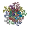 7yevC  7yezC  7yf0C 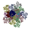 7yfeC M: map data used to model this data C: citing same article ( |
|---|---|
| Similar structure data |


-Links
 PDBj
PDBj


- Assembly
| Deposited unit | 
|
|---|---|
| 1 |
|
-Components
-Protein , 4 types, 22 molecules 12345ABCDEabcdeHIJKLRU
| #1: Protein | Helicase / Lambda 1 Mass: 141801.297 Da / Num. of mol.: 15 / Source method: isolated from a natural source / Source: (natural) Mammalian orthoreovirus 3 / References: UniProt: C9E874, RNA helicase#2: Protein | Mass: 143963.438 Da / Num. of mol.: 5 / Source method: isolated from a natural source / Source: (natural) Mammalian orthoreovirus 3 / References: UniProt: C9E871#5: Protein | | RNA-dependent RNA polymeraseMass: 142472.953 Da / Num. of mol.: 1 / Source method: isolated from a natural source / Source: (natural) Mammalian orthoreovirus 3 / References: UniProt: C9E870#7: Protein | | Mass: 83434.266 Da / Num. of mol.: 1 / Source method: isolated from a natural source / Source: (natural) Mammalian orthoreovirus 3 / References: UniProt: C9E872 |
|---|
-RNA chain , 3 types, 3 molecules MNT
| #3: RNA chain | Mass: 6637.965 Da / Num. of mol.: 1 / Source method: obtained synthetically / Details: Transcript RNA / Source: (synth.) Mammalian orthoreovirus 3 |
|---|---|
| #4: RNA chain | Mass: 11391.729 Da / Num. of mol.: 1 / Source method: obtained synthetically / Details: Encoding RNA / Source: (synth.) Mammalian orthoreovirus 3 |
| #6: RNA chain | Mass: 11391.729 Da / Num. of mol.: 1 / Source method: obtained synthetically / Details: Template RNA / Source: (synth.) Mammalian orthoreovirus 3 |
-Non-polymers , 5 types, 26 molecules 
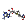


| #8: Chemical | ChemComp-ZN /  Mass: 65.409 Da / Num. of mol.: 8 / Source method: obtained synthetically / Formula: Zn / Feature type: SUBJECT OF INVESTIGATION Mass: 65.409 Da / Num. of mol.: 8 / Source method: obtained synthetically / Formula: Zn / Feature type: SUBJECT OF INVESTIGATION#9: Chemical | ChemComp-SAM / S-Adenosyl methionine Mass: 398.437 Da / Num. of mol.: 10 / Source method: obtained synthetically / Formula: C15H22N6O5S Mass: 398.437 Da / Num. of mol.: 10 / Source method: obtained synthetically / Formula: C15H22N6O5S#10: Chemical | ChemComp-GTP / Guanosine triphosphate Mass: 523.180 Da / Num. of mol.: 5 / Source method: obtained synthetically / Formula: C10H16N5O14P3 / Comment: GTP, energy-carrying molecule*YM Mass: 523.180 Da / Num. of mol.: 5 / Source method: obtained synthetically / Formula: C10H16N5O14P3 / Comment: GTP, energy-carrying molecule*YM#11: Chemical | ChemComp-UTP / | Uridine triphosphate Mass: 484.141 Da / Num. of mol.: 1 / Source method: obtained synthetically / Formula: C9H15N2O15P3 / Comment: UTP*YM Mass: 484.141 Da / Num. of mol.: 1 / Source method: obtained synthetically / Formula: C9H15N2O15P3 / Comment: UTP*YM#12: Chemical |  Mass: 24.305 Da / Num. of mol.: 2 / Source method: obtained synthetically / Formula: Mg Mass: 24.305 Da / Num. of mol.: 2 / Source method: obtained synthetically / Formula: Mg |
|---|
-Details
| Has ligand of interest | Y |
|---|
-Experimental details
-Experiment
| Experiment | Method: ELECTRON MICROSCOPY |
|---|---|
| EM experiment | Aggregation state: PARTICLE / 3D reconstruction method: single particle reconstruction |
- Sample preparation
| Component |
| ||||||||||||||||||||||||
|---|---|---|---|---|---|---|---|---|---|---|---|---|---|---|---|---|---|---|---|---|---|---|---|---|---|
| Source (natural) | Organism: Mammalian orthoreovirus 3 | ||||||||||||||||||||||||
| Details of virus | Empty: NO / Enveloped: NO / Isolate: STRAIN / Type: VIRION | ||||||||||||||||||||||||
| Buffer solution | pH: 8 | ||||||||||||||||||||||||
| Specimen | Embedding applied: NO / Shadowing applied: NO / Staining applied: NO / Vitrification applied: YES | ||||||||||||||||||||||||
| Vitrification | Cryogen name: ETHANE |
- Electron microscopy imaging
| Experimental equipment |  Model: Titan Krios / Image courtesy: FEI Company |
|---|---|
| Microscopy | Model: FEI TITAN KRIOS |
| Electron gun | Electron source: FIELD EMISSION GUN / Accelerating voltage: 300 kV / Illumination mode: FLOOD BEAM |
| Electron lens | Mode: BRIGHT FIELDBright-field microscopy / Nominal defocus max: 1800 nm / Nominal defocus min: 1500 nm |
| Image recording | Electron dose: 30 e/Å2 / Film or detector model: GATAN K2 SUMMIT (4k x 4k) |
- Processing
| Software | Name: PHENIX / Version: (1.13_2998:phenix.real_space_refine) / Classification: refinement |
|---|---|
| CTF correction | Type: PHASE FLIPPING AND AMPLITUDE CORRECTION |
| 3D reconstruction | Resolution: 3 Å / Resolution method: FSC 0.143 CUT-OFF / Num. of particles: 100968 / Symmetry type: POINT |
| Refinement | Cross valid method: THROUGHOUT |
| Displacement parameters | Biso max: 316.85 Å2 / Biso mean: 85.9855 Å2 / Biso min: 49.29 Å2 |
