 Movie
Movie Controller

[English] 日本語
 Yorodumi
- PDB-7tau: Refined capsid structure of human adenovirus D26 at 3.4 A resolution -
+ Open data

- Basic information
| Entry | Database: PDB / ID: 7tau | ||||||
|---|---|---|---|---|---|---|---|
| Title | Refined capsid structure of human adenovirus D26 at 3.4 A resolution | ||||||
 Components Components |
| ||||||
 Keywords Keywords |  VIRUS / Human Adenovirus D26 HAdV-D26 Ad26 Hexon Penton base IX VIRUS / Human Adenovirus D26 HAdV-D26 Ad26 Hexon Penton base IX | ||||||
| Function / homology |  Function and homology information Function and homology informationhexon binding / viral capsid, decoration / T=25 icosahedral viral capsid / lysis of host organelle involved in viral entry into host cell / viral procapsid / microtubule-dependent intracellular transport of viral material towards nucleus / adhesion receptor-mediated virion attachment to host cell / viral release from host cell / host cell / endocytosis involved in viral entry into host cell ...hexon binding / viral capsid, decoration / T=25 icosahedral viral capsid / lysis of host organelle involved in viral entry into host cell / viral procapsid / microtubule-dependent intracellular transport of viral material towards nucleus / adhesion receptor-mediated virion attachment to host cell / viral release from host cell / host cell / endocytosis involved in viral entry into host cell / viral capsid / host cell cytoplasm / cell adhesion / symbiont entry into host cell / host cell nucleus / virion attachment to host cell / structural molecule activitySimilarity search - Function | ||||||
| Biological species |  Human adenovirus 26 Human adenovirus 26 | ||||||
| Method | ELECTRON MICROSCOPY / single particle reconstruction / cryo EM / Resolution: 3.38 Å | ||||||
 Authors Authors | Reddy, V.S. / Yu, X. / Barry, M.A. | ||||||
| Funding support |  United States, 1items United States, 1items
| ||||||
 Citation Citation | Journal: Viruses / Year: 2022 Title: Refined Capsid Structure of Human Adenovirus D26 at 3.4 Å Resolution. Authors: Vijay S Reddy / Xiaodi Yu / Michael A Barry / Abstract: Various adenoviruses are being used as viral vectors for the generation of vaccines against chronic and emerging diseases (e.g., AIDS, COVID-19). Here, we report the improved capsid structure for one ...Various adenoviruses are being used as viral vectors for the generation of vaccines against chronic and emerging diseases (e.g., AIDS, COVID-19). Here, we report the improved capsid structure for one of these vectors, human adenovirus D26 (HAdV-D26), at 3.4 Å resolution, by reprocessing the previous cryo-electron microscopy dataset and obtaining a refined model. In addition to overall improvements in the model, the highlights of the structure include (1) locating a segment of the processed peptide of VIII that was previously believed to be released from the mature virions, (2) reorientation of the helical appendage domain (APD) of IIIa situated underneath the vertex region relative to its counterpart observed in the cleavage defective () mutant of HAdV-C5 that resulted in the loss of interactions between the APD and hexon bases, and (3) the revised conformation of the cleaved N-terminal segments of pre-protein VI (pVIn), located in the hexon cavities, is highly conserved, with notable stacking interactions between the conserved His13 and Phe18 residues. Taken together, the improved model of HAdV-D26 capsid provides a better understanding of protein-protein interactions in HAdV capsids and facilitates the efforts to modify and/or design adenoviral vectors with altered properties. Last but not least, we provide some insights into clotting factors (e.g., FX and PF4) binding to AdV vectors. #1: Journal: Sci Adv / Year: 2017Title: Cryo-EM structure of human adenovirus D26 reveals the conservation of structural organization among human adenoviruses. Authors: Xiaodi Yu / David Veesler / Melody G Campbell / Mary E Barry / Francisco J Asturias / Michael A Barry / Vijay S Reddy / Abstract: Human adenoviruses (HAdVs) cause acute respiratory, ocular, and gastroenteric diseases and are also frequently used as gene and vaccine delivery vectors. Unlike the archetype human adenovirus C5 ...Human adenoviruses (HAdVs) cause acute respiratory, ocular, and gastroenteric diseases and are also frequently used as gene and vaccine delivery vectors. Unlike the archetype human adenovirus C5 (HAdV-C5), human adenovirus D26 (HAdV-D26) belongs to species-D HAdVs, which target different cellular receptors, and is differentially recognized by immune surveillance mechanisms. HAdV-D26 is being championed as a lower seroprevalent vaccine and oncolytic vector in preclinical and human clinical studies. To understand the molecular basis for their distinct biological properties and independently validate the structures of minor proteins, we determined the first structure of species-D HAdV at 3.7 Å resolution by cryo-electron microscopy. All the hexon hypervariable regions (HVRs), including HVR1, have been identified and exhibit a distinct organization compared to those of HAdV-C5. Despite the differences in the arrangement of helices in the coiled-coil structures, protein IX molecules form a continuous hexagonal network on the capsid exterior. In addition to the structurally conserved region (3 to 300) of IIIa, we identified an extra helical domain comprising residues 314 to 390 that further stabilizes the vertex region. Multiple (two to three) copies of the cleaved amino-terminal fragment of protein VI (pVIn) are observed in each hexon cavity, suggesting that there could be ≥480 copies of VI present in HAdV-D26. In addition, a localized asymmetric reconstruction of the vertex region provides new details of the three-pronged "claw hold" of the trimeric fiber and its interactions with the penton base. These observations resolve the previous conflicting assignments of the minor proteins and suggest the likely conservation of their organization across different HAdVs. | ||||||
| History |
|
- Structure visualization
| Movie |
Movie viewer |
|---|---|
| Structure viewer | Molecule: MolmilJmol/JSmol |
- Downloads & links
-Download
| PDBx/mmCIF format | 7tau.cif.gz | 2.2 MB | Display | PDBx/mmCIF format |
|---|---|---|---|---|
| PDB format | pdb7tau.ent.gz | Display | PDB format | |
| PDBx/mmJSON format | 7tau.json.gz | Tree view | PDBx/mmJSON format | |
| Others |  Other downloads Other downloads |
-Validation report
| Arichive directory | https://data.pdbj.org/pub/pdb/validation_reports/ta/7tauftp://data.pdbj.org/pub/pdb/validation_reports/ta/7tau | HTTPS FTP |
|---|
-Related structure data
| Related structure data | 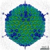 25786MC M: map data used to model this data C: citing same article ( |
|---|---|
| Similar structure data |


-Links
 PDBj
PDBj


- Assembly
| Deposited unit | 
|
|---|---|
| 1 | x 60
|
| 2 |
|
| 3 | x 5
|
| 4 | x 6
|
| 5 |
|
| Symmetry | Point symmetry: (Schoenflies symbol: I (icosahedral)) |
-Components
-Protein , 7 types, 30 molecules ABCDEFGHIJKLNOMPQRSUV123456789
| #1: Protein | / CP-H / Protein II Mass: 107218.703 Da / Num. of mol.: 12 / Source method: isolated from a natural source / Source: (natural) Human adenovirus 26 / References: UniProt: Q3S8B3#2: Protein | | / CP-P / Penton base protein / Protein IIIMass: 58647.703 Da / Num. of mol.: 1 / Source method: isolated from a natural source / Source: (natural) Human adenovirus 26 / References: UniProt: A4ZKL2#3: Protein | | Mass: 41039.184 Da / Num. of mol.: 1 / Source method: isolated from a natural source / Source: (natural) Human adenovirus 26 / References: UniProt: A4ZKM1#4: Protein | | Mass: 62201.371 Da / Num. of mol.: 1 / Source method: isolated from a natural source / Source: (natural) Human adenovirus 26 / References: UniProt: A4ZKL1#5: Protein | Mass: 13800.377 Da / Num. of mol.: 4 / Source method: isolated from a natural source / Source: (natural) Human adenovirus 26 / References: UniProt: A4ZKK7#6: Protein | Mass: 24633.643 Da / Num. of mol.: 2 / Source method: isolated from a natural source / Source: (natural) Human adenovirus 26 / References: UniProt: A4ZKM0#7: Protein | Mass: 25546.086 Da / Num. of mol.: 9 / Source method: isolated from a natural source / Source: (natural) Human adenovirus 26 / References: UniProt: A4ZKL5 |
|---|
-Protein/peptide , 1 types, 1 molecules X
| #8: Protein/peptide | Mass: 1294.587 Da / Num. of mol.: 1 / Source method: isolated from a natural source / Source: (natural) Human adenovirus 26 |
|---|
-Experimental details
-Experiment
| Experiment | Method: ELECTRON MICROSCOPY |
|---|---|
| EM experiment | Aggregation state: PARTICLE / 3D reconstruction method: single particle reconstruction |
- Sample preparation
| Component | Name: Human adenovirus 26Adenoviridae / Type: TISSUE / Details: Adenovirus / Entity ID: all / Source: NATURAL |
|---|---|
| Source (natural) | Organism: Human adenovirus 26 |
| Buffer solution | pH: 8.1 / Details: 40 mM Tris pH 8.1 300 mM NaCl 10 mM CaCl2 |
| Buffer component | Conc.: 40 mM / Name: Tris / Formula: Tris |
| Specimen | Conc.: 5 mg/ml / Embedding applied: NO / Shadowing applied: NO / Staining applied: NO / Vitrification applied: YES / Details: Gradient purified virus |
| Specimen support | Grid material: COPPER / Grid type: C-flat-1.2/1.3 |
| Vitrification | Instrument: GATAN CRYOPLUNGE 3 / Cryogen name: ETHANE / Humidity: 90 % / Chamber temperature: 298 K / Details: Blot for 3 seconds before plunging |
- Electron microscopy imaging
| Microscopy | Model: FEI TITAN |
|---|---|
| Electron gun | Electron source: FIELD EMISSION GUN / Accelerating voltage: 300 kV / Illumination mode: FLOOD BEAM |
| Electron lens | Mode: BRIGHT FIELDBright-field microscopy / Nominal magnification: 22500 X / Nominal defocus max: 3000 nm / Nominal defocus min: 800 nm / Cs: 2.7 mm / C2 aperture diameter: 100 µm / Alignment procedure: COMA FREE |
| Specimen holder | Cryogen: NITROGEN / Specimen holder model: FEI TITAN KRIOS AUTOGRID HOLDER |
| Image recording | Electron dose: 53 e/Å2 / Detector mode: COUNTING / Film or detector model: GATAN K2 SUMMIT (4k x 4k) / Num. of grids imaged: 2 / Num. of real images: 2000 |
| Image scans | Movie frames/image: 38 |
- Processing
| EM software |
| ||||||||||||||||||||||||||||||||
|---|---|---|---|---|---|---|---|---|---|---|---|---|---|---|---|---|---|---|---|---|---|---|---|---|---|---|---|---|---|---|---|---|---|
| CTF correction | Type: NONE | ||||||||||||||||||||||||||||||||
| Particle selection | Num. of particles selected: 37026 | ||||||||||||||||||||||||||||||||
| Symmetry | Point symmetry: I (icosahedral) | ||||||||||||||||||||||||||||||||
| 3D reconstruction | Resolution: 3.38 Å / Resolution method: FSC 0.143 CUT-OFF / Num. of particles: 30834 / Algorithm: FOURIER SPACE / Num. of class averages: 1 / Symmetry type: POINT | ||||||||||||||||||||||||||||||||
| Atomic model building | B value: 150 / Protocol: RIGID BODY FIT / Space: REAL / Target criteria: Correlation coefficient | ||||||||||||||||||||||||||||||||
| Atomic model building | PDB-ID: 5TX1 Accession code: 5TX1 / Source name: PDB / Type: experimental model |

