 Movie
Movie Controller

[English] 日本語
 Yorodumi
- PDB-7e7d: Cryo-EM structure of the SARS-CoV-2 wild-type S-Trimer from a sub... -
+ Open data

- Basic information
| Entry | Database: PDB / ID: 7e7d | ||||||
|---|---|---|---|---|---|---|---|
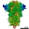
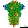
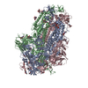
| Title | Cryo-EM structure of the SARS-CoV-2 wild-type S-Trimer from a subunit vaccine candidate | ||||||
 Components Components | Spike glycoprotein,Collagen alpha-1(I) chain | ||||||
 Keywords Keywords |  VIRAL PROTEIN / spike protein / COVID-19 / vaccine VIRAL PROTEIN / spike protein / COVID-19 / vaccine | ||||||
| Function / homology |  Function and homology information Function and homology informationEnhanced cleavage of VWF variant by ADAMTS13 / Defective VWF cleavage by ADAMTS13 variant / Defective VWF binding to collagen type I / cellular response to fluoride / collagen type I trimer / tooth mineralization / cellular response to vitamin E / collagen type IV trimer / bone trabecula formation / Anchoring fibril formation ...Enhanced cleavage of VWF variant by ADAMTS13 / Defective VWF cleavage by ADAMTS13 variant / Defective VWF binding to collagen type I / cellular response to fluoride / collagen type I trimer / tooth mineralization / cellular response to vitamin E / collagen type IV trimer / bone trabecula formation / Anchoring fibril formation / Enhanced binding of GP1BA variant to VWF multimer:collagen / Defective binding of VWF variant to GPIb:IX:V / Crosslinking of collagen fibrils / collagen biosynthetic process / Collagen chain trimerization / platelet-derived growth factor binding / extracellular matrix structural constituent conferring tensile strength / intramembranous ossification / Extracellular matrix organization / embryonic skeletal system development / Collagen biosynthesis and modifying enzymes / cartilage development involved in endochondral bone morphogenesis / skin morphogenesis / collagen-activated tyrosine kinase receptor signaling pathway / Platelet Adhesion to exposed collagen / endochondral ossification / cellular response to fibroblast growth factor stimulus / collagen fibril organization / negative regulation of cell-substrate adhesion / response to steroid hormone / face morphogenesis / Scavenging by Class A Receptors / skin development / MET activates PTK2 signaling / Assembly of collagen fibrils and other multimeric structures / Syndecan interactions / GP1b-IX-V activation signalling / blood vessel development / RUNX2 regulates osteoblast differentiation / Platelet Aggregation (Plug Formation) / Collagen degradation / protein localization to nucleus / Non-integrin membrane-ECM interactions / ECM proteoglycans / response to hyperoxia / Integrin cell surface interactions / positive regulation of epithelial to mesenchymal transition / response to mechanical stimulus / cellular response to retinoic acid / cellular response to epidermal growth factor stimulus / GPVI-mediated activation cascade / response to cAMP / cellular response to transforming growth factor beta stimulus / visual perception / extracellular matrix organization / ossification / secretory granule / skeletal system development / cellular response to glucose stimulus / Cell surface interactions at the vascular wall / cellular response to amino acid stimulus / sensory perception of sound / response to insulin / response to hydrogen peroxide / osteoblast differentiation / cellular response to mechanical stimulus / positive regulation of canonical Wnt signaling pathway / Immunoregulatory interactions between a Lymphoid and a non-Lymphoid cell / protein transport / response to estradiol / cellular response to tumor necrosis factor / Maturation of spike protein / collagen-containing extracellular matrix / viral translation / Translation of Structural Proteins / Virion Assembly and Release / host cell surface / host extracellular space / suppression by virus of host tetherin activity / Induction of Cell-Cell Fusion / protease binding / structural constituent of virion / entry receptor-mediated virion attachment to host cell / host cell endoplasmic reticulum-Golgi intermediate compartment membrane / receptor-mediated endocytosis of virus by host cell / Attachment and Entry / membrane fusion / positive regulation of viral entry into host cell / receptor-mediated virion attachment to host cell / receptor ligand activity / host cell surface receptor binding / positive regulation of cell migration / response to xenobiotic stimulus / fusion of virus membrane with host plasma membrane / endoplasmic reticulum lumen / fusion of virus membrane with host endosome membrane / viral envelope / virion attachment to host cell / SARS-CoV-2 activates/modulates innate and adaptive immune responses / host cell plasma membraneSimilarity search - Function | ||||||
| Biological species |   Severe acute respiratory syndrome coronavirus 2 Severe acute respiratory syndrome coronavirus 2 Homo sapiens (human) Homo sapiens (human) | ||||||
| Method | ELECTRON MICROSCOPY / single particle reconstruction / cryo EM / Resolution: 3.2 Å | ||||||
 Authors Authors | Zheng, S. / Ma, J. | ||||||
| Funding support |  China, 1items China, 1items
| ||||||
 Citation Citation | Journal: J Virol / Year: 2021 Title: Cryo-EM structure of S-Trimer, a subunit vaccine candidate for COVID-19. Authors: Jiahao Ma / Danmei Su / Yinyan Sun / Xueqin Huang / Ying Liang / Linqiang Fang / Yan Ma / Wenhui Li / Peng Liang / Sanduo Zheng / Abstract: Within a year after its emergence, the severe acute respiratory syndrome coronavirus 2 (SARS-CoV-2) has infected over 100 million people worldwide with a death toll over 2 million. Vaccination ...Within a year after its emergence, the severe acute respiratory syndrome coronavirus 2 (SARS-CoV-2) has infected over 100 million people worldwide with a death toll over 2 million. Vaccination remains the best hope to ultimately put this pandemic to an end. Here, using Trimer-Tag technology, we produced both wild-type (WT) and furin site mutant (MT) S-Trimers for COVID-19 vaccine studies. Cryo-EM structures of the WT and MT S-Trimers, determined at 3.2 Å and 2.6 Å respectively, revealed that both antigens adopt a tightly closed conformation and their structures are essentially identical to that of the previously solved full-length WT S protein in detergent. The tightly closed conformation is stabilized by fatty acid and polysorbate 80 binding at the receptor binding domains (RBDs) and the N terminal domains (NTDs) respectively. Additionally, we identified an important pH switch in the WT S-Trimer that shows dramatic conformational change and accounts for its increased stability at lower pH. These results validate Trimer-Tag as a platform technology in production of metastable WT S-Trimer as a candidate for COVID-19 subunit vaccine.Effective vaccine against SARS-CoV-2 is critical to end the COVID-19 pandemic. Here, using Trimer-Tag technology, we are able to produce stable and large quantities of WT S-Trimer, a subunit vaccine candidate for COVID-19 with high safety and efficacy from animal and Phase 1 clinical trial studies. Cryo-EM structures of the S-Trimer subunit vaccine candidate show that it predominately adopts tightly closed pre-fusion state, and resembles that of the native and full-length spike in detergent, confirming its structural integrity. WT S-Trimer is currently being evaluated in global Phase 2/3 clinical trial. Combining with published structures of the S protein, we also propose a model to dissect the conformation change of the spike protein before receptor binding. | ||||||
| History |
|
- Structure visualization
| Movie |
Movie viewer |
|---|---|
| Structure viewer | Molecule: MolmilJmol/JSmol |
- Downloads & links
-Download
| PDBx/mmCIF format | 7e7d.cif.gz | 598.4 KB | Display | PDBx/mmCIF format |
|---|---|---|---|---|
| PDB format | pdb7e7d.ent.gz | 491.2 KB | Display | PDB format |
| PDBx/mmJSON format | 7e7d.json.gz | Tree view | PDBx/mmJSON format | |
| Others |  Other downloads Other downloads |
-Validation report
| Arichive directory | https://data.pdbj.org/pub/pdb/validation_reports/e7/7e7dftp://data.pdbj.org/pub/pdb/validation_reports/e7/7e7d | HTTPS FTP |
|---|
-Related structure data
| Related structure data | 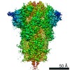 30999MC  7e7bC C: citing same article ( M: map data used to model this data |
|---|---|
| Similar structure data |


-Links
 PDBj
PDBj


- Assembly
| Deposited unit | 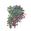
|
|---|---|
| 1 |
|
-Components
| #1: Protein | Mass: 168164.328 Da / Num. of mol.: 3 Source method: isolated from a genetically manipulated source Details: Chimeric protein Source: (gene. exp.) Severe acute respiratory syndrome coronavirus 2, (gene. exp.) Homo sapiens (human)Gene: S, 2, COL1A1 / Production host:   Cricetulus griseus (Chinese hamster) / References: UniProt: P0DTC2, UniProt: P02452 Cricetulus griseus (Chinese hamster) / References: UniProt: P0DTC2, UniProt: P02452#2: Polysaccharide | 2-acetamido-2-deoxy-beta-D-glucopyranose-(1-4)-2-acetamido-2-deoxy-beta-D-glucopyranose / Mass: 424.401 Da / Num. of mol.: 32 / Source method: obtained synthetically#3: Sugar | ChemComp-NAG / N-Acetylglucosamine  Type: D-saccharide, beta linking / Mass: 221.208 Da / Num. of mol.: 19 / Source method: obtained synthetically / Formula: C8H15NO6 Type: D-saccharide, beta linking / Mass: 221.208 Da / Num. of mol.: 19 / Source method: obtained synthetically / Formula: C8H15NO6#4: Chemical | Elaidic acid  Mass: 282.461 Da / Num. of mol.: 3 / Source method: obtained synthetically / Formula: C18H34O2 Mass: 282.461 Da / Num. of mol.: 3 / Source method: obtained synthetically / Formula: C18H34O2Has ligand of interest | N | |
|---|
-Experimental details
-Experiment
| Experiment | Method: ELECTRON MICROSCOPY |
|---|---|
| EM experiment | Aggregation state: PARTICLE / 3D reconstruction method: single particle reconstruction |
- Sample preparation
| Component | Name: SARS-CoV-2 spike protein fused to the C-terminal region of human type 1a collagen Type: COMPLEX / Entity ID: #1 / Source: RECOMBINANT | ||||||||||||
|---|---|---|---|---|---|---|---|---|---|---|---|---|---|
| Molecular weight | Experimental value: NO | ||||||||||||
| Source (natural) |
| ||||||||||||
| Source (recombinant) | Organism: Cricetulus griseus (Chinese hamster) | ||||||||||||
| Buffer solution | pH: 7.4 | ||||||||||||
| Specimen | Conc.: 0.3 mg/ml / Embedding applied: NO / Shadowing applied: NO / Staining applied: NO / Vitrification applied: YES / Details: monodisperse | ||||||||||||
| Specimen support | Grid type: Quantifoil R1.2/1.3 | ||||||||||||
| Vitrification | Instrument: FEI VITROBOT MARK I / Cryogen name: ETHANE / Humidity: 100 % / Chamber temperature: 282 K Details: blot time 2 seconds, blot force 4, waiting time 8 seconds |
- Electron microscopy imaging
| Experimental equipment |  Model: Titan Krios / Image courtesy: FEI Company |
|---|---|
| Microscopy | Model: TFS KRIOS |
| Electron gun | Electron source: FIELD EMISSION GUN / Accelerating voltage: 300 kV / Illumination mode: FLOOD BEAM |
| Electron lens | Mode: BRIGHT FIELDBright-field microscopy / Nominal magnification: 64000 X / Calibrated defocus min: 600 nm / Calibrated defocus max: 2800 nm / Cs: 0 mm / Alignment procedure: BASIC |
| Specimen holder | Cryogen: NITROGEN / Specimen holder model: FEI TITAN KRIOS AUTOGRID HOLDER |
| Image recording | Electron dose: 50 e/Å2 / Film or detector model: GATAN K3 BIOQUANTUM (6k x 4k) / Num. of grids imaged: 2 / Num. of real images: 2000 |
- Processing
| Software | Name: PHENIX / Version: 1.16_3549: / Classification: refinement | ||||||||||||||||||||||||||||||
|---|---|---|---|---|---|---|---|---|---|---|---|---|---|---|---|---|---|---|---|---|---|---|---|---|---|---|---|---|---|---|---|
| EM software |
| ||||||||||||||||||||||||||||||
| CTF correction | Type: PHASE FLIPPING AND AMPLITUDE CORRECTION | ||||||||||||||||||||||||||||||
| Particle selection | Num. of particles selected: 1504770 | ||||||||||||||||||||||||||||||
| 3D reconstruction | Resolution: 3.2 Å / Resolution method: FSC 0.143 CUT-OFF / Num. of particles: 173288 / Symmetry type: POINT | ||||||||||||||||||||||||||||||
| Atomic model building | Space: REAL | ||||||||||||||||||||||||||||||
| Atomic model building | PDB-ID: 6VXX | ||||||||||||||||||||||||||||||
| Refinement | Highest resolution: 3.2 Å | ||||||||||||||||||||||||||||||
| Refine LS restraints |
|

