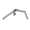
 Movie
Movie Controller

[English] 日本語
 Yorodumi
- PDB-6rko: Cryo-EM structure of the E. coli cytochrome bd-I oxidase at 2.68 ... -
+ Open data

- Basic information
| Entry | Database: PDB / ID: 6rko | ||||||
|---|---|---|---|---|---|---|---|
| Title | Cryo-EM structure of the E. coli cytochrome bd-I oxidase at 2.68 A resolution | ||||||
 Components Components |
| ||||||
 Keywords Keywords |  MEMBRANE PROTEIN / Oxidoreductase Cytochrome bd oxidase bd oxidase Oxidase MEMBRANE PROTEIN / Oxidoreductase Cytochrome bd oxidase bd oxidase Oxidase | ||||||
| Function / homology |  Function and homology information Function and homology informationquinol oxidase (electrogenic, proton-motive force generating) / oxidoreductase activity, acting on diphenols and related substances as donors / cytochrome complex / aerobic electron transport chain / outer membrane / oxidoreductase activity, acting on diphenols and related substances as donors, oxygen as acceptor / oxidative phosphorylation / cellular response to hypoxia / membrane => GO:0016020 / electron transfer activity ...quinol oxidase (electrogenic, proton-motive force generating) / oxidoreductase activity, acting on diphenols and related substances as donors / cytochrome complex / aerobic electron transport chain / outer membrane / oxidoreductase activity, acting on diphenols and related substances as donors, oxygen as acceptor / oxidative phosphorylation / cellular response to hypoxia / membrane => GO:0016020 / electron transfer activity / heme binding / membrane / metal ion binding / plasma membraneSimilarity search - Function | ||||||
| Biological species |  Escherichia coli (E. coli) Escherichia coli (E. coli) | ||||||
| Method | ELECTRON MICROSCOPY / single particle reconstruction / cryo EM / Resolution: 2.68 Å | ||||||
 Authors Authors | Safarian, S. / Hahn, A. / Kuehlbrandt, W. / Michel, H. | ||||||
| Funding support |  Germany, 1items Germany, 1items
| ||||||
 Citation Citation | Journal: Science / Year: 2019 Title: Active site rearrangement and structural divergence in prokaryotic respiratory oxidases. Authors: S Safarian / A Hahn / D J Mills / M Radloff / M L Eisinger / A Nikolaev / J Meier-Credo / F Melin / H Miyoshi / R B Gennis / J Sakamoto / J D Langer / P Hellwig / W Kühlbrandt / H Michel /    Abstract: Cytochrome bd-type quinol oxidases catalyze the reduction of molecular oxygen to water in the respiratory chain of many human-pathogenic bacteria. They are structurally unrelated to mitochondrial ...Cytochrome bd-type quinol oxidases catalyze the reduction of molecular oxygen to water in the respiratory chain of many human-pathogenic bacteria. They are structurally unrelated to mitochondrial cytochrome c oxidases and are therefore a prime target for the development of antimicrobial drugs. We determined the structure of the cytochrome bd-I oxidase by single-particle cryo-electron microscopy to a resolution of 2.7 angstroms. Our structure contains a previously unknown accessory subunit CydH, the L-subfamily-specific Q-loop domain, a structural ubiquinone-8 cofactor, an active-site density interpreted as dioxygen, distinct water-filled proton channels, and an oxygen-conducting pathway. Comparison with another cytochrome bd oxidase reveals structural divergence in the family, including rearrangement of high-spin hemes and conformational adaption of a transmembrane helix to generate a distinct oxygen-binding site. | ||||||
| History |
|
- Structure visualization
| Movie |
Movie viewer |
|---|---|
| Structure viewer | Molecule: MolmilJmol/JSmol |
- Downloads & links
-Download
| PDBx/mmCIF format | 6rko.cif.gz | 179.3 KB | Display | PDBx/mmCIF format |
|---|---|---|---|---|
| PDB format | pdb6rko.ent.gz | 137.3 KB | Display | PDB format |
| PDBx/mmJSON format | 6rko.json.gz | Tree view | PDBx/mmJSON format | |
| Others |  Other downloads Other downloads |
-Validation report
| Arichive directory | https://data.pdbj.org/pub/pdb/validation_reports/rk/6rkoftp://data.pdbj.org/pub/pdb/validation_reports/rk/6rko | HTTPS FTP |
|---|
-Related structure data
| Related structure data |  4908MC M: map data used to model this data C: citing same article ( |
|---|---|
| Similar structure data |


-Links
 PDBj
PDBj


- Assembly
| Deposited unit | 
|
|---|---|
| 1 |
|
-Components
-Cytochrome bd-I ubiquinol oxidase subunit ... , 3 types, 3 molecules BAX
| #1: Protein | Mass: 42479.828 Da / Num. of mol.: 1 Source method: isolated from a genetically manipulated source Source: (gene. exp.) Escherichia coli (strain K12) (bacteria)Strain: K12 / Gene: cydB, cyd-2, b0734, JW0723 / Production host: Escherichia coli (E. coli) / Strain (production host): C43 / Variant (production host): delta bo3References: UniProt: P0ABK2, quinol oxidase (electrogenic, proton-motive force generating) |
|---|---|
| #2: Protein | Mass: 58251.723 Da / Num. of mol.: 1 Source method: isolated from a genetically manipulated source Source: (gene. exp.) Escherichia coli (strain K12) (bacteria)Strain: K12 / Gene: cydA, cyd-1, b0733, JW0722 / Production host: Escherichia coli (E. coli) / Strain (production host): C43 / Variant (production host): delta bo3References: UniProt: P0ABJ9, quinol oxidase (electrogenic, proton-motive force generating) |
| #4: Protein/peptide | Mass: 4043.663 Da / Num. of mol.: 1 Source method: isolated from a genetically manipulated source Source: (gene. exp.) Escherichia coli (strain K12) (bacteria)Strain: K12 / Gene: cydX, ybgT, b4515, JW0724 / Production host: Escherichia coli (E. coli) / Strain (production host): C43 / Variant (production host): delta bo3References: UniProt: P56100, quinol oxidase (electrogenic, proton-motive force generating) |
-Protein/peptide , 1 types, 1 molecules H
| #3: Protein/peptide | Mass: 2999.651 Da / Num. of mol.: 1 Source method: isolated from a genetically manipulated source Source: (gene. exp.) Escherichia coli (strain K12) (bacteria)Strain: K12 / Gene: ynhF, b4602, JW1649.1 / Production host: Escherichia coli (E. coli) / Strain (production host): C43 / Variant (production host): delta bo3 / References: UniProt: A5A618 |
|---|
-Non-polymers , 6 types, 40 molecules 


| #5: Chemical | ChemComp-UQ8 /  Mass: 727.109 Da / Num. of mol.: 1 / Source method: obtained synthetically / Formula: C49H74O4 Mass: 727.109 Da / Num. of mol.: 1 / Source method: obtained synthetically / Formula: C49H74O4 | ||||||||
|---|---|---|---|---|---|---|---|---|---|
| #6: Chemical | ChemComp-POV / ( POPC Mass: 760.076 Da / Num. of mol.: 4 / Source method: obtained synthetically / Formula: C42H82NO8P / Comment: phospholipid*YM Mass: 760.076 Da / Num. of mol.: 4 / Source method: obtained synthetically / Formula: C42H82NO8P / Comment: phospholipid*YM#7: Chemical | ChemComp-HDD / | Heme Mass: 632.487 Da / Num. of mol.: 1 / Source method: obtained synthetically / Formula: C34H32FeN4O5 Mass: 632.487 Da / Num. of mol.: 1 / Source method: obtained synthetically / Formula: C34H32FeN4O5#8: Chemical |  Mass: 618.503 Da / Num. of mol.: 2 / Source method: obtained synthetically / Formula: C34H34FeN4O4 Mass: 618.503 Da / Num. of mol.: 2 / Source method: obtained synthetically / Formula: C34H34FeN4O4#9: Chemical | ChemComp-OXY / | Oxygen Mass: 31.999 Da / Num. of mol.: 1 / Source method: obtained synthetically / Formula: O2 Mass: 31.999 Da / Num. of mol.: 1 / Source method: obtained synthetically / Formula: O2#10: Water | ChemComp-HOH / | WaterMass: 18.015 Da / Num. of mol.: 31 / Source method: isolated from a natural source / Formula: H2O |
-Experimental details
-Experiment
| Experiment | Method: ELECTRON MICROSCOPY |
|---|---|
| EM experiment | Aggregation state: PARTICLE / 3D reconstruction method: single particle reconstruction |
- Sample preparation
| Component | Name: Cytochrome bd-I oxidase from E. coliUbiquinol oxidase (H+-transporting) Type: COMPLEX / Entity ID: #1-#4 / Source: RECOMBINANT | ||||||||||||
|---|---|---|---|---|---|---|---|---|---|---|---|---|---|
| Molecular weight | Value: 0.1077 MDa / Experimental value: NO | ||||||||||||
| Source (natural) | Organism: Escherichia coli K-12 (bacteria) | ||||||||||||
| Source (recombinant) | Organism: Escherichia coli (E. coli) / Strain: C43 / Plasmid: pet17b_cydABX-StrepII | ||||||||||||
| Buffer solution | pH: 7.5 | ||||||||||||
| Buffer component |
| ||||||||||||
| Specimen | Conc.: 7 mg/ml / Embedding applied: NO / Shadowing applied: NO / Staining applied: NO / Vitrification applied: YES | ||||||||||||
| Specimen support | Grid material: COPPER / Grid mesh size: 200 divisions/in. / Grid type: Quantifoil R1.2/1.3 | ||||||||||||
| Vitrification | Instrument: FEI VITROBOT MARK IV / Cryogen name: ETHANE / Humidity: 100 % / Chamber temperature: 277 K |
- Electron microscopy imaging
| Experimental equipment |  Model: Titan Krios / Image courtesy: FEI Company |
|---|---|
| Microscopy | Model: FEI TITAN KRIOS |
| Electron gun | Electron source: FIELD EMISSION GUN / Accelerating voltage: 300 kV / Illumination mode: FLOOD BEAM |
| Electron lens | Mode: BRIGHT FIELDBright-field microscopy / Nominal magnification: 96000 X / Calibrated magnification: 96000 X / Nominal defocus max: 2500 nm / Nominal defocus min: 500 nm / Calibrated defocus min: 500 nm / Calibrated defocus max: 2500 nm / Cs: 2.7 mm / C2 aperture diameter: 70 µm / Alignment procedure: COMA FREE |
| Specimen holder | Cryogen: NITROGEN / Specimen holder model: FEI TITAN KRIOS AUTOGRID HOLDER / Temperature (max): 70 K / Temperature (min): 70 K |
| Image recording | Average exposure time: 2 sec. / Electron dose: 40 e/Å2 / Detector mode: COUNTING / Film or detector model: FEI FALCON III (4k x 4k) / Num. of grids imaged: 1 / Num. of real images: 5463 |
- Processing
| Software | Name: PHENIX / Version: 1.15.2_3472: / Classification: refinement | ||||||||||||||||||||||||||||||||||||
|---|---|---|---|---|---|---|---|---|---|---|---|---|---|---|---|---|---|---|---|---|---|---|---|---|---|---|---|---|---|---|---|---|---|---|---|---|---|
| EM software |
| ||||||||||||||||||||||||||||||||||||
| CTF correction | Type: NONE | ||||||||||||||||||||||||||||||||||||
| Particle selection | Num. of particles selected: 3500000 | ||||||||||||||||||||||||||||||||||||
| Symmetry | Point symmetry: C1 (asymmetric) | ||||||||||||||||||||||||||||||||||||
| 3D reconstruction | Resolution: 2.68 Å / Resolution method: FSC 0.143 CUT-OFF / Num. of particles: 170000 / Algorithm: BACK PROJECTION / Num. of class averages: 3 / Symmetry type: POINT | ||||||||||||||||||||||||||||||||||||
| Atomic model building | B value: 120 / Protocol: AB INITIO MODEL / Space: REAL / Target criteria: Cross-correlation coefficient |
