 Movie
Movie Controller

[English] 日本語
 Yorodumi
- PDB-6g1k: Electron cryo-microscopy structure of the canonical TRPC4 ion channel -
+ Open data

- Basic information
| Entry | Database: PDB / ID: 6g1k | |||||||||
|---|---|---|---|---|---|---|---|---|---|---|
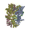
| Title | Electron cryo-microscopy structure of the canonical TRPC4 ion channel | |||||||||
 Components Components | Transient receptor potential cation channel subfamily c member 4a | |||||||||
 Keywords Keywords |  TRANSPORT PROTEIN / ION CHANNEL / TRPC4 / ANKYRIN REPEATS / CIRB DOMAIN / MEMBRANE PROTEIN TRANSPORT PROTEIN / ION CHANNEL / TRPC4 / ANKYRIN REPEATS / CIRB DOMAIN / MEMBRANE PROTEIN | |||||||||
| Function / homology |  Function and homology informationstore-operated calcium channel activity / cation channel complex / inositol 1,4,5 trisphosphate binding / monoatomic cation transport / monoatomic cation channel activity / regulation of cytosolic calcium ion concentration / calcium ion transmembrane transport / plasma membrane Function and homology informationstore-operated calcium channel activity / cation channel complex / inositol 1,4,5 trisphosphate binding / monoatomic cation transport / monoatomic cation channel activity / regulation of cytosolic calcium ion concentration / calcium ion transmembrane transport / plasma membraneSimilarity search - Function | |||||||||
| Biological species |  Danio rerio (zebrafish) Danio rerio (zebrafish) | |||||||||
| Method | ELECTRON MICROSCOPY / single particle reconstruction / cryo EM / Resolution: 3.6 Å | |||||||||
 Authors Authors | Vinayagam, D. / Mager, T. / Apelbaum, A. / Bothe, A. / Merino, F. / Hofnagel, O. / Gatsogiannis, C. / Raunser, S. | |||||||||
| Funding support |  Germany, 2items Germany, 2items
| |||||||||
 Citation Citation | Journal: Elife / Year: 2018 Title: Electron cryo-microscopy structure of the canonical TRPC4 ion channel. Authors: Deivanayagabarathy Vinayagam / Thomas Mager / Amir Apelbaum / Arne Bothe / Felipe Merino / Oliver Hofnagel / Christos Gatsogiannis / Stefan Raunser / Abstract: Canonical transient receptor channels (TRPC) are non-selective cation channels. They are involved in receptor-operated Ca signaling and have been proposed to act as store-operated channels (SOC). ...Canonical transient receptor channels (TRPC) are non-selective cation channels. They are involved in receptor-operated Ca signaling and have been proposed to act as store-operated channels (SOC). Their malfunction is related to cardiomyopathies and their modulation by small molecules has been shown to be effective against renal cancer cells. The molecular mechanism underlying the complex activation and regulation is poorly understood. Here, we report the electron cryo-microscopy structure of zebrafish TRPC4 in its unliganded (apo), closed state at an overall resolution of 3.6 Å. The structure reveals the molecular architecture of the cation conducting pore, including the selectivity filter and lower gate. The cytoplasmic domain contains two key hubs that have been shown to interact with modulating proteins. Structural comparisons with other TRP channels give novel insights into the general architecture and domain organization of this superfamily of channels and help to understand their function and pharmacology. | |||||||||
| History |
|
- Structure visualization
| Movie |
Movie viewer |
|---|---|
| Structure viewer | Molecule: MolmilJmol/JSmol |
- Downloads & links
-Download
| PDBx/mmCIF format | 6g1k.cif.gz | 475.2 KB | Display | PDBx/mmCIF format |
|---|---|---|---|---|
| PDB format | pdb6g1k.ent.gz | 395.1 KB | Display | PDB format |
| PDBx/mmJSON format | 6g1k.json.gz | Tree view | PDBx/mmJSON format | |
| Others |  Other downloads Other downloads |
-Validation report
| Arichive directory | https://data.pdbj.org/pub/pdb/validation_reports/g1/6g1kftp://data.pdbj.org/pub/pdb/validation_reports/g1/6g1k | HTTPS FTP |
|---|
-Related structure data
| Related structure data |  4339MC M: map data used to model this data C: citing same article ( |
|---|---|
| Similar structure data |


-Links
 PDBj
PDBj

- Assembly
| Deposited unit | 
|
|---|---|
| 1 |
|
-Components
| #1: Protein | Mass: 105934.500 Da / Num. of mol.: 4 Source method: isolated from a genetically manipulated source Source: (gene. exp.) Danio rerio (zebrafish) / Gene: trpc4b, trpc4a / Plasmid: pEG BacMam / Details (production host): mammalian expression vector / Cell line (production host): HEK293S GnTI- / Production host:  Homo sapiens (human) / Tissue (production host): embryonic kidney / References: UniProt: U3N7D8 Homo sapiens (human) / Tissue (production host): embryonic kidney / References: UniProt: U3N7D8#2: Chemical | ChemComp-Y01 /   Mass: 486.726 Da / Num. of mol.: 4 / Source method: obtained synthetically / Formula: C31H50O4 Mass: 486.726 Da / Num. of mol.: 4 / Source method: obtained synthetically / Formula: C31H50O4#3: Chemical | ChemComp-44E / (   Mass: 368.360 Da / Num. of mol.: 4 / Source method: obtained synthetically / Formula: C15H29O8P / Comment: phospholipid*YM Mass: 368.360 Da / Num. of mol.: 4 / Source method: obtained synthetically / Formula: C15H29O8P / Comment: phospholipid*YM |
|---|
-Experimental details
-Experiment
| Experiment | Method: ELECTRON MICROSCOPY |
|---|---|
| EM experiment | Aggregation state: PARTICLE / 3D reconstruction method: single particle reconstruction |
- Sample preparation
| Component | Name: TRPC4 / Type: ORGANELLE OR CELLULAR COMPONENT / Details: Membrane protein / Entity ID: #1 / Source: RECOMBINANT |
|---|---|
| Molecular weight | Value: 0.432 MDa / Experimental value: NO |
| Source (natural) | Organism: Danio rerio (zebrafish) |
| Source (recombinant) | Organism: Homo sapiens (human) / Cell: HEK293 GnTI- / Plasmid: pEG BacMam |
| Buffer solution | pH: 7.4 |
| Buffer component | Name: PBS |
| Specimen | Conc.: 0.25 mg/ml / Embedding applied: NO / Shadowing applied: NO / Staining applied: NO / Vitrification applied: YES / Details: The sample was monodisperse |
| Specimen support | Grid material: COPPER / Grid mesh size: 300 divisions/in. / Grid type: Quantifoil R1.2/1.3 |
| Vitrification | Instrument: FEI VITROBOT MARK III / Cryogen name: ETHANE / Humidity: 100 % / Chamber temperature: 277.15 K |
- Electron microscopy imaging
| Experimental equipment |  Model: Titan Krios / Image courtesy: FEI Company |
|---|---|
| Microscopy | Model: FEI TITAN KRIOS |
| Electron gun | Electron source: FIELD EMISSION GUN / Accelerating voltage: 300 kV / Illumination mode: OTHER |
| Electron lens | Mode: BRIGHT FIELDBright-field microscopy / Nominal defocus max: 2400 nm / Nominal defocus min: 1200 nm / Calibrated defocus min: 844 nm / Calibrated defocus max: 2931 nm |
| Specimen holder | Cryogen: NITROGEN / Specimen holder model: FEI TITAN KRIOS AUTOGRID HOLDER |
| Image recording | Average exposure time: 12 sec. / Electron dose: 70 e/Å2 / Detector mode: COUNTING / Film or detector model: GATAN K2 SUMMIT (4k x 4k) / Num. of grids imaged: 4 / Num. of real images: 3890 |
| EM imaging optics | Spherical aberration corrector: CS corrected Microscope |
- Processing
| EM software |
| |||||||||||||||||||||||||||||||||||||||||||||||||||||||
|---|---|---|---|---|---|---|---|---|---|---|---|---|---|---|---|---|---|---|---|---|---|---|---|---|---|---|---|---|---|---|---|---|---|---|---|---|---|---|---|---|---|---|---|---|---|---|---|---|---|---|---|---|---|---|---|---|
| CTF correction | Type: PHASE FLIPPING AND AMPLITUDE CORRECTION | |||||||||||||||||||||||||||||||||||||||||||||||||||||||
| Particle selection | Num. of particles selected: 426377 | |||||||||||||||||||||||||||||||||||||||||||||||||||||||
| Symmetry | Point symmetry: C4 (4 fold cyclic) | |||||||||||||||||||||||||||||||||||||||||||||||||||||||
| 3D reconstruction | Resolution: 3.6 Å / Resolution method: FSC 0.143 CUT-OFF / Num. of particles: 132622 / Algorithm: FOURIER SPACE / Num. of class averages: 6 / Symmetry type: POINT | |||||||||||||||||||||||||||||||||||||||||||||||||||||||
| Atomic model building | B value: 90.04 / Protocol: OTHER / Space: REAL / Target criteria: Real-space correlation | |||||||||||||||||||||||||||||||||||||||||||||||||||||||
| Atomic model building |
|


