 Movie
Movie Controller


+ Open data

- Basic information
| Entry | Database: PDB / ID: 4xnv | ||||||
|---|---|---|---|---|---|---|---|
| Title | The human P2Y1 receptor in complex with BPTU | ||||||
 Components Components | P2Y purinoceptor 1, Rubredoxin, P2Y purinoceptor 1 | ||||||
 Keywords Keywords | TRANSPORT PROTEIN/INHIBITOR / human P2Y1 receptor /  G protein coupled receptor / platelet activation / membrane protein / inhibitor complex / TRANSPORT PROTEIN-INHIBITOR complex / PSI-Biology / Structural Genomics / GPCR Network / GPCR G protein coupled receptor / platelet activation / membrane protein / inhibitor complex / TRANSPORT PROTEIN-INHIBITOR complex / PSI-Biology / Structural Genomics / GPCR Network / GPCR | ||||||
| Function / homology |  Function and homology information Function and homology informationG protein-coupled ATP receptor activity / relaxation of muscle / A1 adenosine receptor binding / G protein-coupled ADP receptor activity / P2Y receptors / cellular response to purine-containing compound / G protein-coupled purinergic nucleotide receptor activity / positive regulation of inositol trisphosphate biosynthetic process / positive regulation of monoatomic ion transport / positive regulation of penile erection ...G protein-coupled ATP receptor activity / relaxation of muscle / A1 adenosine receptor binding / G protein-coupled ADP receptor activity / P2Y receptors / cellular response to purine-containing compound / G protein-coupled purinergic nucleotide receptor activity / positive regulation of inositol trisphosphate biosynthetic process / positive regulation of monoatomic ion transport / positive regulation of penile erection / negative regulation of norepinephrine secretion / glial cell migration / regulation of presynaptic cytosolic calcium ion concentration / G protein-coupled adenosine receptor signaling pathway / positive regulation of hormone secretion / signal transduction involved in regulation of gene expression / response to growth factor / cellular response to ATP / regulation of synaptic vesicle exocytosis / eating behavior / monoatomic ion transport / presynaptic active zone membrane / response to mechanical stimulus / adenylate cyclase-inhibiting G protein-coupled receptor signaling pathway / blood vessel diameter maintenance / protein localization to plasma membrane / establishment of localization in cell / ADP binding / cilium / platelet activation / ADP signalling through P2Y purinoceptor 1 / signaling receptor activity / cell body / phospholipase C-activating G protein-coupled receptor signaling pathway / regulation of cell shape / positive regulation of cytosolic calcium ion concentration / scaffold protein binding / G alpha (q) signalling events / postsynaptic membrane / basolateral plasma membrane / postsynaptic density / electron transfer activity / cell surface receptor signaling pathway / positive regulation of ERK1 and ERK2 cascade / positive regulation of protein phosphorylation / iron ion binding / apical plasma membrane / G protein-coupled receptor signaling pathway / protein heterodimerization activity / glutamatergic synapse / dendrite / cell surface / positive regulation of transcription by RNA polymerase II / ATP binding / plasma membraneSimilarity search - Function | ||||||
| Biological species |  Homo sapiens (human) Homo sapiens (human) Clostridium pasteurianum (bacteria) Clostridium pasteurianum (bacteria) | ||||||
| Method | X-RAY DIFFRACTION / SYNCHROTRON / Resolution: 2.2 Å | ||||||
 Authors Authors | Zhang, D. / Gao, Z. / Jacobson, K. / Han, G.W. / Stevens, R. / Zhao, Q. / Wu, B. / GPCR Network (GPCR) | ||||||
 Citation Citation | Journal: Nature / Year: 2015 Title: Two disparate ligand-binding sites in the human P2Y1 receptor Authors: Zhang, D. / Gao, Z.G. / Zhang, K. / Kiselev, E. / Crane, S. / Wang, J. / Paoletta, S. / Yi, C. / Ma, L. / Zhang, W. / Han, G.W. / Liu, H. / Cherezov, V. / Katritch, V. / Jiang, H. / Stevens, ...Authors: Zhang, D. / Gao, Z.G. / Zhang, K. / Kiselev, E. / Crane, S. / Wang, J. / Paoletta, S. / Yi, C. / Ma, L. / Zhang, W. / Han, G.W. / Liu, H. / Cherezov, V. / Katritch, V. / Jiang, H. / Stevens, R.C. / Jacobson, K.A. / Zhao, Q. / Wu, B. | ||||||
| History |
|
- Structure visualization
| Structure viewer | Molecule: MolmilJmol/JSmol |
|---|
- Downloads & links
-Download
| PDBx/mmCIF format | 4xnv.cif.gz | 150.3 KB | Display | PDBx/mmCIF format |
|---|---|---|---|---|
| PDB format | pdb4xnv.ent.gz | 123.6 KB | Display | PDB format |
| PDBx/mmJSON format | 4xnv.json.gz | Tree view | PDBx/mmJSON format | |
| Others |  Other downloads Other downloads |
-Validation report
| Arichive directory | https://data.pdbj.org/pub/pdb/validation_reports/xn/4xnvftp://data.pdbj.org/pub/pdb/validation_reports/xn/4xnv | HTTPS FTP |
|---|
-Related structure data


-Links
 PDBj
PDBj


- Assembly
| Deposited unit | 
| ||||||||
|---|---|---|---|---|---|---|---|---|---|
| 1 |
| ||||||||
| Unit cell |
|
-Components
-Protein , 1 types, 1 molecules A
| #1: Protein | Mass: 47468.289 Da / Num. of mol.: 1 / Mutation: D320N Source method: isolated from a genetically manipulated source Details: Chimera protein of P2Y purinoceptor 1 (P2RY1_HUMA) with Rubredoxin (RUBR_CLOPA) inserted into ICL3 domain between residues 247LYS and 253PRO, and replaced residues 248ASN to 252SER. Source: (gene. exp.) Homo sapiens (human), (gene. exp.) Clostridium pasteurianum (bacteria)Gene: P2RY1 / Production host:  Spodoptera (butterflies/moths) / References: UniProt: P47900, UniProt: P00268 Spodoptera (butterflies/moths) / References: UniProt: P47900, UniProt: P00268 |
|---|
-Non-polymers , 7 types, 45 molecules 
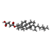


| #2: Chemical | ChemComp-BUR /  Mass: 445.434 Da / Num. of mol.: 1 / Source method: obtained synthetically / Formula: C23H22F3N3O3 Mass: 445.434 Da / Num. of mol.: 1 / Source method: obtained synthetically / Formula: C23H22F3N3O3 | ||||||||
|---|---|---|---|---|---|---|---|---|---|
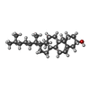
| #3: Chemical | ChemComp-CLR / Cholesterol Mass: 386.654 Da / Num. of mol.: 1 / Source method: obtained synthetically / Formula: C27H46O Mass: 386.654 Da / Num. of mol.: 1 / Source method: obtained synthetically / Formula: C27H46O | ||||||||
| #4: Chemical |  Mass: 486.726 Da / Num. of mol.: 3 / Source method: obtained synthetically / Formula: C31H50O4 Mass: 486.726 Da / Num. of mol.: 3 / Source method: obtained synthetically / Formula: C31H50O4#5: Chemical | ChemComp-OLC / (  Mass: 356.540 Da / Num. of mol.: 8 / Source method: obtained synthetically / Formula: C21H40O4 Mass: 356.540 Da / Num. of mol.: 8 / Source method: obtained synthetically / Formula: C21H40O4#6: Chemical | ChemComp-1PE / | Polyethylene glycol Mass: 238.278 Da / Num. of mol.: 1 / Source method: obtained synthetically / Formula: C10H22O6 / Comment: precipitant*YM Mass: 238.278 Da / Num. of mol.: 1 / Source method: obtained synthetically / Formula: C10H22O6 / Comment: precipitant*YM#7: Chemical | ChemComp-ZN / |  Mass: 65.409 Da / Num. of mol.: 1 / Source method: obtained synthetically / Formula: Zn Mass: 65.409 Da / Num. of mol.: 1 / Source method: obtained synthetically / Formula: Zn#8: Water | ChemComp-HOH / | WaterMass: 18.015 Da / Num. of mol.: 30 / Source method: isolated from a natural source / Formula: H2O |
-Experimental details
-Experiment
| Experiment | Method: X-RAY DIFFRACTION / Number of used crystals: 1 |
|---|
- Sample preparation
| Crystal | Density Matthews: 2.11 Å3/Da / Density % sol: 41.8 % |
|---|---|
| Crystal grow | Temperature: 293 K / Method: lipidic cubic phase / Details: PEG2000MME Sodium Citrate |
-Data collection
| Diffraction | Mean temperature: 100 K |
|---|---|
| Diffraction source | Source: SYNCHROTRON / Site: SPring-8  / Beamline: BL41XU / Wavelength: 1 Å / Beamline: BL41XU / Wavelength: 1 Å |
| Detector | Type: DECTRIS PILATUS 6M / Detector: PIXEL / Date: Jun 2, 2014 |
| Radiation | Protocol: SINGLE WAVELENGTH / Monochromatic (M) / Laue (L): M / Scattering type: x-ray |
| Radiation wavelength | Wavelength: 1 Å / Relative weight: 1 |
| Reflection | Resolution: 2.2→50 Å / Num. obs: 19881 / % possible obs: 99.9 % / Redundancy: 7.2 % / Biso Wilson estimate: 42.95 Å2 / Net I/σ(I): 12.1 |
- Processing
| Software |
| ||||||||||||||||||||||||||||||||||||||||||||||||||||||||||||||||||||||||||||||||||||||||||||||||||||||||||||||||||
|---|---|---|---|---|---|---|---|---|---|---|---|---|---|---|---|---|---|---|---|---|---|---|---|---|---|---|---|---|---|---|---|---|---|---|---|---|---|---|---|---|---|---|---|---|---|---|---|---|---|---|---|---|---|---|---|---|---|---|---|---|---|---|---|---|---|---|---|---|---|---|---|---|---|---|---|---|---|---|---|---|---|---|---|---|---|---|---|---|---|---|---|---|---|---|---|---|---|---|---|---|---|---|---|---|---|---|---|---|---|---|---|---|---|---|---|
| Refinement | Resolution: 2.2→41.4 Å / Cor.coef. Fo:Fc: 0.9412 / Cor.coef. Fo:Fc free: 0.9312 / SU R Cruickshank DPI: 0.293 / Cross valid method: THROUGHOUT / σ(F): 0 / SU R Blow DPI: 0.265 / SU Rfree Blow DPI: 0.19 / SU Rfree Cruickshank DPI: 0.2
| ||||||||||||||||||||||||||||||||||||||||||||||||||||||||||||||||||||||||||||||||||||||||||||||||||||||||||||||||||
| Displacement parameters | Biso mean: 63.8 Å2
| ||||||||||||||||||||||||||||||||||||||||||||||||||||||||||||||||||||||||||||||||||||||||||||||||||||||||||||||||||
| Refine analyze | Luzzati coordinate error obs: 0.369 Å | ||||||||||||||||||||||||||||||||||||||||||||||||||||||||||||||||||||||||||||||||||||||||||||||||||||||||||||||||||
| Refinement step | Cycle: LAST / Resolution: 2.2→41.4 Å
| ||||||||||||||||||||||||||||||||||||||||||||||||||||||||||||||||||||||||||||||||||||||||||||||||||||||||||||||||||
| Refine LS restraints |
| ||||||||||||||||||||||||||||||||||||||||||||||||||||||||||||||||||||||||||||||||||||||||||||||||||||||||||||||||||
| LS refinement shell | Resolution: 2.2→2.32 Å / Total num. of bins used: 10
| ||||||||||||||||||||||||||||||||||||||||||||||||||||||||||||||||||||||||||||||||||||||||||||||||||||||||||||||||||
| Refinement TLS params. | Method: refined / Origin x: -11.1074 Å / Origin y: 17.9445 Å / Origin z: 9.7233 Å
| ||||||||||||||||||||||||||||||||||||||||||||||||||||||||||||||||||||||||||||||||||||||||||||||||||||||||||||||||||
| Refinement TLS group | Selection details: { A|39 - A|334 } |
