[English] 日本語
Yorodumi
- PDB-4gfh: Topoisomerase II-DNA-AMPPNP complex -

+
Open data


ID or keywords:

Loading...

-
Basic information

Entry
Database: PDB / ID: 4gfh
TitleTopoisomerase II-DNA-AMPPNP complex
Components
  • DNA (5'-D(*CP*GP*CP*GP*AP*AP*TP*CP*GP*TP*CP*AP*TP*CP*C)-3')
  • DNA (5'-D(*CP*GP*CP*GP*GP*TP*AP*GP*CP*AP*GP*TP*AP*GP*G)-3')
  • DNA (5'-D(P*CP*CP*TP*AP*CP*TP*GP*CP*TP*AP*C)-3')
  • DNA (5'-D(P*GP*GP*AP*TP*GP*AP*CP*GP*AP*TP*(TSP))-3')
  • DNA topoisomerase 2Topoisomerase
KeywordsIsomerase/DNA / TOPOISOMERASE / PROTEIN-DNA COMPLEX / DNA SUPERCOILING / DNA REPLICATION / ATP-BINDING / DNA-BINDING / ISOMERASE / NUCLEOTIDE-BINDING / NUCLEUS / PHOSPHOPROTEIN / ISOMERASE-DNA COMPLEX
Function / homology
Function and homology information


replication fork progression beyond termination site / DNA replication termination region / chromatin remodeling at centromere / regulation of mitotic recombination / sister chromatid segregation / resolution of meiotic recombination intermediates / SUMOylation of DNA replication proteins / synaptonemal complex / reciprocal meiotic recombination / DNA topoisomerase type II (double strand cut, ATP-hydrolyzing) activity ...replication fork progression beyond termination site / DNA replication termination region / chromatin remodeling at centromere / regulation of mitotic recombination / sister chromatid segregation / resolution of meiotic recombination intermediates / SUMOylation of DNA replication proteins / synaptonemal complex / reciprocal meiotic recombination / DNA topoisomerase type II (double strand cut, ATP-hydrolyzing) activity / DNA topoisomerase (ATP-hydrolysing) / DNA strand elongation involved in DNA replication / rRNA transcription / DNA topological change / chromatin organization / mitochondrion / DNA binding / ATP binding / identical protein binding / metal ion binding / nucleus
Similarity search - Function
DNA topoisomerase 2, TOPRIM domain / DNA topoisomerase II, eukaryotic-type / C-terminal associated domain of TOPRIM / C-terminal associated domain of TOPRIM / Topoisomerase (Topo) IIA-type catalytic domain profile. / DNA topoisomerase, type IIA, alpha-helical domain superfamily / DNA topoisomerase, type IIA, domain A / DNA topoisomerase, type IIA, domain A, alpha-beta / DNA gyrase/topoisomerase IV, subunit A / DNA Topoisomerase IV ...DNA topoisomerase 2, TOPRIM domain / DNA topoisomerase II, eukaryotic-type / C-terminal associated domain of TOPRIM / C-terminal associated domain of TOPRIM / Topoisomerase (Topo) IIA-type catalytic domain profile. / DNA topoisomerase, type IIA, alpha-helical domain superfamily / DNA topoisomerase, type IIA, domain A / DNA topoisomerase, type IIA, domain A, alpha-beta / DNA gyrase/topoisomerase IV, subunit A / DNA Topoisomerase IV / DNA topoisomerase, type IIA, subunit B, domain 2 / DNA gyrase B / DNA topoisomerase, type IIA / DNA topoisomerase, type IIA, conserved site / DNA topoisomerase II signature. / TopoisomeraseII / DNA topoisomerase, type IIA, subunit B, C-terminal / Toprim domain / DNA topoisomerase, type IIA-like domain superfamily / Toprim domain profile. / TOPRIM domain / Histidine kinase-, DNA gyrase B-, and HSP90-like ATPase / Histidine kinase-like ATPases / Histidine kinase/HSP90-like ATPase / Histidine kinase/HSP90-like ATPase superfamily / Ribosomal protein S5 domain 2-type fold, subgroup / Ribosomal protein S5 domain 2-type fold
Similarity search - Domain/homology
PHOSPHOAMINOPHOSPHONIC ACID-ADENYLATE ESTER / DNA / DNA (> 10) / DNA topoisomerase 2
Similarity search - Component
Biological speciesSaccharomyces cerevisiae (brewer's yeast)
MethodX-RAY DIFFRACTION / SYNCHROTRON / MOLECULAR REPLACEMENT / Resolution: 4.408 Å
AuthorsSchmidt, B.H. / Osheroff, N. / Berger, J.M.
CitationJournal: Nat.Struct.Mol.Biol. / Year: 2012
Title: Structure of a topoisomerase II-DNA-nucleotide complex reveals a new control mechanism for ATPase activity.
Authors: Schmidt, B.H. / Osheroff, N. / Berger, J.M.
History
DepositionAug 3, 2012Deposition site: RCSB / Processing site: RCSB
Revision 1.0Oct 3, 2012Provider: repository / Type: Initial release
Revision 1.1Dec 12, 2012Group: Database references
Revision 1.2Jul 31, 2019Group: Data collection / Derived calculations / Category: chem_comp / struct_conn / Item: _chem_comp.type / _struct_conn.pdbx_leaving_atom_flag

-
Structure visualization

Structure viewerMolecule:
MolmilJmol/JSmol

Downloads & links

-
Assembly

Deposited unit
A: DNA topoisomerase 2
B: DNA (5'-D(P*CP*CP*TP*AP*CP*TP*GP*CP*TP*AP*C)-3')
C: DNA (5'-D(*CP*GP*CP*GP*GP*TP*AP*GP*CP*AP*GP*TP*AP*GP*G)-3')
D: DNA (5'-D(P*GP*GP*AP*TP*GP*AP*CP*GP*AP*TP*(TSP))-3')
E: DNA (5'-D(*CP*GP*CP*GP*AP*AP*TP*CP*GP*TP*CP*AP*TP*CP*C)-3')
F: DNA topoisomerase 2
G: DNA (5'-D(P*CP*CP*TP*AP*CP*TP*GP*CP*TP*AP*C)-3')
H: DNA (5'-D(*CP*GP*CP*GP*GP*TP*AP*GP*CP*AP*GP*TP*AP*GP*G)-3')
I: DNA (5'-D(P*GP*GP*AP*TP*GP*AP*CP*GP*AP*TP*(TSP))-3')
J: DNA (5'-D(*CP*GP*CP*GP*AP*AP*TP*CP*GP*TP*CP*AP*TP*CP*C)-3')
hetero molecules


Theoretical massNumber of molelcules
Total (without water)305,57814
Polymers304,51710
Non-polymers1,0614
Water0
1


  • Idetical with deposited unit
  • defined by author
TypeNameSymmetry operationNumber
identity operation1_555x,y,z1
Unit cell
Length a, b, c (Å)169.135, 169.884, 169.212
Angle α, β, γ (deg.)90.00, 90.00, 90.00
Int Tables number19
Space group name H-MP212121
DetailsAS PER THE AUTHORS THE BIOLOGICAL ASSEMBLY IS A DIMER

-
Components

-
Protein , 1 types, 2 molecules AF

#1: Protein DNA topoisomerase 2 / Topoisomerase / DNA topoisomerase II


Mass: 136355.125 Da / Num. of mol.: 2 / Fragment: UNP RESIDUES 1-1177
Source method: isolated from a genetically manipulated source
Source: (gene. exp.) Saccharomyces cerevisiae (brewer's yeast)
Strain: ATCC 204508 / S288c / Gene: TOP2, TOR3, YNL088W, N2244 / References: UniProt: P06786, EC: 5.99.1.3

-
DNA chain , 4 types, 8 molecules BGCHDIEJ

#2: DNA chain DNA (5'-D(P*CP*CP*TP*AP*CP*TP*GP*CP*TP*AP*C)-3')


Mass: 3269.149 Da / Num. of mol.: 2 / Source method: obtained synthetically
#3: DNA chain DNA (5'-D(*CP*GP*CP*GP*GP*TP*AP*GP*CP*AP*GP*TP*AP*GP*G)-3')


Mass: 4675.035 Da / Num. of mol.: 2 / Source method: obtained synthetically
#4: DNA chain DNA (5'-D(P*GP*GP*AP*TP*GP*AP*CP*GP*AP*TP*(TSP))-3')


Mass: 3429.312 Da / Num. of mol.: 2 / Source method: obtained synthetically
#5: DNA chain DNA (5'-D(*CP*GP*CP*GP*AP*AP*TP*CP*GP*TP*CP*AP*TP*CP*C)-3')


Mass: 4529.949 Da / Num. of mol.: 2 / Source method: obtained synthetically

-
Non-polymers , 2 types, 4 molecules

#6: Chemical ChemComp-MG / MAGNESIUM ION


Mass: 24.305 Da / Num. of mol.: 2 / Source method: obtained synthetically / Formula: Mg
#7: Chemical ChemComp-ANP / PHOSPHOAMINOPHOSPHONIC ACID-ADENYLATE ESTER


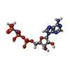
Mass: 506.196 Da / Num. of mol.: 2 / Source method: obtained synthetically / Formula: C10H17N6O12P3 / Comment: AMP-PNP, energy-carrying molecule analogue*YM

-
Experimental details

-
Experiment

ExperimentMethod: X-RAY DIFFRACTION / Number of used crystals: 1

-
Sample preparation

CrystalDensity Matthews: 3.99 Å3/Da / Density % sol: 69.18 %
Crystal growTemperature: 291 K / Method: vapor diffusion, hanging drop / pH: 8
Details: 23% PEG 300, 100 mM Tris (8.0), VAPOR DIFFUSION, HANGING DROP, temperature 291K

-
Data collection

DiffractionMean temperature: 100 K
Diffraction sourceSource: SYNCHROTRON / Site: ALS / Beamline: 8.3.1 / Wavelength: 1.1159 Å
DetectorType: ADSC QUANTUM 315r / Detector: CCD / Date: Aug 21, 2010
RadiationMonochromator: DOUBLE FLAT CRYSTAL, SI (111) / Protocol: SINGLE WAVELENGTH / Monochromatic (M) / Laue (L): M / Scattering type: x-ray
Radiation wavelengthWavelength: 1.1159 Å / Relative weight: 1
ReflectionResolution: 4.408→50 Å / Num. all: 28660 / Num. obs: 28660 / % possible obs: 91.7 % / Observed criterion σ(F): 0 / Observed criterion σ(I): 0
Reflection shellResolution: 4.408→4.57 Å / Redundancy: 4.2 % / Mean I/σ(I) obs: 2 / Rsym value: 0.703 / % possible all: 92.9

-
Processing

Software
NameVersionClassification
ADSCQuantumdata collection
PHASERphasing
PHENIX(phenix.refine: 1.7.3_928)refinement
HKL-2000data reduction
HKL-2000data scaling
RefinementMethod to determine structure: MOLECULAR REPLACEMENT / Resolution: 4.408→46.988 Å / SU ML: 0.54 / σ(F): 1.33 / Phase error: 30.39 / Stereochemistry target values: ML
RfactorNum. reflection% reflectionSelection details
Rfree0.2754 1450 5.06 %random
Rwork0.2391 ---
obs0.241 28660 91.22 %-
all-28660 --
Solvent computationShrinkage radii: 0.86 Å / VDW probe radii: 1.1 Å / Solvent model: FLAT BULK SOLVENT MODEL / Bsol: 187.65 Å2 / ksol: 0.312 e/Å3
Displacement parameters
Baniso -1Baniso -2Baniso -3
1--8.3384 Å2-0 Å20 Å2
2--2.357 Å2-0 Å2
3---5.9815 Å2
Refinement stepCycle: LAST / Resolution: 4.408→46.988 Å
ProteinNucleic acidLigandSolventTotal
Num. atoms18054 2118 64 0 20236
Refine LS restraints
Refine-IDTypeDev idealNumber
X-RAY DIFFRACTIONf_bond_d0.00420878
X-RAY DIFFRACTIONf_angle_d0.84228615
X-RAY DIFFRACTIONf_dihedral_angle_d15.7948059
X-RAY DIFFRACTIONf_chiral_restr0.0543120
X-RAY DIFFRACTIONf_plane_restr0.0043269
LS refinement shell
Resolution (Å)Rfactor RfreeNum. reflection RfreeRfactor RworkNum. reflection RworkRefine-ID% reflection obs (%)
4.408-4.56520.42171180.36962690X-RAY DIFFRACTION90
4.5652-4.74780.37391410.33092707X-RAY DIFFRACTION93
4.7478-4.96370.33031480.29662718X-RAY DIFFRACTION92
4.9637-5.22510.33131450.28172722X-RAY DIFFRACTION93
5.2251-5.55190.32671420.26552749X-RAY DIFFRACTION92
5.5519-5.97980.34731460.27192738X-RAY DIFFRACTION92
5.9798-6.58010.33711440.26722692X-RAY DIFFRACTION91
6.5801-7.52890.24621650.22312690X-RAY DIFFRACTION90
7.5289-9.47290.24391470.18062745X-RAY DIFFRACTION91
9.4729-46.9880.22211540.20742759X-RAY DIFFRACTION88
Refinement TLS params.

Method: refined / Refine-ID: X-RAY DIFFRACTION

IDL112)L122)L132)L222)L232)L332)S11 (Å °)S12 (Å °)S13 (Å °)S21 (Å °)S22 (Å °)S23 (Å °)S31 (Å °)S32 (Å °)S33 (Å °)T112)T122)T132)T222)T232)T332)Origin x (Å)Origin y (Å)Origin z (Å)
12.87330.8298-0.02913.152-0.79220.41720.2541-1.3744-0.36670.9153-0.5053-0.2056-0.42211.07660.18221.7641-0.1245-0.17741.78270.31290.99827.892821.619175.5921
25.38052.78330.98075.03893.33434.16160.521-0.2556-0.31240.1966-0.78260.4485-0.4555-0.20770.2522.0154-0.0320.36271.49870.12692.0684-5.312311.292450.8516
33.9642.04251.64973.02763.06314.5942-0.29810.43020.1071-1.06450.3945-0.0497-0.85250.9176-0.01791.861-0.1578-0.34792.06480.07531.9909-12.106129.1108-6.0183
42.57380.58351.4621.281.06432.95430.0752-0.41360.30220.2244-0.445-0.07830.22750.0530.43661.87670.0013-0.16571.5481-0.13871.7032-48.81643.06171.794
53.62532.58674.81042.78191.95098.5573-0.74690.31571.2027-0.6636-0.65680.56051.09090.17441.18751.50710.1149-0.25591.8246-0.03521.8533-22.892742.689115.0696
63.14951.29380.20685.2606-3.11382.64680.56651.1084-0.24821.84340.20311.96870.41210.7006-0.71862.2195-0.23510.22081.4538-0.17711.5332-24.029439.262316.5929
74.8443-3.12764.8932.8248-4.28776.29331.1707-0.22961.2729-0.4032-0.96580.65991.01621.4999-0.09491.7557-0.0889-0.34172.2403-0.01451.2271-27.1749-0.245519.4257
81.57092.17690.03733.21430.49732.4254-0.1550.16550.1724-0.79710.31290.2052-0.6133-1.08220.07982.26790.2553-0.08961.81440.20032.3839-25.69143.135218.2475
91.12250.7442-0.24613.2783-1.58941.92740.4277-1.1111-0.4625-0.1922-0.612-0.8802-0.11671.1460.19120.9798-0.2805-0.22821.7320.0861.82633.300220.892250.2072
103.6033-2.59591.20345.0811-1.68645.18740.1898-0.0651-0.4335-0.3233-0.4895-0.6295-0.77940.32280.55472.2196-0.17930.36051.40770.18412.00619.214231.346237.1597
113.2841-1.44381.76211.4558-1.95113.3358-0.1747-0.7443-0.50510.67640.38850.7421-0.1051-0.3267-0.252.31230.0921-0.38742.43160.07862.2724-48.295613.347730.1698
123.8392-1.63811.71191.2971-0.51523.20550.4004-0.09830.3988-0.0479-0.3988-0.42450.21910.38380.02931.56980.0776-0.1931.2787-0.04761.7945-40.4565-0.5927-6.5379
Refinement TLS group
IDRefine-IDRefine TLS-IDSelection details
1X-RAY DIFFRACTION1chain A and (resseq 7:240)
2X-RAY DIFFRACTION2chain A and (resseq 241:408)
3X-RAY DIFFRACTION3chain A and (resseq 421:690)
4X-RAY DIFFRACTION4chain A and (resseq 691:1177)
5X-RAY DIFFRACTION5chain B or chain D
6X-RAY DIFFRACTION6chain C or chain E
7X-RAY DIFFRACTION7chain G or chain I
8X-RAY DIFFRACTION8chain H or chain J
9X-RAY DIFFRACTION9chain F and (resseq 7:240)
10X-RAY DIFFRACTION10chain F and (resseq 241:404)
11X-RAY DIFFRACTION11chain F and (resseq 421:690)
12X-RAY DIFFRACTION12chain F and (resseq 691:1177)

+
About Yorodumi

-
News

-
Feb 9, 2022. New format data for meta-information of EMDB entries

New format data for meta-information of EMDB entries

  • Version 3 of the EMDB header file is now the official format.
  • The previous official version 1.9 will be removed from the archive.

Related info.:EMDB header

External links:wwPDB to switch to version 3 of the EMDB data model

-
Aug 12, 2020. Covid-19 info

Covid-19 info

URL: https://pdbj.org/emnavi/covid19.php

New page: Covid-19 featured information page in EM Navigator.

Related info.:Covid-19 info / Mar 5, 2020. Novel coronavirus structure data

+
Mar 5, 2020. Novel coronavirus structure data

Novel coronavirus structure data

Related info.:Yorodumi Speices / Aug 12, 2020. Covid-19 info

External links:COVID-19 featured content - PDBj / Molecule of the Month (242):Coronavirus Proteases

+
Jan 31, 2019. EMDB accession codes are about to change! (news from PDBe EMDB page)

EMDB accession codes are about to change! (news from PDBe EMDB page)

  • The allocation of 4 digits for EMDB accession codes will soon come to an end. Whilst these codes will remain in use, new EMDB accession codes will include an additional digit and will expand incrementally as the available range of codes is exhausted. The current 4-digit format prefixed with “EMD-” (i.e. EMD-XXXX) will advance to a 5-digit format (i.e. EMD-XXXXX), and so on. It is currently estimated that the 4-digit codes will be depleted around Spring 2019, at which point the 5-digit format will come into force.
  • The EM Navigator/Yorodumi systems omit the EMD- prefix.

Related info.:Q: What is EMD? / ID/Accession-code notation in Yorodumi/EM Navigator

External links:EMDB Accession Codes are Changing Soon! / Contact to PDBj

+
Jul 12, 2017. Major update of PDB

Major update of PDB

  • wwPDB released updated PDB data conforming to the new PDBx/mmCIF dictionary.
  • This is a major update changing the version number from 4 to 5, and with Remediation, in which all the entries are updated.
  • In this update, many items about electron microscopy experimental information are reorganized (e.g. em_software).
  • Now, EM Navigator and Yorodumi are based on the updated data.

External links:wwPDB Remediation / Enriched Model Files Conforming to OneDep Data Standards Now Available in the PDB FTP Archive

-
Yorodumi

Thousand views of thousand structures

  • Yorodumi is a browser for structure data from EMDB, PDB, SASBDB, etc.
  • This page is also the successor to EM Navigator detail page, and also detail information page/front-end page for Omokage search.
  • The word "yorodu" (or yorozu) is an old Japanese word meaning "ten thousand". "mi" (miru) is to see.

Related info.:EMDB / PDB / SASBDB / Comparison of 3 databanks / Yorodumi Search / Aug 31, 2016. New EM Navigator & Yorodumi / Yorodumi Papers / Jmol/JSmol / Function and homology information / Changes in new EM Navigator and Yorodumi

Read more