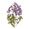
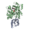
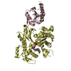
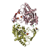
[English] 日本語
Yorodumi
- PDB-2pav: Ternary complex of Profilin-Actin with the Last Poly-Pro of Human VASP -

+
Open data


ID or keywords:

Loading...

-
Basic information

Entry
Database: PDB / ID: 2pav
TitleTernary complex of Profilin-Actin with the Last Poly-Pro of Human VASP
Components
  • Actin, alpha skeletal muscle
  • Profilin-1
  • Vasodilator-stimulated phosphoprotein
KeywordsSTRUCTURAL PROTEIN / Ternary complex / Profilin / Actin / VASP / Poly-Proline / Loading Poly-Pro Site
Function / homology
Function and homology information


synapse maturation / modification of postsynaptic actin cytoskeleton / negative regulation of actin filament bundle assembly / adenyl-nucleotide exchange factor activity / profilin binding / positive regulation of actin filament bundle assembly / negative regulation of actin filament polymerization / Signaling by ROBO receptors / Cell-extracellular matrix interactions / regulation of actin filament polymerization ...synapse maturation / modification of postsynaptic actin cytoskeleton / negative regulation of actin filament bundle assembly / adenyl-nucleotide exchange factor activity / profilin binding / positive regulation of actin filament bundle assembly / negative regulation of actin filament polymerization / Signaling by ROBO receptors / Cell-extracellular matrix interactions / regulation of actin filament polymerization / actin polymerization or depolymerization / positive regulation of ATP-dependent activity / PCP/CE pathway / filopodium membrane / proline-rich region binding / positive regulation of ruffle assembly / negative regulation of stress fiber assembly / cytoskeletal motor activator activity / tropomyosin binding / myosin heavy chain binding / positive regulation of actin filament polymerization / mesenchyme migration / positive regulation of epithelial cell migration / troponin I binding / actin filament bundle / filamentous actin / skeletal muscle thin filament assembly / actin filament bundle assembly / lamellipodium membrane / striated muscle thin filament / skeletal muscle myofibril / actin monomer binding / Generation of second messenger molecules / bicellular tight junction / skeletal muscle fiber development / stress fiber / titin binding / phosphatidylinositol-4,5-bisphosphate binding / actin filament polymerization / phosphotyrosine residue binding / filopodium / neural tube closure / actin filament / axon guidance / RHO GTPases Activate Formins / Hydrolases; Acting on acid anhydrides; Acting on acid anhydrides to facilitate cellular and subcellular movement / modulation of chemical synaptic transmission / small GTPase binding / SH3 domain binding / calcium-dependent protein binding / actin cytoskeleton / Platelet degranulation / lamellipodium / cell body / actin binding / cell cortex / actin cytoskeleton organization / protein homotetramerization / blood microparticle / protein stabilization / cytoskeleton / hydrolase activity / cadherin binding / protein domain specific binding / focal adhesion / glutamatergic synapse / calcium ion binding / positive regulation of gene expression / regulation of transcription by RNA polymerase II / magnesium ion binding / RNA binding / extracellular exosome / ATP binding / membrane / identical protein binding / nucleus / plasma membrane / cytosol / cytoplasm
Similarity search - Function
Vasodilator-stimulated phosphoprotein/ENA/VASP-like / Profilin1/2/3, vertebrate / VASP tetramerisation / VASP tetramerisation domain superfamily / VASP tetramerisation domain / : / Profilin conserved site / Profilin signature. / Profilin / WH1/EVH1 domain ...Vasodilator-stimulated phosphoprotein/ENA/VASP-like / Profilin1/2/3, vertebrate / VASP tetramerisation / VASP tetramerisation domain superfamily / VASP tetramerisation domain / : / Profilin conserved site / Profilin signature. / Profilin / WH1/EVH1 domain / WH1 domain / WH1 domain profile. / WASP homology region 1 / Profilin / Profilin / Profilin superfamily / Dynein light chain 2a, cytoplasmic / ATPase, substrate binding domain, subdomain 4 / Actin; Chain A, domain 4 / ATPase, nucleotide binding domain / Actins signature 1. / Actin, conserved site / Actins signature 2. / Actin/actin-like conserved site / Actins and actin-related proteins signature. / Beta-Lactamase / Actin / Actin family / Actin / ATPase, nucleotide binding domain / Nucleotidyltransferase; domain 5 / PH-like domain superfamily / Alpha-Beta Complex / 2-Layer Sandwich / Alpha Beta
Similarity search - Domain/homology
ADENOSINE-5'-TRIPHOSPHATE / Profilin-1 / Vasodilator-stimulated phosphoprotein / Actin, alpha skeletal muscle
Similarity search - Component
Biological speciesHomo sapiens (human)
Oryctolagus cuniculus (rabbit)
MethodX-RAY DIFFRACTION / SYNCHROTRON / MOLECULAR REPLACEMENT / Resolution: 1.8 Å
AuthorsFerron, F. / Rebowski, G. / Dominguez, R.
CitationJournal: Embo J. / Year: 2007
Title: Structural basis for the recruitment of profilin-actin complexes during filament elongation by Ena/VASP.
Authors: Ferron, F. / Rebowski, G. / Lee, S.H. / Dominguez, R.
History
DepositionMar 27, 2007Deposition site: RCSB / Processing site: RCSB
Revision 1.0Oct 23, 2007Provider: repository / Type: Initial release
Revision 1.1Jul 13, 2011Group: Version format compliance
Revision 1.2Mar 6, 2013Group: Advisory / Other
Revision 1.3Aug 30, 2023Group: Data collection / Database references ...Data collection / Database references / Derived calculations / Refinement description
Category: chem_comp_atom / chem_comp_bond ...chem_comp_atom / chem_comp_bond / database_2 / pdbx_initial_refinement_model / pdbx_struct_conn_angle / struct_conn / struct_conn_type / struct_ref_seq_dif / struct_sheet / struct_site
Item: _database_2.pdbx_DOI / _database_2.pdbx_database_accession ..._database_2.pdbx_DOI / _database_2.pdbx_database_accession / _pdbx_struct_conn_angle.ptnr1_auth_seq_id / _pdbx_struct_conn_angle.ptnr3_auth_seq_id / _pdbx_struct_conn_angle.value / _struct_conn.conn_type_id / _struct_conn.id / _struct_conn.pdbx_dist_value / _struct_conn.pdbx_leaving_atom_flag / _struct_conn.ptnr1_auth_comp_id / _struct_conn.ptnr1_auth_seq_id / _struct_conn.ptnr1_label_asym_id / _struct_conn.ptnr1_label_atom_id / _struct_conn.ptnr1_label_comp_id / _struct_conn.ptnr1_label_seq_id / _struct_conn.ptnr2_auth_comp_id / _struct_conn.ptnr2_auth_seq_id / _struct_conn.ptnr2_label_asym_id / _struct_conn.ptnr2_label_atom_id / _struct_conn.ptnr2_label_comp_id / _struct_conn.ptnr2_label_seq_id / _struct_conn_type.id / _struct_ref_seq_dif.details / _struct_sheet.number_strands / _struct_site.pdbx_auth_asym_id / _struct_site.pdbx_auth_comp_id / _struct_site.pdbx_auth_seq_id

-
Structure visualization

Structure viewerMolecule:
MolmilJmol/JSmol

Downloads & links

-
Assembly

Deposited unit
A: Actin, alpha skeletal muscle
P: Profilin-1
V: Vasodilator-stimulated phosphoprotein
hetero molecules


Theoretical massNumber of molelcules
Total (without water)58,7175
Polymers58,1703
Non-polymers5472
Water9,764542
1


  • Idetical with deposited unit
  • defined by author
TypeNameSymmetry operationNumber
identity operation1_555x,y,z1
Unit cell
Length a, b, c (Å)37.467, 75.460, 180.741
Angle α, β, γ (deg.)90.00, 90.00, 90.00
Int Tables number19
Space group name H-MP212121

-
Components

-
Protein , 2 types, 2 molecules AP

#1: Protein Actin, alpha skeletal muscle / / Alpha-actin-1


Mass: 41875.633 Da / Num. of mol.: 1 / Source method: isolated from a natural source / Details: alpha skeletal muscle / Source: (natural) Oryctolagus cuniculus (rabbit) / References: UniProt: P68135
#2: Protein Profilin-1 / / Profilin I


Mass: 14940.021 Da / Num. of mol.: 1
Source method: isolated from a genetically manipulated source
Source: (gene. exp.) Homo sapiens (human) / Gene: PFN1 / Plasmid: pET29 / Species (production host): Escherichia coli / Production host: Escherichia coli BL21(DE3) (bacteria) / Strain (production host): BL21(DE3) / References: UniProt: P07737

-
Protein/peptide , 1 types, 1 molecules V

#3: Protein/peptide Vasodilator-stimulated phosphoprotein / / VASP


Mass: 1354.509 Da / Num. of mol.: 1 / Source method: obtained synthetically / Details: This sequence occurs naturally in humans / References: UniProt: P50552

-
Non-polymers , 3 types, 544 molecules

#4: Chemical ChemComp-CA / CALCIUM ION


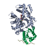
Mass: 40.078 Da / Num. of mol.: 1 / Source method: obtained synthetically / Formula: Ca
#5: Chemical ChemComp-ATP / ADENOSINE-5'-TRIPHOSPHATE / Adenosine triphosphate


Mass: 507.181 Da / Num. of mol.: 1 / Source method: obtained synthetically / Formula: C10H16N5O13P3 / Comment: ATP, energy-carrying molecule*YM
#6: Water ChemComp-HOH / water / Water


Mass: 18.015 Da / Num. of mol.: 542 / Source method: isolated from a natural source / Formula: H2O

-
Experimental details

-
Experiment

ExperimentMethod: X-RAY DIFFRACTION / Number of used crystals: 1

-
Sample preparation

CrystalDensity Matthews: 2.19 Å3/Da / Density % sol: 43.94 %
Crystal growTemperature: 277.15 K / Method: vapor diffusion, sitting drop / pH: 7.4
Details: 200 mM sodium formate, 20% PEG 3350, pH 7.4, VAPOR DIFFUSION, SITTING DROP, temperature 277.15K

-
Data collection

DiffractionMean temperature: 200 K
Diffraction sourceSource: SYNCHROTRON / Site: APS / Beamline: 14-BM-C / Wavelength: 0.9002 Å
DetectorType: ADSC QUANTUM 315 / Detector: CCD / Date: Dec 19, 2005
RadiationMonochromator: Bent conical Si-mirror (Rh coated) / Protocol: SINGLE WAVELENGTH / Monochromatic (M) / Laue (L): M / Scattering type: x-ray
Radiation wavelengthWavelength: 0.9002 Å / Relative weight: 1
ReflectionResolution: 1.8→50 Å / Num. all: 47947 / Num. obs: 47947 / % possible obs: 98.1 % / Observed criterion σ(F): 0 / Observed criterion σ(I): 0 / Redundancy: 7 % / Biso Wilson estimate: 15 Å2 / Rmerge(I) obs: 0.076 / Rsym value: 0.076 / Net I/σ(I): 28.1
Reflection shellResolution: 1.8→1.86 Å / Redundancy: 5.9 % / Rmerge(I) obs: 0.345 / Mean I/σ(I) obs: 5.7 / Num. unique all: 4691 / Rsym value: 0.345 / % possible all: 97.7

-
Processing

Software
NameVersionClassificationNB
REFMAC5.2.0019refinement
PDB_EXTRACT2data extraction
SCALEPACKdata scaling
AMoREphasing
RefinementMethod to determine structure: MOLECULAR REPLACEMENT
Starting model: PDB ENTRY 2BTF
Resolution: 1.8→47.09 Å / Cor.coef. Fo:Fc: 0.966 / Cor.coef. Fo:Fc free: 0.945 / SU B: 4.503 / SU ML: 0.076 / TLS residual ADP flag: LIKELY RESIDUAL / Cross valid method: THROUGHOUT / σ(F): 0 / ESU R: 0.124 / ESU R Free: 0.124 / Stereochemistry target values: MAXIMUM LIKELIHOOD / Details: HYDROGENS HAVE BEEN ADDED IN THE RIDING POSITIONS
RfactorNum. reflection% reflectionSelection details
Rfree0.208 2401 5 %RANDOM
Rwork0.158 ---
all0.16 45338 --
obs0.16 47739 98.14 %-
Solvent computationIon probe radii: 0.8 Å / Shrinkage radii: 0.8 Å / VDW probe radii: 1.4 Å / Solvent model: MASK
Displacement parametersBiso mean: 15.002 Å2
Baniso -1Baniso -2Baniso -3
1-0.09 Å20 Å20 Å2
2---0.1 Å20 Å2
3---0 Å2
Refinement stepCycle: LAST / Resolution: 1.8→47.09 Å
ProteinNucleic acidLigandSolventTotal
Num. atoms3968 0 32 542 4542
Refine LS restraints
Refine-IDTypeDev idealDev ideal targetNumber
X-RAY DIFFRACTIONr_bond_refined_d0.0190.0224351
X-RAY DIFFRACTIONr_angle_refined_deg1.6631.9825969
X-RAY DIFFRACTIONr_dihedral_angle_1_deg5.925588
X-RAY DIFFRACTIONr_dihedral_angle_2_deg35.53224.153183
X-RAY DIFFRACTIONr_dihedral_angle_3_deg14.16915761
X-RAY DIFFRACTIONr_dihedral_angle_4_deg14.341527
X-RAY DIFFRACTIONr_chiral_restr0.1870.2669
X-RAY DIFFRACTIONr_gen_planes_refined0.0080.023321
X-RAY DIFFRACTIONr_nbd_refined0.210.22262
X-RAY DIFFRACTIONr_nbtor_refined0.3120.23016
X-RAY DIFFRACTIONr_xyhbond_nbd_refined0.1970.2458
X-RAY DIFFRACTIONr_metal_ion_refined0.0360.24
X-RAY DIFFRACTIONr_symmetry_vdw_refined0.2480.240
X-RAY DIFFRACTIONr_symmetry_hbond_refined0.2040.231
X-RAY DIFFRACTIONr_mcbond_it1.0961.52784
X-RAY DIFFRACTIONr_mcangle_it1.6124413
X-RAY DIFFRACTIONr_scbond_it2.7331798
X-RAY DIFFRACTIONr_scangle_it4.094.51520
LS refinement shellResolution: 1.8→1.847 Å / Total num. of bins used: 20
RfactorNum. reflection% reflection
Rfree0.243 170 -
Rwork0.189 3273 -
obs-3443 97.51 %
Refinement TLS params.

Method: refined / Refine-ID: X-RAY DIFFRACTION

IDL112)L122)L132)L222)L232)L332)S11 (Å °)S12 (Å °)S13 (Å °)S21 (Å °)S22 (Å °)S23 (Å °)S31 (Å °)S32 (Å °)S33 (Å °)T112)T122)T132)T222)T232)T332)Origin x (Å)Origin y (Å)Origin z (Å)
10.2876-0.0216-0.10050.54430.04850.8485-0.03230.027-0.02580.02060.01560.05220.1288-0.04970.0167-0.1012-0.00870.0025-0.04190.0021-0.0514.2318.43966.404
21.13490.22460.33512.5944-0.43571.70960.0657-0.03920.02470.3214-0.0577-0.0437-0.29220.0758-0.0081-0.0411-0.0240.0106-0.0771-0.0165-0.090216.09649.0477.82
38.7444-3.10235.067711.5629-6.90285.4278-0.30690.24190.76670.2084-0.4350.5532-0.99420.11930.74180.0002-0.0021-0.0004-0.00050.0020.000620.36263.03369.215
Refinement TLS group

Refine-ID: X-RAY DIFFRACTION / Selection: ALL

IDRefine TLS-IDAuth asym-IDLabel asym-IDAuth seq-IDLabel seq-ID
11AA5 - 325 - 32
21AA33 - 7033 - 70
31AA71 - 13071 - 130
41AA131 - 148131 - 148
51AA149 - 181149 - 181
61AA182 - 262182 - 262
71AA263 - 274263 - 274
81AA275 - 336275 - 336
91AA337 - 375337 - 375
102PB1 - 151 - 15
112PB16 - 3816 - 38
122PB39 - 9239 - 92
132PB93 - 11693 - 116
142PB117 - 139117 - 139
153VC201 - 2064 - 9
163VC207 - 21310 - 16

+
About Yorodumi

-
News

-
Feb 9, 2022. New format data for meta-information of EMDB entries

New format data for meta-information of EMDB entries

  • Version 3 of the EMDB header file is now the official format.
  • The previous official version 1.9 will be removed from the archive.

Related info.:EMDB header

External links:wwPDB to switch to version 3 of the EMDB data model

-
Aug 12, 2020. Covid-19 info

Covid-19 info

URL: https://pdbj.org/emnavi/covid19.php

New page: Covid-19 featured information page in EM Navigator.

Related info.:Covid-19 info / Mar 5, 2020. Novel coronavirus structure data

+
Mar 5, 2020. Novel coronavirus structure data

Novel coronavirus structure data

Related info.:Yorodumi Speices / Aug 12, 2020. Covid-19 info

External links:COVID-19 featured content - PDBj / Molecule of the Month (242):Coronavirus Proteases

+
Jan 31, 2019. EMDB accession codes are about to change! (news from PDBe EMDB page)

EMDB accession codes are about to change! (news from PDBe EMDB page)

  • The allocation of 4 digits for EMDB accession codes will soon come to an end. Whilst these codes will remain in use, new EMDB accession codes will include an additional digit and will expand incrementally as the available range of codes is exhausted. The current 4-digit format prefixed with “EMD-” (i.e. EMD-XXXX) will advance to a 5-digit format (i.e. EMD-XXXXX), and so on. It is currently estimated that the 4-digit codes will be depleted around Spring 2019, at which point the 5-digit format will come into force.
  • The EM Navigator/Yorodumi systems omit the EMD- prefix.

Related info.:Q: What is EMD? / ID/Accession-code notation in Yorodumi/EM Navigator

External links:EMDB Accession Codes are Changing Soon! / Contact to PDBj

+
Jul 12, 2017. Major update of PDB

Major update of PDB

  • wwPDB released updated PDB data conforming to the new PDBx/mmCIF dictionary.
  • This is a major update changing the version number from 4 to 5, and with Remediation, in which all the entries are updated.
  • In this update, many items about electron microscopy experimental information are reorganized (e.g. em_software).
  • Now, EM Navigator and Yorodumi are based on the updated data.

External links:wwPDB Remediation / Enriched Model Files Conforming to OneDep Data Standards Now Available in the PDB FTP Archive

-
Yorodumi

Thousand views of thousand structures

  • Yorodumi is a browser for structure data from EMDB, PDB, SASBDB, etc.
  • This page is also the successor to EM Navigator detail page, and also detail information page/front-end page for Omokage search.
  • The word "yorodu" (or yorozu) is an old Japanese word meaning "ten thousand". "mi" (miru) is to see.

Related info.:EMDB / PDB / SASBDB / Comparison of 3 databanks / Yorodumi Search / Aug 31, 2016. New EM Navigator & Yorodumi / Yorodumi Papers / Jmol/JSmol / Function and homology information / Changes in new EM Navigator and Yorodumi

Read more