 Movie
Movie Controller


+ Open data

- Basic information
| Entry | Database: PDB / ID: 1q45 | ||||||
|---|---|---|---|---|---|---|---|
| Title | 12-0xo-phytodienoate reductase isoform 3 | ||||||
 Components Components | 12-oxophytodienoate-10,11-reductase | ||||||
 Keywords Keywords |  OXIDOREDUCTASE / Flavoprotein / flavoenzyme / xenobiotic reductase / old yellow enzyme / secondary messenger / Structural Genomics / PSI / Protein Structure Initiative / Center for Eukaryotic Structural Genomics / CESG OXIDOREDUCTASE / Flavoprotein / flavoenzyme / xenobiotic reductase / old yellow enzyme / secondary messenger / Structural Genomics / PSI / Protein Structure Initiative / Center for Eukaryotic Structural Genomics / CESG | ||||||
| Function / homology |  Function and homology information Function and homology informationstamen development / 12-oxophytodienoate reductase / 12-oxophytodienoate reductase activity / jasmonic acid biosynthetic process / oxylipin biosynthetic process / response to ozone / response to fungus / peroxisome / FMN bindingSimilarity search - Function | ||||||
| Biological species |  Arabidopsis thaliana (thale cress) Arabidopsis thaliana (thale cress) | ||||||
| Method | X-RAY DIFFRACTION / SYNCHROTRON / MOLECULAR REPLACEMENT / Resolution: 2 Å | ||||||
 Authors Authors | Phillips Jr., G.N. / Johnson, K.A. / Bingman, C.A. / Smith, D.W. / Center for Eukaryotic Structural Genomics (CESG) | ||||||
 Citation Citation | Journal: Proteins / Year: 2005 Title: X-ray structure of Arabidopsis At2g06050, 12-oxophytodienoate reductase isoform 3 Authors: Malone, T.E. / Madson, S.E. / Wrobel, R.L. / Jeon, W.B. / Rosenberg, N.S. / Johnson, K.A. / Bingman, C.A. / Smith, D.W. / Phillips Jr., G.N. / Markley, J.L. / Fox, B.G. | ||||||
| History |
|
- Structure visualization
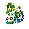
| Structure viewer | Molecule: MolmilJmol/JSmol |
|---|
- Downloads & links
-Download
| PDBx/mmCIF format | 1q45.cif.gz | 160.3 KB | Display | PDBx/mmCIF format |
|---|---|---|---|---|
| PDB format | pdb1q45.ent.gz | 126.1 KB | Display | PDB format |
| PDBx/mmJSON format | 1q45.json.gz | Tree view | PDBx/mmJSON format | |
| Others |  Other downloads Other downloads |
-Validation report
| Arichive directory | https://data.pdbj.org/pub/pdb/validation_reports/q4/1q45ftp://data.pdbj.org/pub/pdb/validation_reports/q4/1q45 | HTTPS FTP |
|---|
-Related structure data
| Related structure data | |
|---|---|
| Similar structure data | |
| Other databases |


-Links
 PDBj
PDBj- Assembly
| Deposited unit | 
| ||||||||||
|---|---|---|---|---|---|---|---|---|---|---|---|
| 1 | 
| ||||||||||
| 2 | 
| ||||||||||
| Unit cell |
|
-Components
| #1: Protein | Mass: 42741.160 Da / Num. of mol.: 2 Source method: isolated from a genetically manipulated source Source: (gene. exp.) Arabidopsis thaliana (thale cress) / Gene: At2g06050 / Plasmid: PVP13 / Production host:  Escherichia coli (E. coli) / Strain (production host): Rosetta / References: UniProt: Q9FUP0, 12-oxophytodienoate reductase Escherichia coli (E. coli) / Strain (production host): Rosetta / References: UniProt: Q9FUP0, 12-oxophytodienoate reductase#2: Chemical | Flavin mononucleotide  Mass: 456.344 Da / Num. of mol.: 2 / Source method: obtained synthetically / Formula: C17H21N4O9P Mass: 456.344 Da / Num. of mol.: 2 / Source method: obtained synthetically / Formula: C17H21N4O9P#3: Water | ChemComp-HOH / | Water Mass: 18.015 Da / Num. of mol.: 410 / Source method: isolated from a natural source / Formula: H2O Mass: 18.015 Da / Num. of mol.: 410 / Source method: isolated from a natural source / Formula: H2O |
|---|
-Experimental details
-Experiment
| Experiment | Method: X-RAY DIFFRACTION / Number of used crystals: 1 |
|---|
- Sample preparation
| Crystal | Density Matthews: 2.37 Å3/Da / Density % sol: 47.6 % | ||||||||||||||||||||||||||||||||||||||||||||||||||||||||
|---|---|---|---|---|---|---|---|---|---|---|---|---|---|---|---|---|---|---|---|---|---|---|---|---|---|---|---|---|---|---|---|---|---|---|---|---|---|---|---|---|---|---|---|---|---|---|---|---|---|---|---|---|---|---|---|---|---|
| Crystal grow | Temperature: 297 K / Method: vapor diffusion, hanging drop / pH: 7.5 Details: Glycine, MEPEG 5000, triethanolamine, pH 7.5, VAPOR DIFFUSION, HANGING DROP, temperature 297K | ||||||||||||||||||||||||||||||||||||||||||||||||||||||||
| Crystal grow | *PLUS pH: 6 / Method: vapor diffusion, hanging drop | ||||||||||||||||||||||||||||||||||||||||||||||||||||||||
| Components of the solutions | *PLUS
|
-Data collection
| Diffraction | Mean temperature: 90 K |
|---|---|
| Diffraction source | Source: SYNCHROTRON / Site: APS  / Beamline: 14-ID-B / Wavelength: 0.979 Å / Beamline: 14-ID-B / Wavelength: 0.979 Å |
| Detector | Type: MARRESEARCH / Detector: CCD / Date: Jun 20, 2003 / Details: Bent cylindrical Si-mirror (Rh coating) |
| Radiation | Monochromator: Diamond (111) double-crystal monochromator / Protocol: SINGLE WAVELENGTH / Monochromatic (M) / Laue (L): M / Scattering type: x-ray |
| Radiation wavelength | Wavelength: 0.979 Å / Relative weight: 1 |
| Reflection | Resolution: 2→25 Å / Num. all: 55478 / Num. obs: 50628 / % possible obs: 90.5 % / Observed criterion σ(I): -3 / Redundancy: 6.09 % / Biso Wilson estimate: 23.3 Å2 / Rmerge(I) obs: 0.133 / Rsym value: 0.133 / Net I/σ(I): 5.7 |
| Reflection shell | Resolution: 2→2.07 Å / Redundancy: 0.39 % / Rmerge(I) obs: 0.687 / Mean I/σ(I) obs: 1.11 / Num. unique all: 2384 / Rsym value: 0.687 / % possible all: 43.6 |
| Reflection | *PLUS Highest resolution: 2 Å / Lowest resolution: 24.92 Å / Num. obs: 48056 / % possible obs: 91.3 % |
| Reflection shell | *PLUS Highest resolution: 2 Å / % possible obs: 43.6 % |
- Processing
| Software |
| ||||||||||||||||||||||||||||||||||||||||||||||||||||||||||||||||||||||||||||||||||||||||||||||||||||
|---|---|---|---|---|---|---|---|---|---|---|---|---|---|---|---|---|---|---|---|---|---|---|---|---|---|---|---|---|---|---|---|---|---|---|---|---|---|---|---|---|---|---|---|---|---|---|---|---|---|---|---|---|---|---|---|---|---|---|---|---|---|---|---|---|---|---|---|---|---|---|---|---|---|---|---|---|---|---|---|---|---|---|---|---|---|---|---|---|---|---|---|---|---|---|---|---|---|---|---|---|---|
| Refinement | Method to determine structure: MOLECULAR REPLACEMENT / Resolution: 2→24.92 Å / Cor.coef. Fo:Fc: 0.947 / Cor.coef. Fo:Fc free: 0.913 / SU B: 4.88 / SU ML: 0.128 / Cross valid method: THROUGHOUT / σ(F): 0 / σ(I): 0 / ESU R: 0.199 / ESU R Free: 0.176 / Stereochemistry target values: MAXIMUM LIKELIHOOD / Details: HYDROGENS HAVE BEEN ADDED IN THE RIDING POSITIONS
| ||||||||||||||||||||||||||||||||||||||||||||||||||||||||||||||||||||||||||||||||||||||||||||||||||||
| Solvent computation | Ion probe radii: 0.8 Å / Shrinkage radii: 0.8 Å / VDW probe radii: 1.4 Å / Solvent model: BABINET MODEL WITH MASK | ||||||||||||||||||||||||||||||||||||||||||||||||||||||||||||||||||||||||||||||||||||||||||||||||||||
| Displacement parameters | Biso mean: 27.896 Å2
| ||||||||||||||||||||||||||||||||||||||||||||||||||||||||||||||||||||||||||||||||||||||||||||||||||||
| Refinement step | Cycle: LAST / Resolution: 2→24.92 Å
| ||||||||||||||||||||||||||||||||||||||||||||||||||||||||||||||||||||||||||||||||||||||||||||||||||||
| Refine LS restraints |
| ||||||||||||||||||||||||||||||||||||||||||||||||||||||||||||||||||||||||||||||||||||||||||||||||||||
| LS refinement shell | Resolution: 2→2.07 Å / Total num. of bins used: 20
| ||||||||||||||||||||||||||||||||||||||||||||||||||||||||||||||||||||||||||||||||||||||||||||||||||||
| Refinement | *PLUS Highest resolution: 2 Å / % reflection Rfree: 5 % / Rfactor Rfree: 0.236 / Rfactor Rwork: 0.186 | ||||||||||||||||||||||||||||||||||||||||||||||||||||||||||||||||||||||||||||||||||||||||||||||||||||
| Solvent computation | *PLUS | ||||||||||||||||||||||||||||||||||||||||||||||||||||||||||||||||||||||||||||||||||||||||||||||||||||
| Displacement parameters | *PLUS | ||||||||||||||||||||||||||||||||||||||||||||||||||||||||||||||||||||||||||||||||||||||||||||||||||||
| Refine LS restraints | *PLUS
|
