 Movie
Movie Controller

[English] 日本語
 Yorodumi
- PDB-1i24: HIGH RESOLUTION CRYSTAL STRUCTURE OF THE WILD-TYPE PROTEIN SQD1, ... -
+ Open data

- Basic information
| Entry | Database: PDB / ID: 1i24 | ||||||
|---|---|---|---|---|---|---|---|
| Title | HIGH RESOLUTION CRYSTAL STRUCTURE OF THE WILD-TYPE PROTEIN SQD1, WITH NAD AND UDP-GLUCOSE | ||||||
 Components Components | SULFOLIPID BIOSYNTHESIS PROTEIN SQD1 | ||||||
 Keywords Keywords |  BIOSYNTHETIC PROTEIN / SDR / SHORT-CHAIN DEHYDROGENASE/REDUCTASE / ROSSMANN FOLD BIOSYNTHETIC PROTEIN / SDR / SHORT-CHAIN DEHYDROGENASE/REDUCTASE / ROSSMANN FOLD | ||||||
| Function / homology |  Function and homology informationUDP-sulfoquinovose synthase / UDPsulfoquinovose synthase activity / glycolipid biosynthetic process / sulfotransferase activity / cellular response to phosphate starvation / chloroplast / zinc ion binding / plasma membrane Function and homology informationUDP-sulfoquinovose synthase / UDPsulfoquinovose synthase activity / glycolipid biosynthetic process / sulfotransferase activity / cellular response to phosphate starvation / chloroplast / zinc ion binding / plasma membraneSimilarity search - Function | ||||||
| Biological species |  Arabidopsis thaliana (thale cress) Arabidopsis thaliana (thale cress) | ||||||
| Method | X-RAY DIFFRACTION / SYNCHROTRON / MOLECULAR REPLACEMENT / Resolution: 1.2 Å | ||||||
 Authors Authors | Theisen, M.J. / Sanda, S.L. / Ginell, S.L. / Benning, C. / Garavito, R.M. | ||||||
 Citation Citation | Journal: To be Published Title: Characterization of the Active Site of Udp-Sulfoquinovose Synthase: Formation of the Sulfonic Acid Product in the Crystalline State Authors: Theisen, M.J. / Sanda, S.L. / Ginell, S.L. / Benning, C. / Garavito, R.M. | ||||||
| History |
|
- Structure visualization
| Structure viewer | Molecule: MolmilJmol/JSmol |
|---|
- Downloads & links
-Download
| PDBx/mmCIF format | 1i24.cif.gz | 106.2 KB | Display | PDBx/mmCIF format |
|---|---|---|---|---|
| PDB format | pdb1i24.ent.gz | 78.9 KB | Display | PDB format |
| PDBx/mmJSON format | 1i24.json.gz | Tree view | PDBx/mmJSON format | |
| Others |  Other downloads Other downloads |
-Validation report
| Arichive directory | https://data.pdbj.org/pub/pdb/validation_reports/i2/1i24ftp://data.pdbj.org/pub/pdb/validation_reports/i2/1i24 | HTTPS FTP |
|---|
-Related structure data
| Related structure data |  1qrrS S: Starting model for refinement |
|---|---|
| Similar structure data |


-Links
 PDBj
PDBj
- Assembly
| Deposited unit | 
| |||||||||
|---|---|---|---|---|---|---|---|---|---|---|
| 1 | 
| |||||||||
| 2 | 
| |||||||||
| Unit cell |
| |||||||||
| Components on special symmetry positions |
|
-Components
| #1: Protein | Mass: 45509.531 Da / Num. of mol.: 1 Fragment: RECOMBINANT SQD1, LACKING N-TERMINAL PUTATIVE CHLOROPLAST TARGETTING SEQUENCE, AND WITH N-TERMINAL 6X HISTIDINE TAG Source method: isolated from a genetically manipulated source Source: (gene. exp.) Arabidopsis thaliana (thale cress) / Strain: COLUMBIA (COL-2) / Gene: SQD1 / Plasmid: PSQD1 / Production host:  Escherichia coli (E. coli) / References: UniProt: O48917 Escherichia coli (E. coli) / References: UniProt: O48917 | ||||||
|---|---|---|---|---|---|---|---|
| #2: Chemical | Sulfate  Mass: 96.063 Da / Num. of mol.: 2 / Source method: obtained synthetically / Formula: SO4 Mass: 96.063 Da / Num. of mol.: 2 / Source method: obtained synthetically / Formula: SO4#3: Chemical | ChemComp-NAD / | Nicotinamide adenine dinucleotide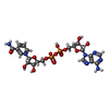  Mass: 663.425 Da / Num. of mol.: 1 / Source method: obtained synthetically / Formula: C21H27N7O14P2 / Comment: NAD*YM Mass: 663.425 Da / Num. of mol.: 1 / Source method: obtained synthetically / Formula: C21H27N7O14P2 / Comment: NAD*YM#4: Chemical | ChemComp-UPG / | Uridine diphosphate glucose  Mass: 566.302 Da / Num. of mol.: 1 / Source method: obtained synthetically / Formula: C15H24N2O17P2 Mass: 566.302 Da / Num. of mol.: 1 / Source method: obtained synthetically / Formula: C15H24N2O17P2#5: Water | ChemComp-HOH / | Water Mass: 18.015 Da / Num. of mol.: 386 / Source method: isolated from a natural source / Formula: H2O Mass: 18.015 Da / Num. of mol.: 386 / Source method: isolated from a natural source / Formula: H2O |
-Experimental details
-Experiment
| Experiment | Method: X-RAY DIFFRACTION / Number of used crystals: 1 |
|---|
- Sample preparation
| Crystal | Density Matthews: 3.5 Å3/Da / Density % sol: 65 % |
|---|---|
| Crystal grow | Temperature: 293 K / Method: vapor diffusion, sitting drop / pH: 6.5 Details: ammonium sulfate, sodium chloride, MES buffer, NAD+, UDP-glucose, pH 6.5, VAPOR DIFFUSION, SITTING DROP, temperature 293K |
-Data collection
| Diffraction | Mean temperature: 100 K |
|---|---|
| Diffraction source | Source: SYNCHROTRON / Site: APS  / Beamline: 19-ID / Wavelength: 1.03321 / Beamline: 19-ID / Wavelength: 1.03321 |
| Detector | Type: CUSTOM-MADE / Detector: CCD / Date: Aug 23, 1999 |
| Radiation | Monochromator: SAGITALLY FOCUSING SI (111) DOUBLE CRYSTAL MONOCHROMATOR AND VERTICALLY FOCUSING MIRROR Protocol: SINGLE WAVELENGTH / Monochromatic (M) / Laue (L): M / Scattering type: x-ray |
| Radiation wavelength | Wavelength: 1.03321 Å / Relative weight: 1 |
| Reflection | Resolution: 1.14→99 Å / Num. obs: 196326 / % possible obs: 86.4 % / Observed criterion σ(I): -3 / Redundancy: 5.9 % / Biso Wilson estimate: 11.59 Å2 / Rmerge(I) obs: 0.097 / Net I/σ(I): 21.4 |
| Reflection shell | Resolution: 1.14→1.19 Å / Redundancy: 1.4 % / Rmerge(I) obs: 0.366 / Mean I/σ(I) obs: 1.7 / % possible all: 38 |
- Processing
| Software |
| ||||||||||||||||||||||||||||||||||||||||||||||||||||||||||||
|---|---|---|---|---|---|---|---|---|---|---|---|---|---|---|---|---|---|---|---|---|---|---|---|---|---|---|---|---|---|---|---|---|---|---|---|---|---|---|---|---|---|---|---|---|---|---|---|---|---|---|---|---|---|---|---|---|---|---|---|---|---|
| Refinement | Method to determine structure: MOLECULAR REPLACEMENT Starting model: PDB ENTRY 1QRR Resolution: 1.2→20 Å / Isotropic thermal model: ISOTROPIC / Cross valid method: THROUGHOUT / σ(F): 0 / Stereochemistry target values: ENGH & HUBER
| ||||||||||||||||||||||||||||||||||||||||||||||||||||||||||||
| Solvent computation | Bsol: 68.07 Å2 / ksol: 0.439 e/Å3 | ||||||||||||||||||||||||||||||||||||||||||||||||||||||||||||
| Displacement parameters | Biso mean: 13.43 Å2 | ||||||||||||||||||||||||||||||||||||||||||||||||||||||||||||
| Refine analyze |
| ||||||||||||||||||||||||||||||||||||||||||||||||||||||||||||
| Refinement step | Cycle: LAST / Resolution: 1.2→20 Å
| ||||||||||||||||||||||||||||||||||||||||||||||||||||||||||||
| Refine LS restraints |
| ||||||||||||||||||||||||||||||||||||||||||||||||||||||||||||
| LS refinement shell | Resolution: 1.2→1.24 Å / Total num. of bins used: 10
|
